


A prospective study on application of human umbilical cord mesenchymal stem cells combined with autologous Meek microskin transplantation in patients with extensive burns
-
摘要:
目的 探讨人脐带间充质干细胞(hUCMSC)联合自体Meek微型皮片移植对大面积烧伤患者的效果。 方法 采用前瞻性自身对照研究方法。2019年5月—2022年6月,解放军联勤保障部队第九九〇医院收治的16例大面积烧伤患者符合入选标准,按照剔除标准剔除3例患者,最终纳入13例患者,其中男10例、女3例,年龄24~61(42±13)岁。共选择受试区20个(创面40个,大小均为10 cm×10 cm)。采用随机数字表法将每个受试区中的2个相邻创面分为涂抹含hUCMSC透明质酸凝胶的hUCMSC+凝胶组及涂抹单纯透明质酸凝胶的单纯凝胶组,每组20个创面,随后2组创面均移植扩展比例为1∶6的自体Meek微型皮片。术后2、3、4周,观察创面愈合情况并计算创面愈合率,记录创面愈合时间。术后,若创面出现脓性分泌物,则采集创面分泌物标本行微生物培养。术后3、6、12个月,采用温哥华瘢痕量表评估创面瘢痕增生情况。术后3个月,取创面组织,行苏木精-伊红(HE)染色观察皮肤组织形态学变化,行免疫组织化学染色观察Ki67阳性和波形蛋白阳性表达情况并统计阳性细胞数。对数据行配对样本t检验及Bonferroni校正。 结果 术后2、3、4周,hUCMSC+凝胶组创面愈合率分别为(80±11)%、(84±12)%、(92±9)%,均明显高于单纯凝胶组的(67±18)%、(74±21)%、(84±16)%(t值分别为4.01、3.52、3.66,P<0.05)。hUCMSC+凝胶组创面愈合时间为(31±11)d,较单纯凝胶组的(36±13)d明显缩短(t=-3.68,P<0.05)。术后,相邻的2组创面分泌物标本微生物培养情况均相同,4个受试区结果为阴性,16个受试区结果为阳性。术后3、6、12个月,单纯凝胶组创面温哥华瘢痕量表评分分别为(7.8±1.9)、(6.7±2.1)、(5.4±1.6)分,均明显高于hUCMSC+凝胶组的(6.8±1.8)、(5.6±1.6)、(4.0±1.4)分(t值分别为-4.79、-4.37、-5.47,P<0.05)。术后3个月,HE染色显示,hUCMSC+凝胶组创面表皮层厚度、表皮嵴等较单纯凝胶组增加;免疫组织化学染色显示,hUCMSC+凝胶组创面Ki67阳性细胞数较单纯凝胶组明显增加(t=4.39,P<0.05),2组创面波形蛋白阳性细胞数无明显差异(P>0.05)。 结论 将含hUCMSC的透明质酸凝胶涂抹于创面的应用方式操作方便,是一种较佳的给药途径。hUCMSC外用可促进大面积烧伤患者自体Meek微型皮片移植术区愈合,缩短创面愈合时间,减轻瘢痕增生,可能与创面表皮层厚度、表皮嵴增加和细胞增殖活跃有关。 Abstract:Objective To investigate the effects of human umbilical cord mesenchymal stem cells (hUCMSCs) combined with autologous Meek microskin transplantation on patients with extensive burns. Methods The prospective self-controlled study was conducted. From May 2019 to June 2022, 16 patients with extensive burns admitted to the 990th Hospital of PLA Joint Logistics Support Force met the inclusion criteria, while 3 patients were excluded according to the exclusion criteria, and 13 patients were finally selected, including 10 males and 3 females, aged 24-61 (42±13) years. A total of 20 trial areas (40 wounds, with area of 10 cm×10 cm in each wound) were selected. Two adjacent wounds in each trial area were divided into hUCMSC+gel group applied with hyaluronic acid gel containing hUCMSCs and gel only group applied with hyaluronic acid gel only according to the random number table, with 20 wounds in each group. Afterwards the wounds in two groups were transplanted with autologous Meek microskin grafts with an extension ratio of 1∶6. In 2, 3, and 4 weeks post operation, the wound healing was observed, the wound healing rate was calculated, and the wound healing time was recorded. The specimen of wound secretion was collected for microorganism culture if there was purulent secretion on the wound post operation. In 3, 6, and 12 months post operation, the scar hyperplasia in wound was assessed using the Vancouver scar scale (VSS). In 3 months post operation, the wound tissue was collected for hematoxylin-eosin (HE) staining to observe the morphological changes and for immunohistochemical staining to observe the positive expressions of Ki67 and vimentin and to count the number of positive cells. Data were statistically analyzed with paired samples t test and Bonferronni correction. Results In 2, 3, and 4 weeks post operation, the wound healing rates in hUCMSC+gel group were (80±11)%, (84±12)%, and (92±9)%, respectively, which were significantly higher than (67±18)%, (74±21)%, and (84±16)% in gel only group (with t values of 4.01, 3.52, and 3.66, respectively, P<0.05). The wound healing time in hUCMSC+gel group was (31±11) d, which was significantly shorter than (36±13) d in gel only group (t=-3.68, P<0.05). The microbiological culture of the postoperative wound secretion specimens from the adjacent wounds in 2 groups was identical, with negative results in 4 trial areas and positive results in 16 trial areas. In 3, 6, and 12 months post operation, the VSS scores of wounds in gel only group were 7.8±1.9, 6.7±2.1, and 5.4±1.6, which were significantly higher than 6.8±1.8, 5.6±1.6, and 4.0±1.4 in hUCMSC+gel group, respectively (with t values of -4.79, -4.37, and -5.47, respectively, P<0.05). In 3 months post operation, HE staining showed an increase in epidermal layer thickness and epidermal crest in wound in hUCMSC+gel group compared with those in gel only group, and immunohistochemical staining showed a significant increase in the number of Ki67 positive cells in wound in hUCMSC+gel group compared with those in gel only group (t=4.39, P<0.05), with no statistically significant difference in the number of vimentin positive cells in wound between the 2 groups (P>0.05). Conclusions The application of hyaluronic acid gel containing hUCMSCs to the wound is simple to perform and is therefore a preferable route. Topical application of hUCMSCs can promote healing of the autologous Meek microskin grafted area in patients with extensive burns, shorten wound healing time, and alleviate scar hyperplasia. The above effects may be related to the increased epidermal thickness and epidermal crest, and active cell proliferation. -
Key words:
- Burns /
- Cicatrix /
- Mesenchymal stem cell transplantation /
- Skin transplantation /
- Meek microskin
-

1 大面积烧伤患者2组创面处理。1A.划定包含2组相邻创面(大小均为10 cm×10 cm)的受试区;1B.hUCMSC+凝胶组涂抹含hUCMSC(细胞总数为3×10⁷个)的透明质酸凝胶,单纯凝胶组涂抹单纯透明质酸凝胶;1C.2组创面均移植扩展比例为1∶6的自体Meek微型皮片
注:标记“+”为人脐带间充质干细胞(hUCMSC)+凝胶组,标记“-”为单纯凝胶组

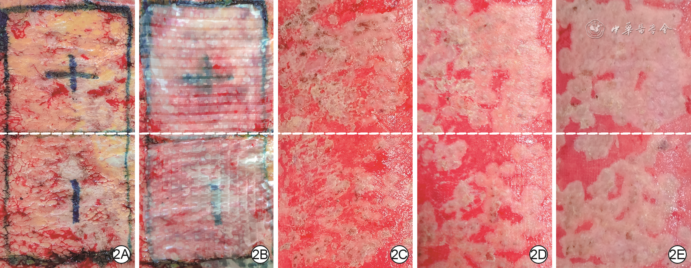
2 大面积烧伤患者2组创面行Meek微型皮片移植前后愈合情况。2A.术前清创后即刻;2B.术中hUCMSC+凝胶组涂抹含hUCMSC(细胞总数为3×10⁷个)的透明质酸凝胶,单纯凝胶组涂抹单纯透明质酸凝胶,且均移植扩展比例为1∶6的自体Meek微型皮片后;2C、2D、2E.分别为术后2、3、4周,hUCMSC+凝胶组创面愈合面积均较单纯凝胶组大
注:白色虚线上方为人脐带间充质干细胞(hUCMSC)+凝胶组,下方为单纯凝胶组

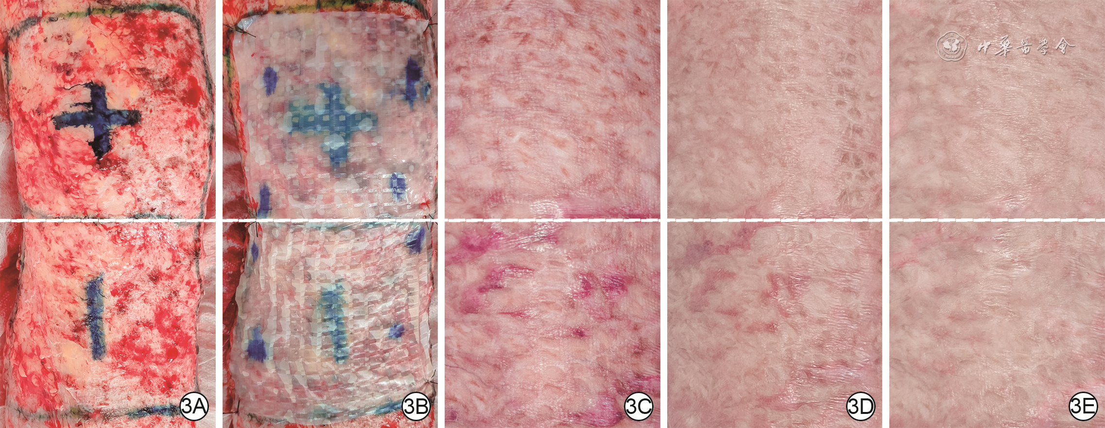
3 大面积烧伤患者2组创面行Meek微型皮片移植前后瘢痕情况。3A.术前清创后即刻;3B.术中hUCMSC+凝胶组涂抹含hUCMSC(细胞总数为3×10⁷个)的透明质酸凝胶,单纯凝胶组涂抹单纯透明质酸凝胶,且均移植扩展比例为1∶6的自体Meek微型皮片后;3C、3D.分别为术后3、6个月,与hUCMSC+凝胶组相比,单纯凝胶组瘢痕高度更高,颜色更深;3E.术后12个月,hUCMSC+凝胶组瘢痕颜色、高度均与正常皮肤相近,单纯凝胶组仍可见凸出的淡粉色瘢痕
注:白色虚线上方为人脐带间充质干细胞(hUCMSC)+凝胶组,下方为单纯凝胶组

4 大面积烧伤患者2组创面行Meek微型皮片移植术后3个月组织学分析。4A. hUCMSC+凝胶组可见表皮嵴,偶见真皮层汗腺、皮脂腺等 苏木精-伊红×100;4B.hUCMSC+凝胶组Ki67大量表达于创面基底层、棘层、颗粒层,Ki67阳性细胞数较多 辣根过氧化物酶-苏木精×100;4C.hUCMSC+凝胶组真皮层可见大量波形蛋白表达 辣根过氧化物酶-苏木精×100;4D.单纯凝胶组表皮层扁平、基底层表皮嵴消失,未见明显皮肤附件 苏木精-伊红×100;4E.单纯凝胶组Ki67主要表达于基底层,Ki67阳性细胞数较少 辣根过氧化物酶-苏木精×100;4F.单纯凝胶组波形蛋白表达与图4C相近 辣根过氧化物酶-苏木精×100
注:人脐带间充质干细胞(hUCMSC)+凝胶组涂抹含hUCMSC(细胞总数为3×10⁷个)的透明质酸凝胶,单纯凝胶组涂抹单纯透明质酸凝胶,随后2组均移植扩展比例为1∶6的自体Meek微型皮片;Ki67和波形蛋白阳性表达均为棕色
表1 大面积烧伤患者2组创面行Meek微型皮片移植术后各时间点愈合率比较(%,
组别 创面数(个) 2周 3周 4周 hUCMSC+凝胶组 20 80±11 84±12 92±9 单纯凝胶组 20 67±18 74±21 84±16 t值 4.01 3.52 3.66 P值 0.020 0.040 0.040 注:人脐带间充质干细胞(hUCMSC)+凝胶组创面涂抹含有hUCMSC(细胞总数为3×10⁷个)的透明质酸凝胶,单纯凝胶组创面涂抹单纯透明质酸凝胶,且均移植扩展比例为1∶6的自体Meek微型皮片  下载: 导出CSV
表2 大面积烧伤患者2组创面行Meek微型皮片移植术后各时间点创面VSS评分比较(分,
组别 创面数(个) 3个月 6个月 12个月 hUCMSC+凝胶组 20 6.8±1.8 5.6±1.6 4.0±1.4 单纯凝胶组 20 7.8±1.9 6.7±2.1 5.4±1.6 t值 -4.79 -4.37 -5.47 P值 <0.001 <0.001 <0.001 注:VSS为温哥华瘢痕量表;人脐带间充质干细胞(hUCMSC)+凝胶组创面涂抹含hUCMSC(细胞总数为3×10⁷个)的透明质酸凝胶,单纯凝胶组创面涂抹单纯透明质酸凝胶,且均移植扩展比例为1∶6的自体Meek微型皮片
下载: 导出CSV
-
[1] QuinteroEC,MachadoJFE,RoblesRAD.Meek micrografting history, indications, technique, physiology and experience: a review article[J].J Wound Care,2018,27(Sup2):S12-18.DOI: 10.12968/jowc.2018.27.Sup2.S12. [2] LeeSZ,HalimAS.Superior long term functional and scar outcome of Meek micrografting compared to conventional split thickness skin grafting in the management of burns[J].Burns,2019,45(6):1386-1400.DOI: 10.1016/j.burns.2019.04.011. [3] 中华医学会烧伤外科学分会MEEK植皮技术中心协作组,海军军医大学第一附属医院烧伤外科,全军烧伤研究所.MEEK微型皮片移植技术临床操作规范[J].中华烧伤杂志,2019,35(8):561-564.DOI: 10.3760/cma.j.issn.1009-2587.2019.08.001. [4] 陈德云 体外诱导人脐带间充质干细胞分化为表皮样细胞 北京 解放军医学院 2015 陈德云. 体外诱导人脐带间充质干细胞分化为表皮样细胞[D].北京:解放军医学院,2015.
[5] 许永安,黄沙,付小兵,等.特定微环境诱导人脐带沃顿胶间充质干细胞向汗腺样细胞分化的实验研究[J].解放军医学杂志,2011,36(3):211-214. [6] SuzdaltsevaY,ZhidkihS,KiselevSL,et al.Locally delivered umbilical cord mesenchymal stromal cells reduce chronic inflammation in long-term nonhealing wounds: a randomized study[J].Stem Cells Int,2020,2020:5308609.DOI: 10.1155/2020/5308609. [7] 中国整形美容协会瘢痕医学分会.瘢痕早期治疗全国专家共识(2020版)[J].中华烧伤杂志,2021,37(2):113-125.DOI: 10.3760/cma.j.cn501120-20200609-00300. [8] Nagamura-InoueT,MukaiT.Umbilical cord is a rich source of mesenchymal stromal cells for cell therapy[J].Curr Stem Cell Res Ther,2016,11(8):634-642.DOI: 10.2174/1574888x10666151026115017. [9] 史浩,胡葵葵.脐带间充质干细胞促进创面愈合的实验研究[J].中国医学工程,2014,22(5):57. [10] 李云霞.脐带间充质干细胞对皮肤创伤愈合影响的实验研究[J].中国美容医学,2016,25(7):51-53. [11] YangY,ZhuS,LiY,et al.Human umbilical cord mesenchymal stem cells ameliorate skin fibrosis development in a mouse model of bleomycin-induced systemic sclerosis[J].Exp Ther Med,2020,20(6):257.DOI: 10.3892/etm.2020.9387. [12] ChenY,HuY,ZhouX,et al.Human umbilical cord-derived mesenchymal stem cells ameliorate psoriasis-like dermatitis by suppressing IL-17-producing γδ T cells[J].Cell Tissue Res,2022,388(3):549-563.DOI: 10.1007/s00441-022-03616-x. [13] BartolucciJ,VerdugoFJ,GonzálezPL,et al.Safety and efficacy of the intravenous infusion of umbilical cord mesenchymal stem cells in patients with heart failure: a phase 1/2 randomized controlled trial (RIMECARD trial [randomized clinical trial of intravenous infusion umbilical cord mesenchymal stem cells on cardiopathy])[J].Circ Res,2017,121(10):1192-1204.DOI: 10.1161/CIRCRESAHA.117.310712. [14] ChinSP,Mohd-ShahrizalMY,LiyanaMZ,et al.High dose of intravenous allogeneic umbilical cord-derived mesenchymal stem cells (CLV-100) infusion displays better immunomodulatory effect among healthy volunteers: a phase 1 clinical study[J].Stem Cells Int,2020,2020:8877003.DOI: 10.1155/2020/8877003. [15] HuP,YangQ,WangQ,et al.Mesenchymal stromal cells-exosomes: a promising cell-free therapeutic tool for wound healing and cutaneous regeneration[J/OL].Burns Trauma,2019,7:38[2022-07-28].https://pubmed.ncbi.nlm.nih.gov/31890717/.DOI: 10.1186/s41038-019-0178-8. [16] HuangD,YiZ,HeX,et al.Distribution of infused umbilical cord mesenchymal stem cells in a rat model of renal interstitial fibrosis[J].Ren Fail,2013,35(8):1146-1150.DOI: 10.3109/0886022X.2013.815109. [17] LeeRH,PulinAA,SeoMJ,et al.Intravenous hMSCs improve myocardial infarction in mice because cells embolized in lung are activated to secrete the anti-inflammatory protein TSG-6[J].Cell Stem Cell,2009,5(1):54-63.DOI: 10.1016/j.stem.2009.05.003. [18] JungJW,KwonM,ChoiJC,et al.Familial occurrence of pulmonary embolism after intravenous, adipose tissue-derived stem cell therapy[J].Yonsei Med J,2013,54(5):1293-1296.DOI: 10.3349/ymj.2013.54.5.1293. [19] FurlaniD,UgurlucanM,OngL,et al.Is the intravascular administration of mesenchymal stem cells safe? Mesenchymal stem cells and intravital microscopy[J].Microvasc Res,2009,77(3):370-376.DOI: 10.1016/j.mvr.2009.02.001. [20] OellerM,Laner-PlambergerS,HochmannS,et al.Selection of tissue factor-deficient cell transplants as a novel strategy for improving hemocompatibility of human bone marrow stromal cells[J].Theranostics,2018,8(5):1421-1434.DOI: 10.7150/thno.21906. [21] DuanJX, LiuWJ, ZengYQ, et al. Umbilical cord mesenchymal stem cells (hUCMSC) for inflammatory regulation after excision and grafting of severe burn wounds in rats[J]. J Burn Care Res, 2020, 42(4):766-773. DOI: 10.1093/jbcr/iraa207. [22] LiuL,YuY,HouY,et al.Human umbilical cord mesenchymal stem cells transplantation promotes cutaneous wound healing of severe burned rats[J].PLoS One,2014,9(2):e88348.DOI: 10.1371/journal.pone.0088348. [23] 李梦芸,刘德伍,毛远桂.干细胞源性外泌体在创面修复中的作用研究进展[J].中华烧伤杂志,2017,33(3):180-184.DOI: 10.3760/cma.j.issn.1009-2587.2017.03.013. [24] LiuSJ,MengMY,HanS,et al.Umbilical cord mesenchymal stem cell-derived exosomes ameliorate HaCaT cell photo-aging[J].Rejuvenation Res,2021,24(4):283-293.DOI: 10.1089/rej.2020.2313. [25] ShoharaR,YamamotoA,TakikawaS,et al.Mesenchymal stromal cells of human umbilical cord Wharton's jelly accelerate wound healing by paracrine mechanisms[J].Cytotherapy,2012,14(10):1171-1181.DOI: 10.3109/14653249.2012.706705. [26] WangM,XuX,LeiX,et al.Mesenchymal stem cell-based therapy for burn wound healing[J/OL].Burns Trauma,2021,9:tkab002[2022-07-28].https://pubmed.ncbi.nlm.nih.gov/34212055/.DOI: 10.1093/burnst/tkab002. [27] 王配合,彭毅志.烧伤创面痂下活组织细菌定量培养与植皮存活率分析[J].中华烧伤杂志,2004,20(1):45-46.DOI: 10.3760/cma.j.issn.1009-2587.2004.01.017. [28] CookN.Methicillin-resistant Staphylococcus aureus versus the burn patient[J].Burns,1998,24(2):91-98.DOI: 10.1016/s0305-4179(97)00114-9. [29] OhH,BooS.Assessment of burn-specific health-related quality of life and patient scar status following burn[J].Burns,2017,43(7):1479-1485.DOI: 10.1016/j.burns.2017.03.023. [30] WangY,BeekmanJ,HewJ,et al.Burn injury: challenges and advances in burn wound healing, infection, pain and scarring[J].Adv Drug Deliv Rev,2018,123:3-17.DOI: 10.1016/j.addr.2017.09.018. [31] 沙焱, 谢英, 陈志军, 等. 脐带间充质干细胞对矽肺大鼠肺纤维化的干预研究 [J].中华劳动卫生职业病杂志,2019,37 (6): 401-407. DOI: 10.3760/cma.j.issn.1001-9391.2019.06.001. [32] HouschyarKS,TapkingC,NietzschmannI,et al.Five years experience with Meek grafting in the management of extensive burns in an adult burn center[J].Plast Surg(Oakv),2019,27(1):44-48.DOI: 10.1177/2292550318800331. -
 精选文章讲读-肖荣.mp4

-


 点击查看大图

计量
- 文章访问数: 1129
- HTML全文浏览量: 47
- PDF下载量: 53
- 被引次数: 0
