Current Issue

2024, Volume 40,  Issue 4

Expert Forum
Regenerative rehabilitation: the effects of physical factors on regeneration
Wu Jun, Wei Yating
2024, 40(4): 301-306. doi: 10.3760/cma.j.cn501225-20231229-00278
Abstract:
Wound regeneration and repair is one of the primary research fields in burn and wound repair surgery. In recent years, with the continuous advancement of treatment concept and technologies in the field of rehabilitation, the connection between rehabilitation treatment and wound regeneration and repair has become closer, forming a new concept "regenerative rehabilitation". This article discussed the concept formation and development status of regenerative rehabilitation, and the future development and potential leading value of regenerative rehabilitation field.
Clinical application of photobiomodulation in trauma repair and medical aesthetics
Yao Min, Zhang Yiqiu
2024, 40(4): 307-313. doi: 10.3760/cma.j.cn501225-20240203-00048
Abstract:
In recent years, with the deepening of researches on the molecular biological mechanisms of photobiomodulation (PBM), PBM has gradually been applied in clinical practice, providing effective treatment methods and approaches for various diseases. Compared with traditional photothermal therapy, PBM has the characteristics of good therapeutic effect, almost no adverse reaction, and simple operation, and its clinical efficacy is becoming increasingly significant. This article provides a detailed explanation on the mechanism of PBM, its application characteristics and development trends in trauma repair and medical aesthetics, in order to provide a theoretical basis for the extensively clinical application of this therapy.
Original Article · Wound Repair and Scar
Effects of applying human umbilical cord mesenchymal stem cell exosomes through different pathways to treat full-thickness skin defect wounds in mice
Wang Hongyu, Ba Te, Zhou Biao, Yan Zengqiang, Wang Ruijia, Liu Lingying
2024, 40(4): 314-322. doi: 10.3760/cma.j.cn501225-20231123-00203
Abstract:
  Objective   To investigate the effects of human umbilical cord mesenchymal stem cell (hUCMSC) exosomes in the treatment of full-thickness skin defect wounds in mice through local wound application, subcutaneous injection at the wound margin, and tail vein injection, and to explore the optimal administration route of hUCMSC exosomes for wound treatment.   Methods   This study was an experimental study. hUCMSC exosomes were extracted from the discarded umbilical cord tissue of three normal delivery women aged 25-35 years in the Department of Obstetrics and Gynecology of Baogang Hospital of Inner Mongolia and successfully identified. Totally 120 male BALB/c mice aged 6-8 weeks were selected, and full-thickness skin defect wounds were prepared on the back of them. According to the random number table, the injured mice were divided into control group (without drug administration), local wound application group, wound margin subcutaneous injection group, and tail vein injection group (with 30 mice in each group). Mice in the latter three groups were given 0.2 mL phosphate buffer solution containing 200 μg hUCMSC exosomes by local wound application, subcutaneous injection at the wound margin, and tail vein injection, respectively. On post injury day (PID) 7, 14, and 21, the general condition of the wound was observed, and the wound healing rate was calculated; the wound tissue was collected, the pathological changes and collagen fibers were observed respectively by hematoxylin-eosin staining and Masson staining, the number of new microvessels was observed by CD31 immunohistochemical staining, and the content of tumor necrosis factor α (TNF-α) and interleukin-6 (IL-6) was detected by enzyme-linked immunosorbent assay. The sample number was 10 in each group at each time point.   Results   On PID 7, 14, and 21, the wounds of mice in the 4 groups all healed gradually, and the wound healing of the mice in wound margin subcutaneous injection group was the best; the wound healing rates of mice in the three administration groups were significantly higher than those in control group ( P<0.05), the wound healing rates of mice in wound margin subcutaneous injection group and tail vein injection group were significantly higher than those in local wound application group ( P<0.05), and the wound healing rates of mice in wound margin subcutaneous injection group were significantly higher than those in tail vein injection group ( P<0.05). On PID 7, 14, and 21, the growth and epithelialization speed of the wound tissue of mice in the three administration groups were significantly accelerated, and the collagen fibers in the wounds of mice in the three administration groups were larger in number and more neatly arranged in comparison with the control group. On PID 7, 14, and 21, under every 200-fold visual field, the number of new microvessels in the wound tissue of mice in local wound application group was 24.1±2.5, 50.7±4.1, and 44.2±2.3, respectively, the number of new microvessels in the wound tissue of mice in wound margin subcutaneous injection group was 32.2±2.9, 67.5±4.9, and 53.6±3.7, respectively, and the number of new microvessels in the wound tissue of mice in tail vein injection group was 27.8±2.4, 59.1±3.7, and 49.6±2.6, respectively, which was significantly more than 20.6±1.7, 46.7±3.4, and 40.9±2.8 in control group ( P<0.05); the number of new microvessels in the wound tissue of mice in wound margin subcutaneous injection group and tail vein injection group was significantly more than that in local wound application group ( P<0.05); the number of new microvessels in the wound tissue of mice in wound margin subcutaneous injection group was significantly more than that in tail vein injection group ( P<0.05). On PID 7, 14, and 21, the content of TNF-α and IL-6 in the wound tissue of mice in the three administration groups was significantly less than that in control group ( P<0.05), the content of TNF-α and IL-6 in the wound tissue of mice in wound margin subcutaneous injection group and tail vein injection group was significantly less than that in local wound application group ( P<0.05), and the content of TNF-α and IL-6 in the wound tissue of mice in wound margin subcutaneous injection group was significantly less than that in tail vein injection group ( P<0.05).   Conclusions   Local wound application, subcutaneous injection at the wound margin, and tail vein injection of hUCMSC exosomes can all promote the wound healing of full-thickness skin defects in mice through alleviating excessive inflammatory response and promoting angiogenesis. Among them, subcutaneous injection at the wound margin has a better therapeutic effect, indicating subcutaneous injection at the wound margin is the optimal administration route for hUCMSC exosomes in wound treatment.
Effects of gelatin methacrylate anhydride hydrogel loaded with small extracellular vesicles derived from human umbilical cord mesenchymal stem cells in the treatment of full-thickness skin defect wounds in mice
Chen Yiqi, Zhou Yingqian, Wei Qian, Xie Xiaoye, Liu Xinzhu, Li Dawei, Shen Chuan'an
2024, 40(4): 323-332. doi: 10.3760/cma.j.cn501225-20231218-00248
Abstract:
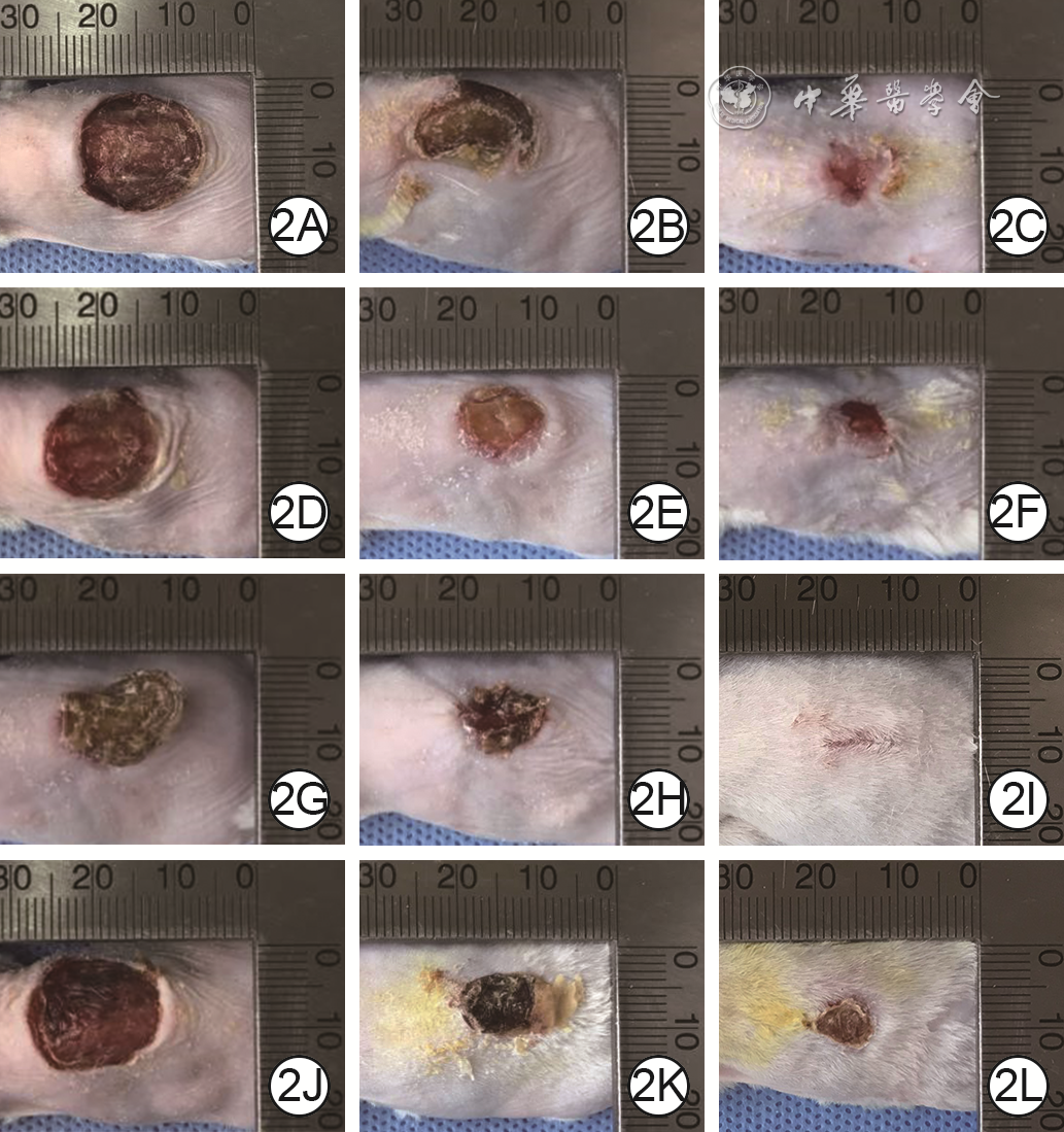
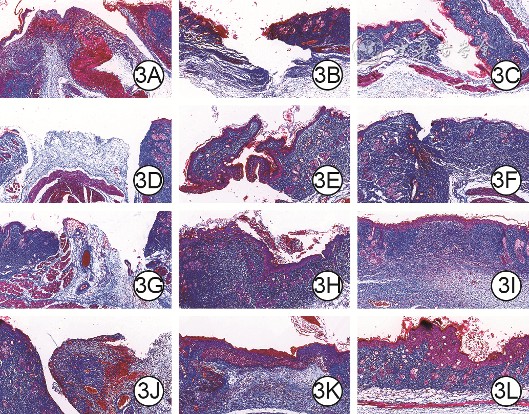
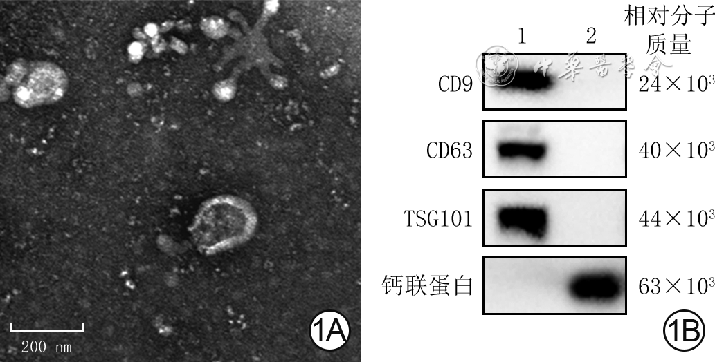
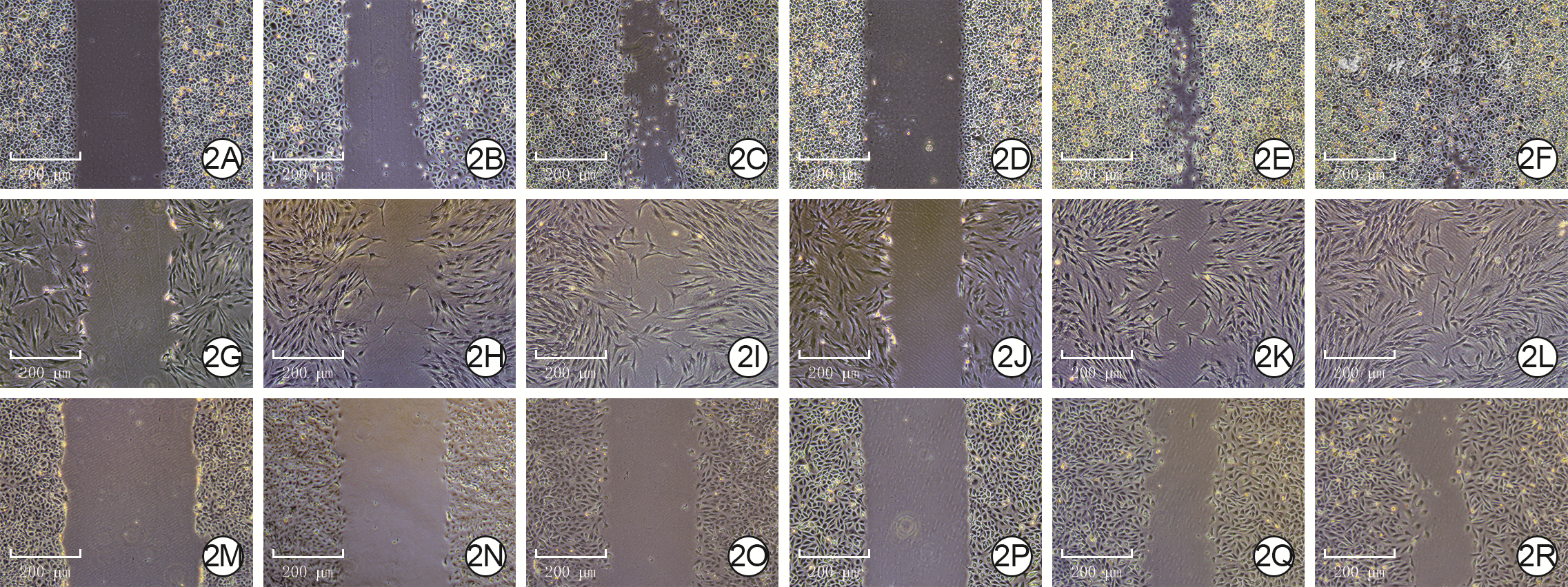
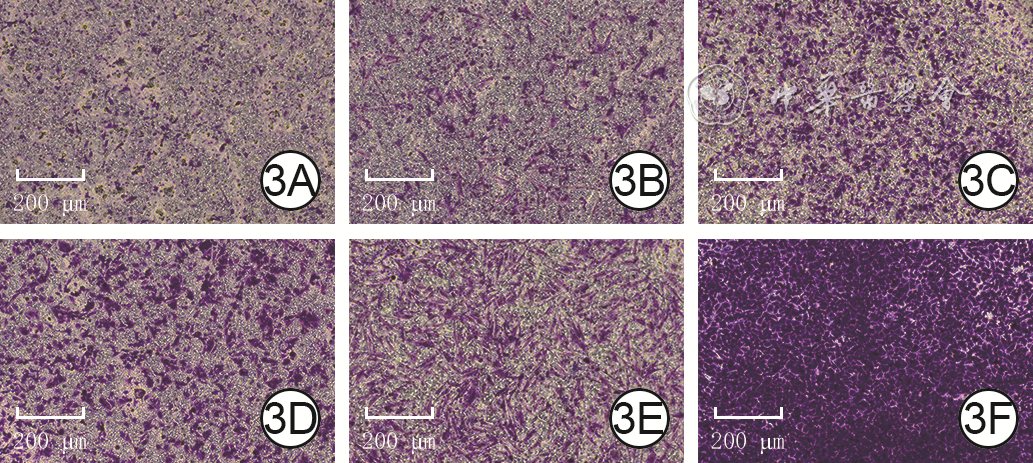
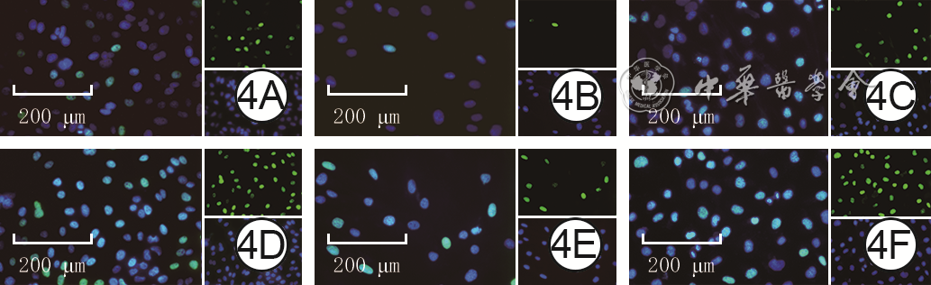
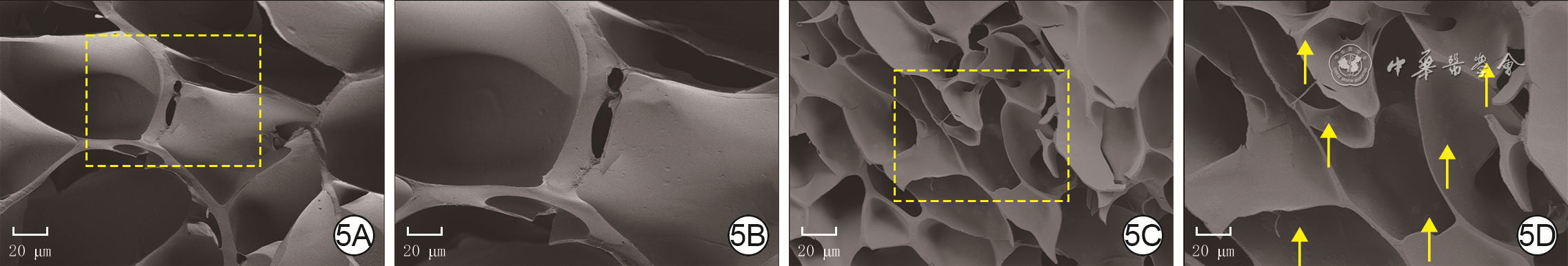
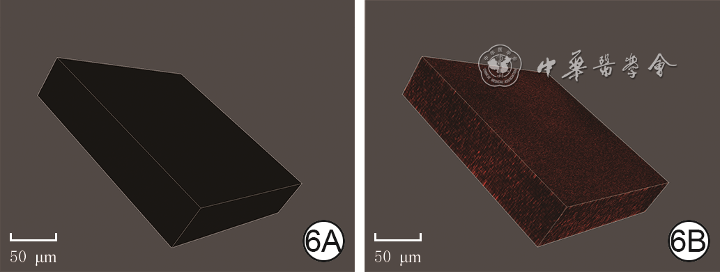
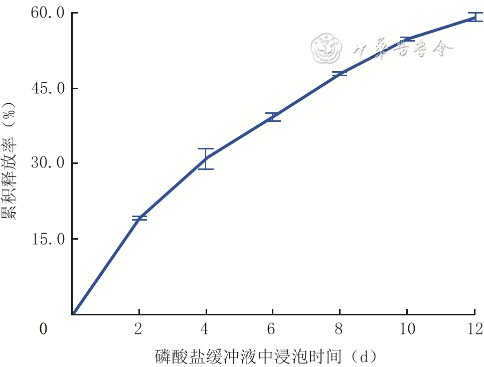
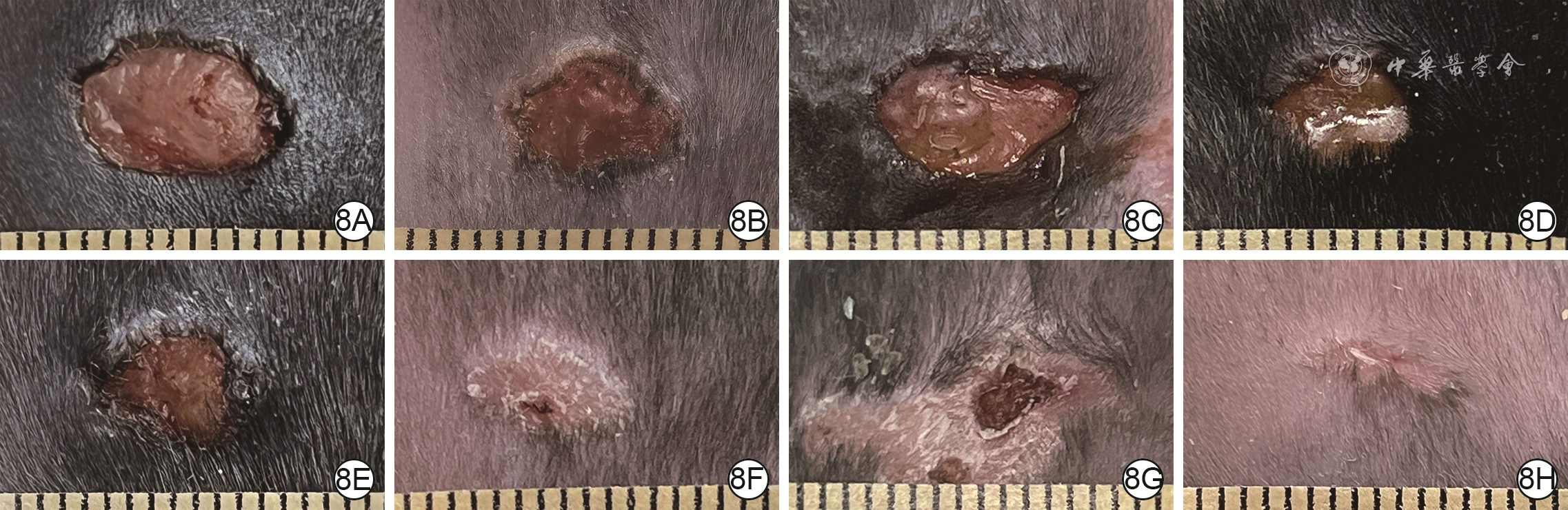
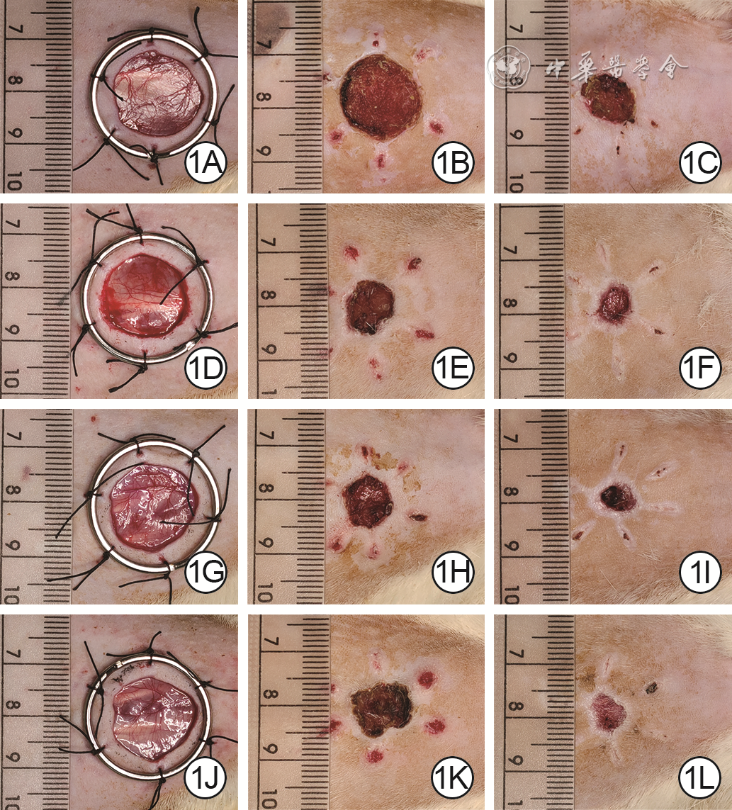
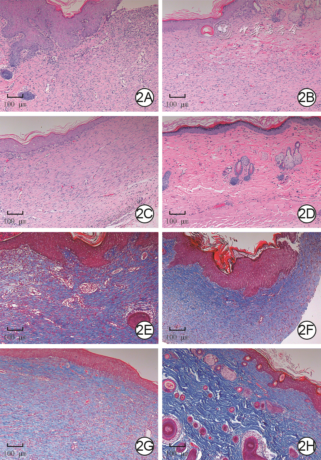
  Objective   To investigate the effects of gelatin methacrylate anhydride (GelMA) hydrogel loaded with small extracellular vesicles derived from human umbilical cord mesenchymal stem cells (hUCMSCs-sEVs) in the treatment of full-thickness skin defect wounds in mice.   Methods   This study was an experimental study. hUCMSCs-sEVs were extracted by ultracentrifugation, their morphology was observed through transmission electron microscope, and the expression of CD9, CD63, tumor susceptibility gene 101 (TSG101), and calnexin was detected by Western blotting. The human umbilical vein endothelial cells (HUVECs), the 3 rd and 4 th passages of human epidermal keratinocytes (HEKs) and human dermal fibroblasts (HDFs) were all divided into blank control group (routinely cultured) and hUCMSC-sEV group (cultured with the cell supernatant containing hUCMSCs-sEVs). The cell scratch test was performed and the cell migration rates at 6, 12, and 24 h after scratching were calculated, the cell Transwell assay was performed and the number of migration cells at 12 h after culture was calculated, and the proportion of proliferating cells was detected by 5-acetylidene-2'-deoxyuridine and Hoechst staining at 24 h after culture, with sample numbers being all 3. The simple GelMA hydrogel and the GelMA hydrogel loaded with hUCMSCs-sEVs (hereinafter referred to as hUCMSC-sEV/GelMA hydrogel) were prepared. Then the micromorphology of 2 kinds of hydrogels was observed under scanning electron microscope, the distribution of hUCMSCs-sEVs was observed by laser scanning confocal microscope, and the cumulative release rates of hUCMSCs-sEVs at 0 (immediately), 2, 4, 6, 8, 10, and 12 d after soaking hUCMSC-sEV/GelMA hydrogel in phosphate buffer solution (PBS) were measured and calculated by protein colorimetric quantification ( n=3). Twenty-four 6-week-old male C57BL/6J mice were divided into PBS group, hUCMSC-sEV alone group, GelMA hydrogel alone group, and hUCMSC-sEV/GelMA hydrogel group according to the random number table, with 6 mice in each group, and after the full-thickness skin defect wounds on the back of mice in each group were produced, the wounds were performed with PBS injection, hUCMSC-sEV suspenson injection, simple GelMA coverage, and hUCMSC-sEV/GelMA hydrogel coverage, respectively. Wound healing was observed on post injury day (PID) 0 (immediately), 4, 8, and 12, and the wound healing rates on PID 4, 8, and 12 were calculated, and the wound tissue was collected on PID 12 for hematoxylin-eosin staining to observe the structure of new tissue, with sample numbers being both 6.   Results   The extracted hUCMSCs-sEVs showed a cup-shaped structure and expressed CD9, CD63, and TSG101, but barely expressed calnexin. At 6, 12, and 24 h after scratching, the migration rates of HEKs (with t values of 25.94, 20.98, and 20.04, respectively), HDFs (with t values of 3.18, 5.68, and 4.28, respectively), and HUVECs (with t values of 4.32, 19.33, and 4.00, respectively) in hUCMSC-sEV group were significantly higher than those in blank control group ( P<0.05). At 12 h after culture, the numbers of migrated HEKs, HDFs, and HUVECs in hUCMSC-sEV group were 550 ± 23, 235 ± 9, and 856 ± 35, respectively, which were significantly higher than 188 ± 14, 97 ± 6, and 370 ± 32 in blank control group (with t values of 22.95, 23.13, and 17.84, respectively , P<0.05). At 24 h after culture, the proportions of proliferating cells of HEKs, HDFs, and HUVECs in hUCMSC-sEV group were significantly higher than those in blank control group (with t values of 22.00, 13.82, and 32.32, respectively, P<0.05). The inside of simple GelMA hydrogel showed a loose and porous sponge-like structure, and hUCMSCs-sEVs was not observed in it. The hUCMSC-sEV/GelMA hydrogel had the same sponge-like structure, and hUCMSCs-sEVs were uniformly distributed in clumps. The cumulative release rate curve of hUCMSCs-sEVs from hUCMSC-sEV/GelMA hydrogel tended to plateau at 2 d after soaking, and the cumulative release rate of hUCMSCs-sEVs was (59.2±1.8)% at 12 d after soaking. From PID 0 to 12, the wound areas of mice in the 4 groups gradually decreased. On PID 4, 8, and 12, the wound healing rates of mice in hUCMSC-sEV/GelMA hydrogel group were significantly higher than those in the other 3 groups ( P<0.05); the wound healing rates of mice in GelMA hydrogel alone group and hUCMSC-sEV alone group were significantly higher than those in PBS group ( P<0.05). On PID 8 and 12, the wound healing rates of mice in hUCMSC-sEV alone group were significantly higher than those in GelMA hydrogel alone group ( P<0.05). On PID 12, the wounds of mice in hUCMSC-sEV/GelMA hydrogel group showed the best wound epithelization, loose and orderly arrangement of dermal collagen, and the least number of inflammatory cells, while the dense arrangement of dermal collagen and varying degrees of inflammatory cell infiltration were observed in the wounds of mice in the other 3 groups.   Conclusions   hUCMSCs-sEVs can promote the migration and proliferation of HEKs, HDFs, and HUVECs which are related to skin wound healing, and slowly release in GelMA hydrogel. The hUCMSC-sEV/GelMA hydrogel as a wound dressing can significantly improve the healing speed of full-thickness skin defect wounds in mice.
Exploring the causality between intestinal flora and hyperplastic scars of human based on two-sample Mendelian randomization analysis
Chen Wentao, Wang Xiaoxiang, Zheng Wenlian, Zhang Weiqiang, Mao Lujia, Zhuo Jianan, Zhou Sitong, Yang Ronghua
2024, 40(4): 333-341. doi: 10.3760/cma.j.cn501225-20231129-00215
Abstract:
  Objective   To investigate the causality between intestinal flora and hypertrophic scars (HS) of human.   Methods   This study was a study based on two-sample Mendelian randomization (TSMR) analysis. The data on intestinal flora ( n=18 473) and HS ( n=208 248) of human were obtained from the genome-wide association study database. Genetically variable genes at five levels (phylum, class, order, family, and genus) of known intestinal flora, i.e., single nucleotide polymorphisms (SNPs), were extracted as instrumental variables for linkage disequilibrium (LD) analysis. Human genotype-phenotype association analysis was performed using PhenoScanner V2 database to exclude SNPs unrelated to HS in intestinal flora and analyze whether the selected SNPs were weak instrumental variables. The causal relationship between intestinal flora SNPs and HS was analyzed through four methods of TSMR analysis, namely inverse variance weighted (IVW), MR-Egger regression, weighted median, and weighted mode. Scatter plots of significant results from the four aforementioned analysis methods were plotted to analyze the correlation between intestinal flora SNPs and HS. Both IVW test and MR-Egger regression test were used to assess the heterogeneity of intestinal flora SNPs, MR-Egger regression test and MR-PRESSO outlier test were used to assess the horizontal multiplicity of intestinal flora SNPs, and leave-one-out sensitivity analysis was used to determine whether HS was caused by a single SNP in the intestinal flora. Reverse TSMR analyses were performed for HS SNPs and genus Intestinimonas or genus Ruminococcus2, respectively, to detect whether there was reverse causality between them.   Results   A total of 196 known intestinal flora, belonging to 9 phyla, 16 classes, 20 orders, 32 families, and 119 genera, were obtained, and multiple SNPs were obtained from each flora as instrumental variables. LD analysis showed that the SNPs of the intestinal flora were consistent with the hypothesis that genetic variation was strongly associated with exposure factors, except for rs1000888, rs12566247, and rs994794. Human genotype-phenotype association analysis showed that none of the selected SNPs after LD analysis was excluded and there were no weak instrumental variables. IVW, MR-Egger regression, weighted median, and weighted mode of TSMR analysis showed that both genus Intestinimonas and genus Ruminococcus2 were causally associated with HS. Among them, forest plots of IVW and MR-Egger regression analyses also showed that 16 SNPs (the same SNPs number of this genus below) of genus Intestinimonas and 15 SNPs (the same SNPs number of this genus below) of genus Ruminococcus2 were protective factors for HS. Further, IVW analysis showed that genus Intestinimonas SNPs (with odds ratio of 0.62, 95% confidence interval of 0.41-0.93, P<0.05) and genus Ruminococcus2 SNPs (with odds ratio of 0.62, 95% confidence interval of 0.40-0.97, P<0.05) were negatively correlated with the risk of HS. Scatter plots showed that SNPs of genus Intestinimonas and genus Ruminococcus2 were protective factors of HS. Both IVW test and MR-Egger regression test showed that SNPs of genus Intestinimonas (with Q values of 5.73 and 5.76, respectively, P>0.05) and genus Ruminococcus2 (with Q values of 13.67 and 15.61, respectively, P>0.05) were not heterogeneous. MR-Egger regression test showed that the SNPs of genus Intestinimonas and genus Ruminococcus2 had no horizontal multiplicity (with intercepts of 0.01 and 0.06, respectively, P>0.05); MR-PRESSO outlier test showed that the SNPs of genus Intestinimonas and genus Ruminococcus2 had no horizontal multiplicity ( P>0.05). Leave-one-out sensitivity analysis showed that no single intestinal flora SNP drove the occurrence of HS. Reverse TSMR analysis showed no reverse causality between HS SNPs and genus Intestinimonas or genus Ruminococcus2 (with odds ratios of 1.01 and 0.99, respectively, 95% confidence intervals of 0.97-1.06 and 0.96-1.04, respectively, P>0.05).   Conclusions   There is a causal relationship between intestinal flora and HS of human, in which genus Intestinimonas and genus Ruminococcus2 have a certain effect on inhibiting HS.
Original Article
A randomized controlled trial on the effect of early eschar dermabrasion combined with antimicrobial soft silicone foam dressing in the treatment of deep partial-thickness burn wounds in children
Shen Yang, He Jun, Liu Junzhang, Zhang Xianfeng, Tan Jie, Tang Wenjun, Yang Hao, Chen Xu, Luo Xingwang
2024, 40(4): 342-347. doi: 10.3760/cma.j.cn501225-20231004-00103
Abstract:
  Objective   To explore the effect of early eschar dermabrasion combined with antimicrobial soft silicone foam dressing (hereinafter referred to as foam dressing) in treating the deep partial-thickness burn wounds in children.   Methods   This study was a randomized controlled trial. From June 2021 to December 2022, 78 pediatric patients with deep partial-thickness burns who met the inclusion criteria were admitted to the Department of Burns in Guiyang Steel Plant Employees Hospital. According to the random number table, the pediatric patients were divided into two groups, with 38 cases left in combined treatment group (with 20 males and 18 females, aged 26.00 (16.75, 39.75) months) and 39 cases in foam dressing group (with 21 males and 18 females, aged 19.00 (14.00, 31.00) months) after the exclusion of one dropped-out child in follow-up. The pediatric patients in combined treatment group underwent eschar dermabrasion of the wound within 48 hours after injury, the wound was covered with foam dressing after operation, and the dressing was replaced once every 7 days; for the pediatric patients in foam dressing group, the wound was sterilized within 48 hours after injury and covered with foam dressing, and the dressing was replaced once every 2 to 3 days. After the wound healing, the children in both groups were routinely applied with silicone gel twice a day for 3 weeks before started wearing elastic sleeves for more than 18 hours a day, and continuously for over than 6 months. The degree of pain during dressing change was evaluated using the children's pain behavior inventory FLACC. The adverse reactions during the treatment period, number of dressing changes, and wound healing time were observed and recorded. Six months after wound healing, the Vancouver scar scale (VSS) was used to evaluate the condition of the wound scar.   Results   When changing dressing, the FLACC score for pain of pediatric patients in combined treatment group was 3.5 (2.0, 5.0), which was significantly lower than 6.0 (5.0, 8.0) in foam dressing group ( Z=-5.40, P<0.05). During the treatment period, no adverse reactions such as wound edema, fluid accumulation, or peripheral skin rash allergies occurred in any pediatric patient in both groups. The number of dressing changes of pediatric patients in combined treatment group was 3 (3, 4) times, which was significantly less than 8 (7, 10) times in foam dressing group ( Z=-7.58, P<0.05). The wound healing time of pediatric patients in combined treatment group was (19±5) days, which was significantly shorter than (25±6) days in foam dressing group ( t=-4.48, P<0.05). Six months after wound healing, the VSS score for scar of pediatric patients in combined treatment group was 5 (2, 8), which was significantly lower than 7 (5, 10) in foam dressing group ( Z=-3.05, P<0.05).   Conclusions   Compared with using foam dressings alone, early eschar dermabrasion combined with foam dressings can reduce the number of dressing changes, alleviate the pain during dressing changes, and shorten the wound healing time in treating children with deep partial-thickness burns, and effectively alleviate scar hyperplasia by combining with anti-scar treatment post burns.
Effects of early debridement and conservative eschar removal followed by wound coverage with acellular dermal matrix in the treatment of children with deep burns
Liang Yan, Shi Wen, Shao Yang, Liu Xinzhuang, Gong Hongmin, Cao Guohui, Gao Cong, Xin Naijun, Song Guodong
2024, 40(4): 348-357. doi: 10.3760/cma.j.cn501225-20230720-00010
Abstract:
  Objective   To explore the effects of early debridement and conservative eschar removal followed by wound coverage with acellular dermal matrix (ADM), i.e., early surgery, in the treatment of children with deep burns.   Methods   This study was a retrospective cohort study. From January 2017 to December 2022, 278 deep burned hospitalized children aged 1-7 years who met the inclusion criteria were admitted to Central Hospital Affiliated to Shandong First Medical University. According to the differences in treatment processes, 134 children who underwent early surgery+routine dressing change were enrolled in eschar removal+dressing change group (77 males and 57 females, aged 1 (1, 2) years), and 144 children who underwent only routine dressing change were enrolled in dressing change alone group (90 males and 54 females, aged 1 (1, 2) years). Fifty-one children without full-thickness burns in eschar removal+dressing change group were enrolled in eschar removal+dressing change group 1 (26 males and 25 females, aged 1 (1, 2) years), and 57 cases of the 83 children with full-thickness burns who did not undergo autologous skin grafting at the same time of early surgery (namely early skin grafting) in eschar removal+dressing change group were included in eschar removal+dressing change group 2 (37 males and 20 females, aged 1 (1, 2) years). Seventy-six children without full-thickness burns in dressing change alone group were included in dressing change alone group 1 (51 males and 25 females, aged 1 (1, 3) years), and 68 children with full-thickness burns in dressing change alone group were included in dressing change alone group 2 (39 males and 29 females, aged 1 (1, 2) years). For deep partial-thickness burn wounds and small full-thickness burn wounds in eschar removal+dressing change group, the eschar removal was performed on the basis of retaining a thin layer of denatured dermis so as to preserve the healthy tissue of the wound base, and ADM was applied to all wounds externally after eschar removal. For larger full-thickness burn wounds in this group, especially those located in the functional part of joints, eschar removal to the plane layer of viable tissue and early autologous skin grafting was needed. When the superficial wounds of children healed or tended to heal, the residual wounds were evaluated, and elective autologous skin grafting was performed if it was difficult to heal within 14 days. The healing time, intervention healing time, times of operation/dressing change, and times of intervention operation/dressing change in children with deep partial-thickness burn wounds of children in eschar removal+dressing change group, dressing change alone group, eschar removal+dressing change group 1, and dressing change alone group 1 were recorded. At the last follow-up (follow-up period was set to 7-12 months), the modified Vancouver scar scale (mVSS) scores of the most severe area of scar hyperplasia of healed deep partial-thickness burn wounds of 54 children in eschar removal+dressing change group and 48 children in dressing change alone group were recorded. The healing time and times of operation/dressing change of all burn wounds of children in eschar removal+dressing change group and dressing change alone group, and the healing time and times of operation/dressing change of full-thickness burn wounds of children in eschar removal+dressing change group 2 and dressing change alone group 2 were recorded. The incidences of wound infection, sepsis, fever, and fever after 5 days of burns in children of eschar removal+dressing change group and dressing change alone group during wound healing.   Results   Compared with those in dressing change alone group, the healing time and intervention healing time were significantly shortened, and the times of operation/dressing change and times of intervention operation/dressing change were significantly reduced in children with deep partial-thickness burn wounds in eschar removal+dressing change group (with Z values of -11.00, -11.33, -12.64, and -11.65, respectively, P<0.05). Compared with those in dressing change alone group 1, the healing time and intervention healing time were significantly shortened, and the times of operation/dressing change and times of intervention operation/dressing change were significantly reduced in children with deep partial-thickness burn wounds in eschar removal+dressing change group 1 (with Z values of 6.57, 6.46, 8.04, and 6.57, respectively, P<0.05). At the last follow-up, the mVSS score of the most severe scar hyperplasia area of healed deep partial-thickness burn wounds of 54 children in eschar removal+dressing change group was 4.00 (3.00,5.00), which was significantly lower than 6.50 (5.00,7.00) of 48 children in dressing change alone group ( Z =-4.67, P<0.05).Compared with those in dressing change alone group, the healing time was significantly shortened, and times of operation/dressing change was significantly reduced in all burn wounds in eschar removal+dressing change group (with Z values of -5.20 and -6.34, respectively, P<0.05). Compared with those in dressing change alone group 2, the healing time was significantly shortened, and times of operation/dressing change was significantly reduced in full-thickness burn wounds in eschar removal+dressing change group 2 (with Z values of -5.22 and -5.73, respectively, P<0.05). During wound healing, the probabilities of fever and fever after 5 days of burns in children of eschar removal+dressing change group were significantly lower than those in dressing change alone group (with χ 2 values of 4.13 and 3.91, respectively, P<0.05); only 1 child in dressing change alone group developed sepsis, and there was no statistically significant difference in the wound infection rate of children in the two groups ( P>0.05).   Conclusions   For children with deep burns, early surgery, and early skin grafting or elective autologous skin grafting as needed, have better short-term and long-term effects than those without early surgery.
Application effects of armor chest straps in patients with sternal dehiscence after repair surgery
Yang Jiao, Wang Ling, Zhang Wanfu, Chen Yang, Guan Hao
2024, 40(4): 358-364. doi: 10.3760/cma.j.cn501225-20230904-00074
Abstract:
  Objective   To investigate the application effects of armor chest straps in patients with sternal dehiscence after repair surgery.   Methods   This study was a retrospective cohort study. The 11 patients who were admitted to the First Affiliated Hospital of Air Force Medical University (hereinafter referred to as the hospital) from March 2020 to March 2021 and used conventional chest straps after sternal dehiscence repair surgery were included in conventional chest strap group. The 12 patients who were admitted to the hospital from April 2021 to March 2022 and used armor chest straps after sternal dehiscence repair surgery were included in armor chest strap group. A special team for sternal dehiscence repair was set up, and the nurses in charge in the team instructed the patients in 2 groups on the correct abdominal breathing method, and the members of the surgical team performed the personalized surgery and wore the corresponding chest straps for the patients in 2 groups. The abdominal breathing frequency and chest breathing frequency on the first day after surgery were recorded. The pain intensity at 6, 24, 48, and 72 h after surgery was self-rated by the patients using numerical rating scale. The time of the first active cough and the time of wound healing after surgery were recorded. At postoperative suture removal, the cutting length of sutures induced by respiratory exercise was recorded. Whether there were complications such as redness, swelling, and exudation in flaps within 2 weeks after surgery were recorded, whether there were complications such as wound dehiscence or infection during follow-up of 3-12 months were recorded, and the incidence proportion of postoperative complications was calculated. At 6 months after surgery, the patients' scar status was evaluated by the Vancouver scar scale.   Results   The abdominal breathing frequency of patients in armor chest strap group was (16.3±1.2) times/min on the first day after surgery, which was significantly higher than (5.3±1.4) times/min in conventional chest strap group ( t=20.00, P<0.05), and the chest breath-ing frequency was (1.2±0.8) times/min, which was significantly lower than (12.4±1.5) times/min in conventional chest strap group ( t=22.36, P<0.05). The pain intensity scores of patients in armor chest strap group at 6, 24, 48, and 72 h after surgery were significantly lower than those in conventional chest strap group (with t values of 15.07, 14.70, 13.66, and 11.03, respectively, P<0.05). The time of the first active cough and the time of wound healing after surgery of patients in armor chest strap group were significantly sooner than those in conventional chest strap group (with t values of 5.51 and 8.90, respectively, P<0.05). At postoperative suture removal, the cutting length of sutures induced by respiratory exercise of patients in conventional chest strap group was 2.0 (0, 5.0) mm, which was significantly longer than 2.0 (1.0, 2.0) mm in armor chest strap group ( Z=4.10, P<0.05). There was no statistically significant difference in the incidence proportion of postoperative complications of patients between the 2 groups ( P>0.05). At 6 months after surgery, the scar score of patients in armor chest strap group was 4.1±1.4, which was significantly lower than 5.6±1.4 in conventional chest strap group ( t=2.71, P<0.05).   Conclusions   The application of armor chest strap in patients with sternal dehiscence after repair surgery can increase the abdominal breathing frequency, reduce the wound cutting force, effectively relieve postoperative pain, increase the first active cough and wound healing speed, and alleviate postoperative scar proliferation, achieving good application effect.
Clinical effects of early rehabilitation treatment after repair surgery of skin and soft tissue defects accompanied by extensor tendon injury on the back of hand
Zhu Chan, He Lin, He Ting, Liang Ying, Zhang Bowen, Zhao Haiyang, Guan Hao, Yang Xuekang, Hu Dahai, Han Juntao, Liu Jiaqi
2024, 40(4): 365-372. doi: 10.3760/cma.j.cn501225-20230820-00057
Abstract:
  Objective   To explore the clinical effects of early rehabilitation treatment after repair surgery of skin and soft tissue defects accompanied by extensor tendon injury on the back of hand.   Methods   This study was a retrospective non-randomized controlled study. From February 2015 to February 2023, 24 patients (15 males and 9 females, aged 12-55 years) with skin and soft tissue defects accompanied by extensor tendon injury on the back of hand, who met the inclusion criteria and were repaired with flap transplantation and tendon grafting or tendon anastomosis, were admitted to the First Affiliated Hospital of Air Force Medical University. According to different intervention time for postoperative rehabilitation treatment of patients, the patients were divided into conventional rehabilitation group and early rehabilitation group, with 12 cases in each group. Patients in early rehabilitation group received rehabilitation treatment immediately after surgery under the rehabilitation guidance of specialized rehabilitation physicians based on the characteristics of different postoperative periods. Patients in conventional rehabilitation group began rehabilitation treatment from the third week after surgery, and their rehabilitation treatment was the same as that of patients in early rehabilitation group from the second week after surgery. The patients in 2 groups were treated in the hospital until the sixth week after surgery. The occurrence of flap vascular crisis and tendon rupture were observed within 6 weeks after surgery. After 6 weeks of surgery, the manual muscle test was used to measure the pinching force between the index finger and thumb, lateral pinching force, three-point pinching force, and grip force of the affected hand; the total action motion method was used to evaluate the finger joint range of motion of the affected hand, and the excellent and good ratio was calculated; the Carroll upper extremity function test was used to score and rate the function of the affected hand.   Results   Within 6 weeks after surgery, only 1 patient in conventional rehabilitation group suffered from venous crisis, and the flap survived after the second surgical exploration and anastomosis of blood vessels; there was no occurrence of tendon rupture in patients of 2 groups. After 6 weeks of surgery, there were no statistically significant differences in pinching force between the index finger and thumb, lateral pinching force, three-point pinching force, or grip force of the affected hand between the two groups of patients ( P>0.05); the excellent and good ratio of the finger joint range of motion of the affected hand of patients in early rehabilitation group was 11/12, which was higher than 7/12 in conventional rehabilitation group, but there was no statistically significant difference ( P>0.05); the affected hand function score of patients in early rehabilitation group was 90±6, which was significantly higher than 83±8 in conventional rehabilitation group ( t=2.41, P<0.05); the function rating of the affected hand of patients in early rehabilitation group was obviously better than that in conventional rehabilitation group ( Z=2.04, P<0.05).   Conclusions   Early rehabilitation treatment for patients with skin and soft tissue defects accompanied by extensor tendon injury on the back of hand after repair surgery can improve hand function, but it would not increase surgery related complications, which is worthy of clinical promotion and application.
A cross-sectional survey and analysis of influencing factors on the occurrence of post-burn psychological stress disorder in preschool children
Lu Xuelan, Liu Yingping, Liu Yiwei, Diao Jielin, Wang Feng, Zhong Fangyu, He Jiale, Chen Lang
2024, 40(4): 373-379. doi: 10.3760/cma.j.cn501225-20230731-00028
Abstract:
  Objective   To explore the occurrence and influencing factors of post-burn psychological stress disorder in preschool children.   Methods   This study was a multi-center cross-sectional survey. From January 2022 to February 2023, 85 preschool children (aged 1 to 6 years) with burns admitted to the Affiliated Hospital of North Sichuan Medical College, Nanchong Central Hospital, Suining Central Hospital, Guang'an People's Hospital, and Guangyuan Central Hospital who met the inclusion criteria were selected as respondents. A self-made general information questionnaire was used to investigate the children's general data including gender, age group, residential area, main caregiver and their education level, and family type, as well as the injury condition including cause of injury and burn severity. The Child Stress Disorders Checklist was used to investigate the occurrence of psychological stress disorder in children at 3 days to 1 month after injury, and the incidence rate was calculated. The children were classified according to their general data and injury condition, and the occurrence of psychological stress disorder in children at 3 days to 1 month after injury was recorded, and the influencing factors for post-burn psychological stress disorder in preschool children were screened.   Results   A total of 85 questionnaires were distributed and 85 valid questionnaires were recovered, with an effective recovery rate of 100%. Among the children, there were 45 boys and 40 girls, with most children aged 1 to 3 years. There were slightly more children in rural areas than in cities. About half of the children were mainly cared for by their parents and grandparents, respectively, and the education level of the main caregivers was mainly high school/technical secondary school. The family type was mainly core family and extended family. The main cause of injury was hydrothermal scald, and the severity of burns was mainly moderate. The incidence rate of psychological stress disorder in this group of children at 3 days to 1 month after injury was 34.12% (29/85). There were statistically significant differences in the occurrence of psychological stress disorder in children with different age groups, causes of injuries, and burn severity at 3 days to 1 month after injury (with χ 2 values​​of 9.18, 7.80, and 25.47, respectively, P<0.05); there were no statistically significant differences in the occurrence of psychological stress disorder in children with different genders, residential area, main caregivers, main caregivers' education levels, or family types at 3 days to 1 month after injury ( P>0.05). Multivariate logistic regression analysis showed that age group and burn severity were independent influencing factors for the occurrence of psychological stress disorder in preschool children after burns (with odds ratios of 8.21 and 33.99, respectively, and 95% confidence intervals of 1.57-43.04 and 5.55-207.93, respectively, P<0.05), the older the child and the more severe the burn, the higher the possibility of the occurrence of psychological stress disorder.   Conclusions   The incidence rate of psychological stress disorder is high in preschool children after burns. Age group and burn severity are independent influencing factors for the occurrence of post-burn psychological stress disorder in this type of children.
Effect of salvia miltiorrhiza combined with roxadustat on wound healing of full-thickness skin defects in diabetic rats and its mechanism
Xia Ruyi, Tang Di, Yang Bin
2024, 40(4): 380-388. doi: 10.3760/cma.j.cn501225-20231020-00124
Abstract:
  Objective   To explore the effect of salvia miltiorrhiza combined with roxadustat on wound healing of full-thickness skin defects in diabetic rats and its mechanism.   Methods   This study was an experimental study. Twenty male 8-week-old Sprague-Dawley rats were used to successfully establish diabetic model, then full-thickness skin defect wounds on their backs were made. The rats were divided into normal saline group, roxadustat alone group, salvia miltiorrhiza alone group, and roxadustat+salvia miltiorrhiza group according to the random number table, with 5 rats in each group. Immediately after injury, the rats in normal saline group were given 5 mL normal saline by gavage, the rats in roxadustat alone group were given 1.5 mg/mL roxadustat suspension by gavage at 25 mg/kg, the rats in salvia miltiorrhiza alone group were given 18 mg/mL salvia miltiorrhiza suspension by gavage at 300 mg/kg, and the rats in roxadustat+salvia miltiorrhiza group were given 19.5 mg/mL roxadustat and salvia miltiorrhiza suspension at roxadustat 25 mg/kg and salvia miltiorrhiza 300 mg/kg. All were administered once a day for 2 weeks. The wounds at 0 (immediately), 4, 8, and 12 d after injury were observed, and the wound healing rates at 4, 8, and 12 d after injury were calculated ( n=5). At 14 d after injury, abdominal aortic blood was collected, and hemoglobin, red cell count, and white blood cell count were detected ( n=5). The wound tissue was collected for hematoxylin-eosin staining to observe inflammatory infiltration, skin tissue structure, and neovascularization, for Masson staining to observe the proportion of collagen fiber ( n=3), for Western blotting to detect the protein expression levels of vascular endothelial growth factor (VEGF), CD31, interleukin 6 (IL-6), tumor necrosis factor α (TNF-α), and IL-1β ( n=3), and for immunohistochemical staining to determine the protein expression levels of epidermal growth factor receptor (EGFR), hypoxia-inducible factor 1α (HIF-1α), and proliferating cell nuclear antigen (PCNA), with sample number of 3.   Results   From 0 to 12 d after injury, the wound areas of rats in 4 groups were gradually decreased. At 4 d after injury, the wound healing rates of rats in salvia miltiorrhiza alone group and roxadustat+salvia miltiorrhiza group were significantly higher than those in normal saline group and roxadustat alone group ( P<0.05). At 8 d after injury, the wound healing rates of rats in roxadustat alone group and salvia miltiorrhiza alone group were significantly higher than the rate in normal saline group ( P<0.05), and the wound healing rate of rats in roxadustat+salvia miltiorrhiza group was significantly higher than the rates in the other 3 groups (with P values all <0.05). At 12 d after injury, the wound healing rates of rats in roxadustat alone group, salvia miltiorrhiza alone group, and roxadustat+salvia miltiorrhiza group were significantly higher than the rate in normal saline group ( P<0.05). At 14 d after injury, there were no statistically significant differences in the hemoglobin or red blood cell count of rats in 4 groups ( P<0.05). The white blood cell count of rats in roxadustat alone group, salvia miltiorrhiza alone group, and roxadustat+salvia miltiorrhiza group were respectively (24.3±1.2)×10 9/L, (26.3±2.4)×10 9/L, and (15.0±0.7)×10 9/L, which were significantly lower than (33.8±2.7)×10 9/L in normal saline group ( P<0.05); the white blood cell count of rats in roxadustat+salvia miltiorrhiza group was significantly lower than that in roxadustat alone group and salvia miltiorrhiza alone group (with Pvalues both <0.05). At 14 d after injury, a large number of inflammatory cell infiltration, disordered skin tissue structure, and few new blood vessels were observed in the wounds of rats in normal saline group; while a small amount of inflammatory cell infiltration, tight skin tissue structure, and rich neovascularization were observed in the wounds of rats in the other 3 groups. There were no statistically significant differences in the proportion of collagen fiber of wounds in rats among the 4 groups ( P>0.05). At 14 d after injury, the protein expression levels of VEGF and CD31 in the wound tissue of rats in roxadustat alone group, salvia miltiorrhiza alone group, and roxadustat+salvia miltiorrhiza group were significantly higher than those in normal saline group ( P<0.05), the protein expression level of CD31 in the wound tissue of rats in roxadustat+salvia miltiorrhiza group was significantly higher than the levels in roxadustat alone group and salvia miltiorrhiza alone group (with P values both <0.05). At 14 d after injury, the protein expression levels of IL-6, TNF-α, and IL-1β in the wound tissue of rats in roxadustat alone group, salvia miltiorrhiza alone group, and roxadustat+salvia miltiorrhiza group were significantly lower than those in normal saline group ( P<0.05); the protein expression levels of IL-6 and IL-1β in the wound tissue of rats in roxadustat+salvia miltiorrhiza group were significantly lower than those in roxadustat alone group and salvia miltiorrhiza alone group ( P<0.05); the protein expression level of TNF-α in the wound tissue of rats in roxadustat+salvia miltiorrhiza group was significantly lower than that in salvia miltiorrhiza alone group ( P<0.05). At 14 d after injury, the protein expression level of EGFR in the wound tissue of rats in roxadustat+salvia miltiorrhiza group was significantly higher than the levels in the other 3 groups (with P values all <0.05); the protein expression levels of HIF-1α in the wound tissue of rats in roxadustat alone group and roxadustat+salvia miltiorrhiza group were significantly higher than the level in normal saline group ( P<0.05), and the protein expression level of HIF-1α in the wound tissue of rats in roxadustat+salvia miltiorrhiza group was significantly higher than that in salvia miltiorrhiza alone group ( P<0.05); there were no statistically significant differences in the protein expression level of PCNA in the wound tissue of rats in 4 groups ( P>0.05).   Conclusions   Roxadustat combined with salvia miltiorrhiza can promote the wound healing of full-thickness skin defects in diabetic rats by promoting blood vessel regeneration and reducing inflammatory response.
Review
Research advances on the role of aerobic glycolysis in skin fibrosis diseases
Zhang Lixia, Hu Dahai
2024, 40(4): 389-394. doi: 10.3760/cma.j.cn501225-20230712-00004
Abstract:
Skin fibrosis diseases mainly include hypertrophic scar, keloid, and systemic sclerosis, etc. The main pathological features are excessive activation of fibroblasts and abnormal deposition of extracellular matrix. In recent years, studies have shown that aerobic glycolysis is closely related to the occurrence and development of skin fibrosis diseases. Drugs targeting aerobic glycolysis has provided new ideas for skin anti-fibrosis treatment. This article reviews the role of enzymes and products related to aerobic glycolysis in the occurrence and development of skin fibrosis diseases and the drugs targeting aerobic glycolysis for the treatment of skin fibrosis diseases.
Research progresses on acute skin failure in children
Yin Xi, Chang Fei, Chen Jie, Gu Lijuan, Ge Yihong, Mao Jiamin
2024, 40(4): 395-400. doi: 10.3760/cma.j.cn501225-20231014-00113
Abstract:
Acute skin failure (ASF) is an inevitable damage to the skin and subcutaneous tissue caused by hemodynamic instability and/or low perfusion. At present, there are some understandings and reports about adult ASF at home and abroad, but there are few reports about children's ASF. This article reviewed the definition, pathophysiological changes, risk factors, clinical manifestations, and management of children's ASF, and put forward suggestions in order to provide ideas for clinical diagnosis and treatment of children's ASF, and promote the further study of children's ASF.