- Medline/PubMed数据库
- Scopus数据库
- PMC数据库
- CSCD
- 北大核心收录期刊
- 统计源期刊
- 我国高质量科技期刊T1级
- 第6届中国精品科技期刊
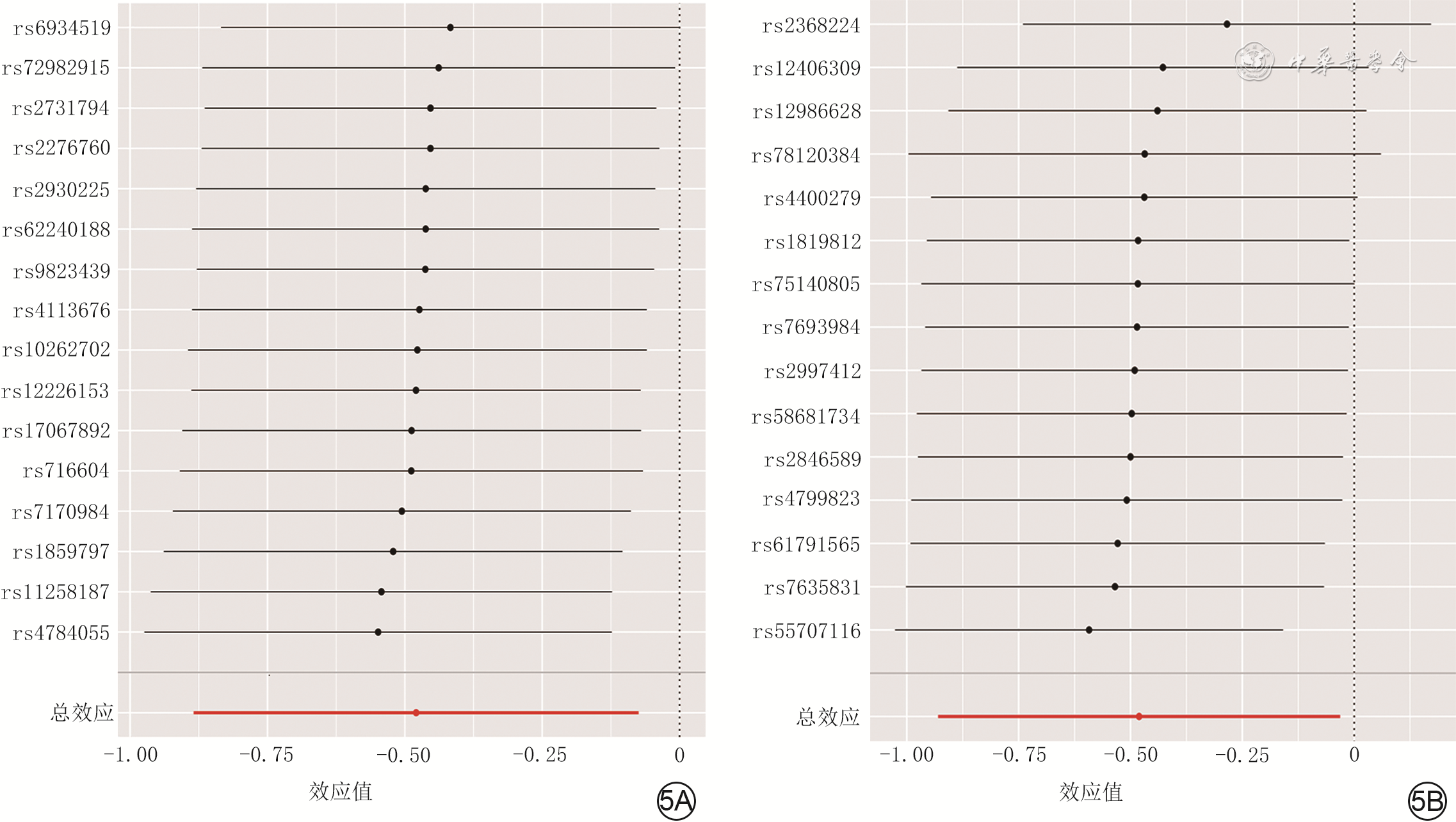
| Citation: | Chen WT,Wang XX,Zheng WL,et al.Exploring the causality between intestinal flora and hyperplastic scars of human based on two-sample Mendelian randomization analysis[J].Chin J Burns Wounds,2024,40(4):333-341.DOI: 10.3760/cma.j.cn501225-20231129-00215.

|

| [1] |
WangZC, ZhaoWY, CaoY, et al. The roles of inflammation in keloid and hypertrophic scars[J]. Front Immunol, 2020,11:603187. DOI: 10.3389/fimmu.2020.603187.
|
| [2] |
ChiangRS, BorovikovaAA, KingK, et al. Current concepts related to hypertrophic scarring in burn injuries[J]. Wound Repair Regen, 2016,24(3):466-477. DOI: 10.1111/wrr.12432.
|
| [3] |
LeeHJ, JangYJ. Recent understandings of biology, prophylaxis and treatment strategies for hypertrophic scars and keloids[J]. Int J Mol Sci, 2018,19(3):711.DOI: 10.3390/ijms19030711.
|
| [4] |
郑建新,张亚军. 二氧化碳点阵激光联合曲安奈德注射治疗增生性瘢痕的疗效[J]. 中国医疗美容,2020,10(5):84-88. DOI: 10.19593/j.issn.2095-0721.2020.05.020.
|
| [5] |
NiuM, ChenP. Crosstalk between gut microbiota and sepsis[J/OL]. Burns Trauma, 2021,9:tkab036[2023-11-29]. https://pubmed.ncbi.nlm.nih.gov/34712743/. DOI: 10.1093/burnst/tkab036.
|
| [6] |
CamposLF, TagliariE, CasagrandeT, et al. Effects of probiotics supplementation on skin wound healing in diabetic rats[J]. Arq Bras Cir Dig, 2020,33(1):e1498. DOI: 10.1590/0102-672020190001e1498.
|
| [7] |
AlamA, NeishA. Role of gut microbiota in intestinal wound healing and barrier function[J]. Tissue Barriers, 2018,6(3):1539595. DOI: 10.1080/21688370.2018.1539595.
|
| [8] |
吴金春, 刘彦民, 苏晓灵. 肠道菌群/肠道微生态与心血管疾病发生的关系研究进展[J].解放军医学杂志,2023,48(7):851-855. DOI: 10.11855/j.issn.0577-7402.2696.2022.0830.
|
| [9] |
WaasdorpM, KromBP, BikkerFJ, et al. The bigger picture: why oral mucosa heals better than skin[J]. Biomolecules, 2021,11(8):1165.DOI: 10.3390/biom11081165.
|
| [10] |
PatelBK, PatelKH, HuangRY, et al. The gut-skin microbiota axis and its role in diabetic wound healing-a review based on current literature[J]. Int J Mol Sci, 2022, 23(4):2375. DOI: 10.3390/ijms23042375.
|
| [11] |
BurgessS, FoleyCN, AllaraE, et al. A robust and efficient method for Mendelian randomization with hundreds of genetic variants[J]. Nat Commun, 2020,11(1):376. DOI: 10.1038/s41467-019-14156-4.
|
| [12] |
LevinMG, JudyR, GillD, et al. Genetics of height and risk of atrial fibrillation: a Mendelian randomization study[J]. PLoS Med, 2020,17(10):e1003288. DOI: 10.1371/journal.pmed.1003288.
|
| [13] |
O'NeillCA, MonteleoneG, McLaughlinJT, et al. The gut-skin axis in health and disease: a paradigm with therapeutic implications[J]. Bioessays, 2016,38(11):1167-1176. DOI: 10.1002/bies.201600008.
|
| [14] |
BelkaidY, HarrisonOJ. Homeostatic immunity and the microbiota[J]. Immunity, 2017,46(4):562-576. DOI: 10.1016/j.immuni.2017.04.008.
|
| [15] |
Polkowska-PruszyńskaB, GerkowiczA, KrasowskaD. The gut microbiome alterations in allergic and inflammatory skin diseases - an update[J]. J Eur Acad Dermatol Venereol, 2020,34(3):455-464. DOI: 10.1111/jdv.15951.
|
| [16] |
BenyacoubJ, BoscoN, BlanchardC, et al. Immune modulation property of Lactobacillus paracasei NCC2461 (ST11) strain and impact on skin defences[J]. Benef Microbes, 2014,5(2):129-136. DOI: 10.3920/BM2013.0014.
|
| [17] |
PeralMC, RachidMM, GobbatoNM, et al. Interleukin-8 production by polymorphonuclear leukocytes from patients with chronic infected leg ulcers treated with Lactobacillus plantarum[J]. Clin Microbiol Infect, 2010,16(3):281-286. DOI: 10.1111/j.1469-0691.2009.02793.x.
|
| [18] |
AfoudaP, DurandGA, LagierJC, et al. Noncontiguous finished genome sequence and description of Intestinimonas massiliensis sp. nov strain GD2T, the second Intestinimonas species cultured from the human gut[J]. Microbiologyopen, 2019,8(1):e00621. DOI: 10.1002/mbo3.621.
|
| [19] |
ZhouJ, LiM, ChenQ, et al. Programmable probiotics modulate inflammation and gut microbiota for inflammatory bowel disease treatment after effective oral delivery[J]. Nat Commun, 2022,13(1):3432. DOI: 10.1038/s41467-022-31171-0.
|
| [20] |
LiuJ, ChangG, HuangJ, et al. Sodium butyrate inhibits the inflammation of lipopolysaccharide-induced acute lung injury in mice by regulating the Toll-like receptor 4/nuclear factor κB signaling pathway[J]. J Agric Food Chem, 2019,67(6):1674-1682. DOI: 10.1021/acs.jafc.8b06359.
|
| [21] |
ArpaiaN, CampbellC, FanX, et al. Metabolites produced by commensal bacteria promote peripheral regulatory T-cell generation[J]. Nature, 2013,504(7480):451-455. DOI: 10.1038/nature12726.
|
| [22] |
HuangC, AkaishiS, HyakusokuH, et al. Are keloid and hypertrophic scar different forms of the same disorder? A fibroproliferative skin disorder hypothesis based on keloid findings[J]. Int Wound J, 2014,11(5):517-522. DOI: 10.1111/j.1742-481X.2012.01118.x.
|
| [23] |
HuangC, OgawaR. Role of inflammasomes in keloids and hypertrophic scars-lessons learned from chronic diabetic wounds and skin fibrosis[J]. Int J Mol Sci, 2022,23(12):6280.DOI: 10.3390/ijms23126820.
|
| [24] |
ZhangT, WangXF, WangZC, et al. Current potential therapeutic strategies targeting the TGF-β/Smad signaling pathway to attenuate keloid and hypertrophic scar formation[J]. Biomed Pharmacother, 2020,129:110287. DOI: 10.1016/j.biopha.2020.110287.
|
| [25] |
La ReauAJ, SuenG. The Ruminococci: key symbionts of the gut ecosystem[J]. J Microbiol, 2018,56(3):199-208. DOI: 10.1007/s12275-018-8024-4.
|
| [26] |
LiY, ZhangSX, YinXF, et al. The gut microbiota and its relevance to peripheral lymphocyte subpopulations and cytokines in patients with rheumatoid arthritis[J]. J Immunol Res, 2021,2021:6665563. DOI: 10.1155/2021/6665563.
|
| [27] |
ZhouN, ShenY, FanL, et al. The characteristics of intestinal-barrier damage in rats with IgA nephropathy[J]. Am J Med Sci, 2020,359(3):168-176. DOI: 10.1016/j.amjms.2019.11.011.
|
| [28] |
WangT, SternesPR, GuoXK, et al. Autoimmune diseases exhibit shared alterations in the gut microbiota[J]. Rheumatology (Oxford), 2024,63(3):856-865. DOI: 10.1093/rheumatology/kead364.
|
| [29] |
Lopez-SilesM, KhanTM, DuncanSH, et al. Cultured representatives of two major phylogroups of human colonic Faecalibacterium prausnitzii can utilize pectin, uronic acids, and host-derived substrates for growth[J]. Appl Environ Microbiol, 2012,78(2):420-428. DOI: 10.1128/AEM.06858-11.
|
| [30] |
MahmudMR, AkterS, TamannaSK, et al. Impact of gut microbiome on skin health: gut-skin axis observed through the lenses of therapeutics and skin diseases[J]. Gut Microbes, 2022,14(1):2096995. DOI: 10.1080/19490976.2022.2096995.
|

Figures(6)
Copyright © Chinese Journal of Burns京ICP备07035254号-14
E-mail:shaoshangzazhi@163.com
Supported by:
Beijing Renhe Information Technology Co. Ltd


 DownLoad: