


Role and mechanism of Vγ4 T cells in impaired wound healing of rapamycin-induced full-thickness skin defects in mice
-
摘要:
目的 探讨Vγ4T细胞在应用雷帕霉素的小鼠全层皮肤缺损创面愈合障碍中的作用及其机制。 方法 采用实验研究方法,选取86只8~12周龄雄性C57BL/6J小鼠(以下简称野生型小鼠)进行后续实验。取5只野生型小鼠,从其腋窝淋巴结分离Vγ4T细胞用于后续实验。取42只野生型小鼠,腹腔注射雷帕霉素建立应用雷帕霉素的小鼠模型,用于后续实验。取18只野生型小鼠,按随机数字表法(分组方法下同)分为不进行任何处理的正常对照组和单纯创伤组、创伤+CC趋化因子配体20(CCL20)抑制剂组(每组6只),将后2组小鼠背部制成全层皮肤缺损创面(创面模型下同),创伤+CCL20抑制剂组小鼠伤后连续3 d于创缘皮下注射CCL20抑制剂,另取6只应用雷帕霉素的小鼠建立创面模型作为雷帕霉素+创伤组,伤后3 d,采用酶消化法提取各创伤小鼠创周皮肤组织的表皮细胞,采用流式细胞仪检测表皮细胞中Vγ4T细胞的百分比。于适宜时间点取正常对照组小鼠背部正常皮肤组织的表皮细胞同前进行检测。取5只野生型小鼠建立创面模型,伤后3 d,提取创周皮肤组织的表皮细胞,采用流式细胞分选仪将细胞群分为Vγ4T细胞、Vγ3T细胞及γδ阴性细胞,分别设为Vγ4T细胞组、Vγ3T细胞组及γδ阴性细胞组(均与B16小鼠黑色素瘤细胞混合),以单纯B16小鼠黑色素瘤细胞为黑色素瘤细胞对照组,采用实时荧光定量反转录PCR(RT-PCR)法检测各组细胞白细胞介素22(IL-22)mRNA表达情况(样本数为6)。取30只应用雷帕霉素的小鼠建立创面模型,伤后即刻分为进行相应注射处理的单纯Vγ4T细胞组与Vγ4T细胞+IL-22抑制剂组以及注射PBS的雷帕霉素对照组(每组10只);另取10只野生型小鼠建立创面模型并注射PBS作为野生型对照组。各组小鼠均连续注射6 d,伤后1、2、3、4、5、6 d于当日注射后计算4组小鼠创面面积百分比。分别取6只野生型小鼠和6只应用雷帕霉素的小鼠建立创面模型,作为野生型组和雷帕霉素组,伤后3 d,分别采用实时荧光定量RT-PCR法及蛋白质印迹法检测2组小鼠创周表皮组织中IL-22、CCL20的mRNA及蛋白的表达情况。取Vγ4T细胞,分为不进行任何处理的正常对照组和用雷帕霉素处理的雷帕霉素组,培养24 h,分别采用实时荧光定量RT-PCR法及蛋白质印迹法检测2组细胞中IL-22的mRNA及蛋白表达情况(样本数为6)。数据分析采用独立样本t检验、重复测量方差分析、单因素方差分析、Bonferroni法、Kruskal-Wallis H检验与Wilcoxon秩和检验。 结果 单纯创伤组小鼠伤后3 d创周皮肤组织的表皮细胞中Vγ4T细胞百分比为0.66%(0.52%,0.81%),明显高于正常对照组小鼠正常皮肤组织的表皮细胞中的0.09%(0.04%,0.14%),Z=4.31,P<0.01;雷帕霉素+创伤组及创伤+CCL20抑制剂组小鼠伤后3 d创周皮肤组织的表皮细胞中Vγ4T细胞百分比分别为0.25%(0.16%,0.37%)、0.24%(0.17%,0.35%),均较单纯创伤组明显降低(Z值分别为2.27、2.25,P<0.05)。Vγ4T细胞组细胞中IL-22 mRNA表达水平明显高于Vγ3T细胞组、γδ阴性细胞组、黑色素瘤细胞对照组(Z值分别为2.96、2.45、3.41,P<0.05或P<0.01)。与野生型对照组比较,雷帕霉素对照组小鼠伤后1~6 d创面面积百分比均明显增大(P<0.01),Vγ4T细胞+IL-22抑制剂组小鼠伤后1 d及伤后3~6 d创面面积百分比均明显增大(P<0.05或P<0.01)。与雷帕霉素对照组比较,单纯Vγ4T细胞组小鼠伤后1~6 d创面面积百分比均明显减小(P<0.05或P<0.01)。与单纯Vγ4T细胞组比较,Vγ4T细胞+IL-22抑制剂组小鼠伤后3~6 d创面面积百分比均明显增大(P<0.05或P<0.01)。伤后3 d,与野生型组比较,雷帕霉素组小鼠创周表皮组织中IL-22蛋白及mRNA的表达水平(t值分别为-7.82、-5.04,P<0.01)、CCL20蛋白及mRNA的表达水平(t值分别为-7.12、-5.73,P<0.01)均显著下降。培养24 h,雷帕霉素组Vγ4T细胞中IL-22蛋白及mRNA的表达水平均显著低于正常对照组(t值分别为-7.75、-6.04,P<0.01)。 结论 在全层皮肤缺损小鼠中,雷帕霉素可能通过抑制CCL20表达使CCL20趋化系统受损导致Vγ4T细胞向表皮的募集减少,并同时抑制Vγ4T细胞分泌IL-22从而减缓创面愈合速度。 Abstract:Objective To investigate the role and mechanism of Vγ4 T cells in impaired wound healing of rapamycin-induced full-thickness skin defects in mice. Methods The experimental research methods were applied. Eighty-six C57BL/6J male mice (hereinafter briefly referred to as wild-type mice) aged 8-12 weeks were selected for the following experiments. Vγ4 T cells were isolated from axillary lymph nodes of five wild-type mice for the following experiments. Intraperitoneal injection of rapamycin for 42 mice was performed to establish rapamycin-treated mice model for the following experiments. Eighteen wild-type mice were divided into normal control group without any treatment, trauma only group, and trauma+CC chemokine ligand 20 (CCL20) inhibitor group according to the random number table (the same grouping method below), with 6 mice in each group. The full-thickness skin defect wound was made on the back of mice in the latter two groups (the same wound model below), and mice in trauma+CCL20 inhibitor group were continuously injected subcutaneously with CCL20 inhibitor at the wound edge for 3 days after injury. Another 6 rapamycin-treated mice were used to establish wound model as rapamycin+trauma group. On post injury day (PID) 3, the epidermal cells of the skin tissue around the wound of each trauma mice were extracted by enzyme digestion, and the percentage of Vγ4 T cells in the epidermal cells was detected by flow cytometry. In normal control group, the epidermal cells of the normal skin tissue in the back of mice were taken at the appropriate time point for detection as above. Five wild-type mice were used to establish wound models. On PID 3, the epidermal cells were extracted from the skin tissue around the wound. The cell populations were divided into Vγ4 T cells, Vγ3 T cells, and γδ negative cells by fluorescence-activated cell sorter, which were set as Vγ4 T cell group, Vγ3 T cell group, and γδ negative cell group (with cells in each group being mixed with B16 mouse melanoma cells), respectively. B16 mouse melanoma cells were used as melanoma cell control group. The expression of interleukin-22 (IL-22) mRNA in cells of each group was detected by real-time fluorescence quantitative reverse transcription polymerase chain reaction (RT-PCR), with the number of samples being 6. Thirty rapamycin-treated mice were used to establish wound models, which were divided into Vγ4 T cell only group and Vγ4 T cell+IL-22 inhibitor group performed with corresponding injections and rapamycin control group injected with phosphate buffer solution (PBS) immediately after injury, with 10 mice in each group. Another 10 wild-type mice were taken to establish wound models and injected with PBS as wild-type control group. Mice in each group were injected continuously for 6 days. The percentage of wound area of mice in the four groups was calculated on PID 1, 2, 3, 4, 5, and 6 after injection on the same day. Six wild-type mice and 6 rapamycin-treated mice were taken respectively to establish wound models as wild-type group and rapamycin group. On PID 3, the mRNA and protein expressions of IL-22 and CCL20 in the peri-wound epidermis tissue of mice in the two groups were detected by real-time fluorescence quantitative RT-PCR and Western blotting, respectively. The Vγ4 T cells were divided into normal control group without any treatment and rapamycin-treated rapamycin group. After being cultured for 24 hours, the mRNA and protein expressions of IL-22 of cells in the two groups were detected by real-time fluorescence quantitative RT-PCR and Western blotting, respectively, with the number of samples being 6. Data were statistically analyzed with independent sample t test, analysis of variance for repeated measurement, one-way analysis of variance, Bonferroni method, Kruskal-Wallis H test, and Wilcoxon rank sum test. Results The percentage of Vγ4 T cells in the epidermal cells of the skin tissue around the wound of mice in trauma only group on PID 3 was 0.66% (0.52%, 0.81%), which was significantly higher than 0.09% (0.04%, 0.14%) in the epidermal cells of the normal skin tissue of mice in normal control group (Z=4.31, P<0.01). The percentages of Vγ4 T cells in the epidermal cells of the skin tissue around the wound of mice in rapamycin+trauma group and trauma+CCL20 inhibitor group on PID 3 were 0.25% (0.16%, 0.37%) and 0.24% (0.17%, 0.35%), respectively, which were significantly lower than that in trauma only group (with Z values of 2.27 and 2.25, respectively, P<0.05). The mRNA expression level of IL-22 of cells in Vγ4 T cell group was significantly higher than that in Vγ3 T cell group, γδ negative cell group, and melanoma cell control group (with Z values of 2.96, 2.45, and 3.41, respectively, P<0.05 or P<0.01). Compared with that in wild-type control group, the percentage of wound area of mice in rapamycin control group increased significantly on PID 1-6 (P<0.01), the percentage of wound area of mice in Vγ4 T cell+IL-22 inhibitor group increased significantly on PID 1 and PID 3-6 (P<0.05 or P<0.01). Compared with that in rapamycin control group, the percentage of wound area of mice in Vγ4 T cell only group decreased significantly on PID 1-6 (P<0.05 or P<0.01). Compared with that in Vγ4 T cell only group, the percentage of wound area of mice in Vγ4 T cell+IL-22 inhibitor group increased significantly on PID 3-6 (P<0.05 or P<0.01). On PID 3, compared with those in wild-type group, the expression levels of IL-22 protein and mRNA (with t values of -7.82 and -5.04, respectively, P<0.01) and CCL20 protein and mRNA (with t values of -7.12 and -5.73, respectively, P<0.01) were decreased significantly in the peri-wound epidermis tissue of mice in rapamycin group. After being cultured for 24 hours, the expression levels of IL-22 protein and mRNA in Vγ4 T cells in rapamycin group were significantly lower than those in normal control group (with t values of -7.75 and -6.04, respectively, P<0.01). Conclusions In mice with full-thickness skin defects, rapamycin may impair the CCL20 chemotactic system by inhibiting the expression of CCL20, leading to a decrease in the recruitment of Vγ4 T cells to the epidermis, and at the same time inhibit the secretion of IL-22 by Vγ4 T cells, thereby slowing the wound healing rate. -
Key words:
- Sirolimus /
- Wound healing /
- Chemokine CCL20 /
- Vγ4 T cells /
- Interleukin-22
-
参考文献
(35) [1] SehgalSN.Sirolimus: its discovery, biological properties, and mechanism of action[J].Transplant Proc,2003,35(3 Suppl):S7-14.DOI: 10.1016/s0041-1345(03)00211-2. [2] HymesLC,WarshawBL.Sirolimus in pediatric patients: results in the first 6 months post-renal transplant[J].Pediatr Transplant,2005,9(4):520-522.DOI: 10.1111/j.1399-3046.2005.00324.x. [3] SchäfferM,SchierR,NapireiM,et al.Sirolimus impairs wound healing[J].Langenbecks Arch Surg,2007,392(3):297-303.DOI: 10.1007/s00423-007-0174-5. [4] MacDonaldAS.Rapamycin in combination with cyclosporine or tacrolimus in liver, pancreas, and kidney transplantation[J].Transplant Proc,2003,35(3 Suppl):S201-208.DOI: 10.1016/s0041-1345(03)00231-8. [5] KuppahallyS,Al-KhaldiA,WeisshaarD,et al.Wound healing complications with de novo sirolimus versus mycophenolate mofetil-based regimen in cardiac transplant recipients[J].Am J Transplant,2006,6(5 Pt 1):986-992.DOI: 10.1111/j.1600-6143.2006.01282.x. [6] DeanPG,LundWJ,LarsonTS,et al.Wound-healing complications after kidney transplantation: a prospective, randomized comparison of sirolimus and tacrolimus[J].Transplantation,2004,77(10):1555-1561.DOI: 10.1097/01.tp.0000123082.31092.53. [7] ValenteJF,HricikD,WeigelK,et al.Comparison of sirolimus vs. mycophenolate mofetil on surgical complications and wound healing in adult kidney transplantation[J].Am J Transplant,2003,3(9):1128-1134.DOI: 10.1034/j.1600-6143.2003.00185.x. [8] MillsRE,TaylorKR,PodshivalovaK,et al.Defects in skin gamma delta T cell function contribute to delayed wound repair in rapamycin-treated mice[J].J Immunol,2008,181(6):3974-3983.DOI: 10.4049/jimmunol.181.6.3974. [9] HaidingerM,PoglitschM,GeyereggerR,et al.A versatile role of mammalian target of rapamycin in human dendritic cell function and differentiation[J].J Immunol,2010,185(7):3919-3931.DOI: 10.4049/jimmunol.1000296. [10] YanezDA,LacherRK,VidyarthiA,et al.The role of macrophages in skin homeostasis[J].Pflugers Arch,2017,469(3/4):455-463.DOI: 10.1007/s00424-017-1953-7. [11] DiegelmannRF,EvansMC.Wound healing: an overview of acute, fibrotic and delayed healing[J].Front Biosci,2004,9:283-289.DOI: 10.2741/1184. [12] SutohY,MohamedRH,KasaharaM.Origin and evolution of dendritic epidermal T cells[J].Front Immunol,2018,9:1059.DOI: 10.3389/fimmu.2018.01059. [13] ChenC,MengZ,RenH,et al.The molecular mechanisms supporting the homeostasis and activation of dendritic epidermal T cell and its role in promoting wound healing[J/OL].Burns Trauma,2021,9:tkab009[2022-03-22]. https://pubmed.ncbi.nlm.nih.gov/34212060/.DOI: 10.1093/burnst/tkab009. [14] MarshallAS,SilvaJR,BannermanCA,et al.Skin-resident γδ T cells exhibit site-specific morphology and activation states[J].J Immunol Res,2019,2019:9020234.DOI: 10.1155/2019/9020234. [15] LiuZ,XuY,ZhangX,et al.Defects in dermal Vγ4 γ δ T cells result in delayed wound healing in diabetic mice[J].Am J Transl Res,2016,8(6):2667-2680. [16] MabuchiT,TakekoshiT,HwangST.Epidermal CCR6+ γδ T cells are major producers of IL-22 and IL-17 in a murine model of psoriasiform dermatitis[J].J Immunol,2011,187(10):5026-5031.DOI: 10.4049/jimmunol.1101817. [17] McGeeHM,SchmidtBA,BoothCJ,et al.IL-22 promotes fibroblast-mediated wound repair in the skin[J].J Invest Dermatol,2013,133(5):1321-1329.DOI: 10.1038/jid.2012.463. [18] AugustineJJ,BodziakKA,HricikDE.Use of sirolimus in solid organ transplantation[J].Drugs,2007,67(3):369-391.DOI: 10.2165/00003495-200767030-00004. [19] RuchinPE,MullerDW,FaddySC,et al.Long-term clinical follow-up of sirolimus-eluting (CYPHER) coronary stents in the treatment of instent restenosis in an unselected population[J].Heart Lung Circ,2007,16(6):440-446.DOI: 10.1016/j.hlc.2007.02.090. [20] WitzigTE,KaufmannSH.Inhibition of the phosphatidylinositol 3-kinase/mammalian target of rapamycin pathway in hematologic malignancies[J].Curr Treat Options Oncol,2006,7(4):285-294.DOI: 10.1007/s11864-006-0038-1. [21] PhungTL,ZivK,DabydeenD,et al.Pathological angiogenesis is induced by sustained Akt signaling and inhibited by rapamycin[J].Cancer Cell,2006,10(2):159-170.DOI: 10.1016/j.ccr.2006.07.003. [22] 贺伟峰.皮肤γδT细胞各亚群在创面再上皮化过程中的调控作用及其相关机制[J].中华烧伤与创面修复杂志,2022,38(2):114-118.DOI: 10.3760/cma.j.cn501120-20211210-00411. [23] 刘勉,朱海杰,杨加彩,等.树突状表皮T淋巴细胞对小鼠创缘表皮细胞增殖和凋亡的影响[J].中华烧伤杂志,2020,36(2):122-130.DOI: 10.3760/cma.j.issn.1009-2587.2020.02.008. [24] 王珏,张小容,贺伟峰,等.树突状表皮T细胞在创面愈合中作用机制的研究进展[J].中华烧伤杂志,2021,37(3):296-300.DOI: 10.3760/cma.j.cn501120-20200226-00092. [25] ZhengY,CollinsSL,LutzMA,et al.A role for mammalian target of rapamycin in regulating T cell activation versus anergy[J].J Immunol,2007,178(4):2163-2170.DOI: 10.4049/jimmunol.178.4.2163. [26] SchutyserE,StruyfS,Van DammeJ.The CC chemokine CCL20 and its receptor CCR6[J].Cytokine Growth Factor Rev,2003,14(5):409-426.DOI: 10.1016/s1359-6101(03)00049-2. [27] HomeyB,Dieu-NosjeanMC,WiesenbornA,et al.Up-regulation of macrophage inflammatory protein-3 alpha/CCL20 and CC chemokine receptor 6 in psoriasis[J].J Immunol,2000,164(12):6621-6632.DOI: 10.4049/jimmunol.164.12.6621. [28] MabuchiT,SinghTP,TakekoshiT,et al.CCR6 is required for epidermal trafficking of γδ-T cells in an IL-23-induced model of psoriasiform dermatitis[J].J Invest Dermatol,2013,133(1):164-171.DOI: 10.1038/jid.2012.260. [29] FurueK,ItoT,TsujiG,et al.The CCL20 and CCR6 axis in psoriasis[J].Scand J Immunol,2020,91(3):e12846.DOI: 10.1111/sji.12846. [30] FujitaH.The role of IL-22 and Th22 cells in human skin diseases[J].J Dermatol Sci,2013,72(1):3-8.DOI: 10.1016/j.jdermsci.2013.04.028. [31] MiyagakiT,SugayaM,SugaH,et al.IL-22, but not IL-17, dominant environment in cutaneous T-cell lymphoma[J].Clin Cancer Res,2011,17(24):7529-7538.DOI: 10.1158/1078-0432.CCR-11-1192. [32] AvitabileS,OdorisioT,MadonnaS,et al.Interleukin-22 promotes wound repair in diabetes by improving keratinocyte pro-healing functions[J].J Invest Dermatol,2015,135(11):2862-2870.DOI: 10.1038/jid.2015.278. [33] KolumamG,WuX,LeeWP,et al.IL-22R ligands IL-20, IL-22, and IL-24 promote wound healing in diabetic db/db mice[J].PLoS One,2017,12(1):e0170639.DOI: 10.1371/journal.pone.0170639. [34] TillackC,EhmannLM,FriedrichM,et al.Anti-TNF antibody- induced psoriasiform skin lesions in patients with inflammatory bowel disease are characterised by interferon-γ-expressing Th1 cells and IL-17A/IL-22-expressing Th17 cells and respond to anti-IL-12/IL-23 antibody treatment[J].Gut,2014,63(4):567-577.DOI: 10.1136/gutjnl-2012-302853. [35] Van BelleAB,de HeuschM,LemaireMM,et al.IL-22 is required for imiquimod-induced psoriasiform skin inflammation in mice[J].J Immunol,2012,188(1):462-469.DOI: 10.4049/jimmunol.1102224. -

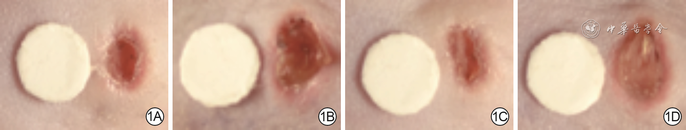
1 4组全层皮肤缺损小鼠伤后3 d创面面积。1A.野生型对照组创面面积较参照物明显缩小;1B.雷帕霉素对照组创面面积明显大于图1A;1C.单纯Vγ4T细胞组创面面积与图1A相近,明显小于图1B;1D.Vγ4T细胞+白细胞介素22抑制剂组创面面积与图1B相近,明显大于图1C
注:除野生型对照组外,其余3组小鼠均应用了雷帕霉素

2 2组全层皮肤缺损小鼠伤后3 d创周表皮组织中IL-22和CCL20的蛋白与mRNA表达。2A.蛋白质印迹法检测IL-22蛋白表达;2B.IL-22蛋白与mRNA的表达;2C.蛋白质印迹法检测CCL20蛋白表达;2D.CCL20蛋白与mRNA的表达
注:条带图上方与条图横坐标下1、2均分别指野生型组、雷帕霉素组;IL-22为白细胞介素22,GAPDH为3-磷酸甘油醛脱氢酶,CCL20为CC趋化因子配体20;与野生型组比较,aP<0.01

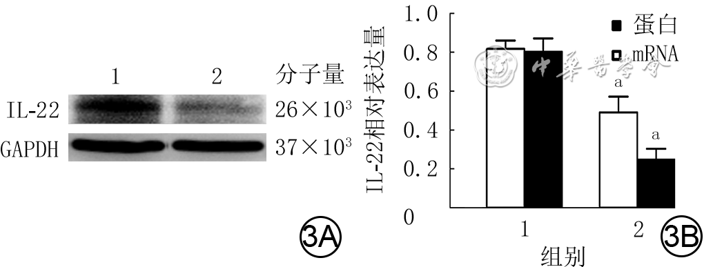
3 2组Vγ4T细胞培养24 h的IL-22蛋白与mRNA表达。3A.蛋白质印迹法检测IL-22的蛋白表达;3B.IL-22蛋白与mRNA的表达
注:条带图上方与条图横坐标下1、2均分别指正常对照组、雷帕霉素组;IL-22为白细胞介素22,GAPDH为3-磷酸甘油醛脱氢酶;与正常对照组比较,aP<0.01
表1 4组全层皮肤缺损小鼠伤后各时间点创面面积百分比比较(%,
组别 样本数 1 d 2 d 3 d 4 d 5 d 6 d 野生型对照组 10 79.3±8.4 65.7±9.6 47.0±9.7 31.5±7.8 21.5±8.1 11.1±4.5 雷帕霉素对照组 10 93.3±2.9a 84.0±8.0a 72.5±9.2a 56.6±9.7a 42.9±7.6a 34.5±7.3a 单纯Vγ4T细胞组 10 85.4±5.1b 70.0±7.9d 54.3±9.4d 37.4±8.3d 27.7±5.3d 16.2±3.9d Vγ4T细胞+白细胞介素22抑制剂组 10 88.3±6.1c 76.6±9.9 68.4±10.2ae 56.2±14.2af 44.0±11.1af 29.9±8.8af F值 9.73 8.07 15.46 15.66 18.26 29.26 P值 <0.001 <0.001 <0.001 <0.001 <0.001 <0.001 注:除野生型对照组外,其余3组小鼠均应用了雷帕霉素;处理因素主效应,F=18.88,P<0.001;时间因素主效应,F=1 126.05,P<0.001;两者交互作用,F=5.05,P<0.001;与野生型对照组比较,aP<0.01,cP<0.05;与雷帕霉素对照组比较,bP<0.05,dP<0.01;与单纯Vγ4T细胞组比较,eP<0.05,fP<0.01  下载: 导出CSV
-

 下载:
下载:


计量
- 文章访问数: 525
- HTML全文浏览量: 183
- PDF下载量: 21
- 被引次数: 0
