


Clinical effects of superficial temporal artery lobulated perforator flaps in repairing skin and soft tissue defects after temporal tumor resection
-
摘要:
目的 探讨应用颞浅动脉分叶穿支皮瓣修复颞区肿瘤切除后皮肤软组织缺损的可行性和临床效果。 方法 采用回顾性观察性研究方法。2017年3月—2022年10月,遵义医科大学附属医院收治颞区皮肤肿瘤患者10例,其中女6例、男4例,年龄42~87岁,鳞状细胞癌者3例、基底细胞癌者7例,病程为6个月~5年。对所有颞区肿瘤均行扩大切除,肿瘤切除后遗留创面面积为5.4 cm×4.2 cm~7.0 cm×4.0 cm。设计颞浅动脉额支皮瓣(面积为5.5 cm×1.2 cm~7.0 cm×1.5 cm)、颞浅动脉下行支皮瓣(面积为4.2 cm×3.5 cm~5.0 cm×4.0 cm)及颞浅动脉顶支皮瓣(面积为4.2 cm×1.0 cm~5.0 cm×1.0 cm)修复创面并重建发际线。将皮瓣供区直接拉拢缝合。术后3~5 d观察皮瓣成活情况,术后5~7 d拆线时观察供受区创面愈合情况。术后随访时,观察患侧颞区外观、瘢痕增生情况、发际线重建情况及肿瘤复发情况。 结果 术后3~5 d所有皮瓣存活良好。术后5~7 d所有供受区创面愈合良好。术后3~6个月随访时,术区切口隐蔽,皮瓣不臃肿且色泽与周围皮肤无明显差异,未见明显瘢痕增生,患侧重建发际线与健侧无明显差异,局部肿瘤均无复发。 结论 针对颞区的大面积皮肤软组织缺损,应用颞浅动脉分叶穿支皮瓣可在分区修复创面的同时Ⅰ期缝合供区,手术操作简便,术后面部外观符合美学要求,局部肿瘤均无复发,修复效果良好。本术式特别适合老年患者颞区大面积皮肤软组织缺损的修复。 Abstract:Objective To explore the feasibility and clinical effects of using superficial temporal artery lobulated perforator flaps in repairing skin and soft tissue defects after tumor resection in the temporal region. Methods A retrospective observational study method was used. From March 2017 to October 2022, ten patients with temporal skin tumors were admitted to the Affiliated Hospital of Zunyi Medical University, including six women and four men, with age ranging from 42 to 87 years. Among them, three patients had squamous cell carcinoma and seven patients had basal cell carcinoma, with disease duration ranging from 6 months to 5 years. All temporal tumors underwent expanded resection, leaving wound areas of 5.4 cm×4.2 cm to 7.0 cm×4.0 cm after tumor resection. Superficial temporal artery frontal branch flaps with areas of 5.5 cm×1.2 cm to 7.0 cm×1.5 cm, superficial temporal artery descending branch flaps with areas of 4.2 cm×3.5 cm to 5.0 cm×4.0 cm, and superficial temporal artery parietal branch flaps with areas of 4.2 cm×1.0 cm to 5.0 cm×1.0 cm were designed to repair the wounds and reconstruct the hairline. The donor areas of the flaps were closed and sutured directly. The survival of the flaps was observed on 3 to 5 days after surgery, and the healing of wounds on the donor and recipient sites was observed when the stitches were removed on 5 to 7 days after surgery. During follow-up after surgery, the appearance of the temporal area, scar hyperplasia, hairline reconstruction, and tumor recurrence were observed in the temporal region on the affected side. Results All the flaps survived well on 3 to 5 days after surgery, and all the donor and recipient site wounds healed well on 5 to 7 days after surgery. During follow-up of 3 to 6 months after surgery, the surgical incisions were concealed; the flaps were not swollen, with a consistent color to the surrounding skin; there were no obvious hypertrophic scars; the reconstructed hairline on the affected side was not significantly different from that of the healthy side; there was no tumor recurrence in the local area. Conclusions For large areas of skin and soft tissue defects in the temporal region, the use of superficial temporal artery lobulated perforator flaps can repair the wounds in different regions and suture the donor sites in the primary stage simultaneously. The surgical operation is simple, and the facial appearance conforms to the aesthetic requirement after surgery with no tumor recurrence in the local area but a good repair effect. This method is particularly suitable for repairing large areas of skin and soft tissue defects in the temporal region in elderly patients. -
Key words:
- Surgical flaps /
- Facial neoplasms /
- Superficial temporal artery /
- Wound repair /
- Temporal region
-
参考文献
(33) [1] HäusslerD,SadickH,RotterN.Facial skin tumors and plastic reconstruction: part 1[J].Laryngorhinootologie,2019,98(2):117-128.DOI: 10.1055/a-0784-9844. [2] Rodríguez-LorenzoA,AudolfssonT,WongC,et al.Vascular perfusion of the facial skin: implications in allotransplantation of facial aesthetic subunits[J].Plast Reconstr Surg,2016,138(5):1073-1079.DOI: 10.1097/PRS.0000000000002701. [3] RighiB,RobottiE,CarminatiM.Combining rotation and V-Y design for lower forehead and temporal region reconstruction with myocutaneous frontal flap: our experience on 27 consecutive cases[J].Ann Plast Surg,2020,85(2):135-140.DOI: 10.1097/SAP.0000000000002256. [4] RichmonJD,YarlagaddaBB,WaxMK,et al.Locoregional and free flap reconstruction of the lateral skull base[J].Head Neck,2015,37(9):1387-1391.DOI: 10.1002/hed.23725. [5] ChowM,SwiftR,HigginsS,et al.Triple advancement flap for the lateral upper forehead and temple[J].J Cutan Med Surg,2018,22(5):533-534.DOI: 10.1177/1203475418777952. [6] HannaTC,GatesJC,KrausDH.Vertical submental island flap for head and neck reconstruction[J].J Oral Maxillofac Surg,2020,78(9):1653.e1-1653.e6.DOI: 10.1016/j.joms.2020.05.010. [7] KoziejM,WnukJ,PolakJ,et al.The superficial temporal artery: a meta-analysis of its prevalence and morphology[J].Clin Anat,2020,33(8):1130-1137.DOI: 10.1002/ca.23550. [8] MinP,LiJ,BrunettiB,et al.Pre-expanded bipedicled visor flap: an ideal option for the reconstruction of upper and lower lip defects postburn in Asian males[J/OL].Burns Trauma,2020,8:tkaa005[2022-08-16].https://pubmed.ncbi.nlm.nih.gov/32341918/. DOI: 10.1093/burnst/tkaa005. [9] KoziejM,TrybusM,HołdaM,et al.The superficial temporal artery: anatomical map for facial reconstruction and aesthetic procedures[J].Aesthet Surg J,2019,39(8):815-823.DOI: 10.1093/asj/sjy287. [10] Jean-PhilippeH,BenoîtB,FrançoiseK,et al.Anatomy and external landmarks of the superficial temporal artery using 3-dimensional computed tomography[J].Surg Radiol Anat,2021,43(2):283-290.DOI: 10.1007/s00276-020-02589-2. [11] HongWJ,LiaoZF,ZengL,et al.Tomography of the forehead arteries and tailored filler injection for forehead volumizing and contouring[J].Dermatol Surg,2020,46(12):1615-1620.DOI: 10.1097/DSS.0000000000002561. [12] MwachakaP, SinkeetS, Ogeng'oJ. Superficial temporal artery among Kenyans: pattern of branching and its relation to pericranial structures[J]. Folia Morphol (Warsz), 2010,69(1):51-53. [13] MedvedF,ManoliT,MedesanR,et al.In vivo analysis of the vascular pattern of the superficial temporal artery based on digital subtraction angiography[J].Microsurgery,2015,35(5):380-386.DOI: 10.1002/micr.22348. [14] RubioRR,LawtonMT,KolaO,et al.The middle temporal artery: surgical anatomy and exposure for cerebral revascularization[J].World Neurosurg,2018,110:e79-e83.DOI: 10.1016/j.wneu.2017.10.100. [15] TalmageGD,SundeJ,WalkerDD,et al.Anatomic basis of the middle temporal artery periosteal rotational flap in otologic surgery[J].Laryngoscope,2016,126(6):1426-1432.DOI: 10.1002/lary.25635. [16] TilottaF,LazarooB,LaujacMH,et al.A study of the vascularization of the auricle by dissection and diaphanization[J].Surg Radiol Anat,2009,31(4):259-265.DOI: 10.1007/s00276-008-0438-y. [17] Al-QattanMM.One pedicled superficial temporal artery hair-bearing flap to reconstruct three different anatomical areas of the burnt face: a personal technique[J].Ann Plast Surg,2021,86(2):159-161.DOI: 10.1097/SAP.0000000000002613. [18] PangXY,RenJ,XuW,et al.Aesthetic eyebrow reconstruction with an expanded scalp island flap pedicled by the superficial temporal artery[J].Aesthetic Plast Surg,2017,41(3):563-567.DOI: 10.1007/s00266-016-0736-4. [19] ZhaoWR,WangHB,LuoCE,et al.Three-dimensional computed tomographic study on the periorbital branches of the ophthalmic artery: arterial variations and clinical relevance[J].Aesthet Surg J,2019,39(10):1109-1117.DOI: 10.1093/asj/sjy228. [20] YamauchiM,YotsuyanagiT,YamashitaK,et al.The reverse superficial temporal artery flap from the preauricular region, for the small facial defects[J].J Plast Reconstr Aesthet Surg,2012,65(2):149-155.DOI: 10.1016/j.bjps.2011.09.013. [21] WeiJ,ChenQ,HerrlerT,et al.Supermicrosurgical reconstruction of nasal tip defects using the preauricular reversed superficial temporal artery flap[J].J Plast Reconstr Aesthet Surg,2020,73(1):58-64.DOI: 10.1016/j.bjps.2019.06.028. [22] GluthMB,SundeJ.Vascularized reconstruction of partial external auditory canal wall defects using the middle temporal artery flap[J].Otol Neurotol,2014,35(1):e31-35.DOI: 10.1097/MAO.0000000000000182. [23] TennaS,BrunettiB,AvetaA,et al.Scalp reconstruction with superficial temporal artery island flap: clinical experience on 30 consecutive cases[J].J Plast Reconstr Aesthet Surg,2013,66(5):660-666.DOI: 10.1016/j.bjps.2013.01.010. [24] 刘梅君,聂开瑜,胡鹏,等.颞浅动脉复合穿支皮瓣修复颞区皮肤软组织缺损并重建鬓角[J].中国修复重建外科杂志,2020,34(4):493-496.DOI: 10.7507/1002-1892.201909046. [25] LebasD, WiartT, GrosC, et al.Use of a rhomboid flap to repair temporal and frontotemporal cutaneous defects: 11 cases[J]. Ann Dermatol Venereol, 2013, 140(3): 170-175. DOI: 10.1016/j.annder.2012.10.604. [26] 陈水莲,林泽旭.面部皮肤鳞状细胞癌扩大切除术联合全厚植皮及放疗的临床效果观察[J].中国医疗美容,2021,11(2):40-43.DOI: 10.19593/j.issn.2095-0721.2021.02.011. [27] LiuA,SuX,JiaoY,et al.Facial defect repair using a flap based on the superficial temporal artery[J].Plast Reconstr Surg Glob Open,2021,9(4):e3541.DOI: 10.1097/GOX.0000000000003541. [28] 夏成德,薛继东,狄海萍,等.额顶部跨中线轴型扩张皮瓣单蒂转移整复面颈部大面积瘢痕畸形的临床效果[J].中华烧伤杂志,2020,36(9):838-844.DOI: 10.3760/cma.j.cn501120-20200311-00149. [29] AhmedM,LohCYY.Frontal branch of the superficial temporal artery advancement flap with burrow's triangles excision for temporal defect reconstruction[J].J Maxillofac Oral Surg,2018,17(4):638-640.DOI: 10.1007/s12663-018-1145-9. [30] 聂开瑜,陈伟,魏在荣,等.颞浅动脉额支皮瓣与逆行耳后动脉皮瓣联合修复耳前皮肤软组织缺损[J].中华整形外科杂志,2017,33(1):8-11.DOI: 10.3760/cma.j.issn.1009-4598.2017.01.003. [31] 苏钰,申卫东,刘军,等.耳颞区不同程度复合组织缺损的修复:附三例报告[J].中华耳鼻咽喉头颈外科杂志,2021,56(5):487-492.DOI: 10.3760/cma.j.cn115330-20200622-00522. [32] 马超,陶然,舒军,等.背阔肌肌皮瓣修复较大软组织缺损的方法及供区继发创面的处理[J].中华烧伤杂志,2020,36(12):1199-1203.DOI: 10.3760/cma.j.cn501120-20191121-00439. [33] NiXD,XuY,WangM,et al.Application of superficial temporal artery flap in wound repairing after the resection of craniofacial malignant tumors[J].J Craniofac Surg,2020,31(2):534-537.DOI: 10.1097/SCS.0000000000006222. -

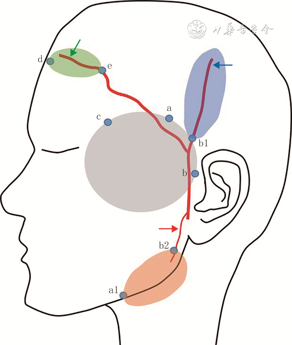
1 颞浅动脉分叶穿支皮瓣设计示意图
注:a点和b点间的连线为颞区发际线,a1点为下行支皮瓣远端,b1点为顶支皮瓣近端,b2点为下行支皮瓣近端,c点为b点斜上方创周的1点,d点为额支皮瓣远端,e点为额支皮瓣近端;灰色圆形示颞区肿瘤切除后创面,蓝色椭圆形示颞浅动脉顶支皮瓣,绿色椭圆形示颞浅动脉额支皮瓣,橙色椭圆形示颞浅动脉下行支皮瓣;蓝色箭头示颞浅动脉顶支,绿色箭头示颞浅动脉额支,红色箭头示颞浅动脉下行支
-

 下载:
下载:


图(3)
计量
- 文章访问数: 474
- HTML全文浏览量: 229
- PDF下载量: 27
- 被引次数: 0
