


Effects and mechanism of hepatocyte growth factor-modified human adipose mesenchymal stem cells on wound healing of full-thickness skin defects in diabetic rats
-
摘要:
目的 探讨肝细胞生长因子(HGF)修饰人脂肪间充质干细胞(ADSC)对糖尿病大鼠全层皮肤缺损创面愈合的影响及其机制。 方法 采用实验研究方法。取2019年12月于空军军医大学第一附属医院整形科行腹部抽脂术的1名35岁健康女性术后弃用腹部脂肪组织,采用胶原酶消化法获取呈长梭形的原代ADSC,其第3代细胞经流式细胞仪鉴定阳性表达ADSC表面标志物CD29、CD90,阴性表达CD34、CD45。取第3代ADSC进行后续实验。采用慢病毒介导的HGF转染ADSC 4 h(获得HGF修饰ADSC)后,常规培养24 h,倒置相差显微镜下观察细胞形态并计算转染率。取81只4周龄雄性SD大鼠,通过高糖高脂饮食联合链脲佐菌素注射诱导为糖尿病大鼠模型后,在每只大鼠背部制作1.5 cm×1.5 cm全层皮肤缺损创面。采用随机数字表法,将致伤大鼠分为磷酸盐缓冲液(PBS)组、单纯ADSC组、HGF修饰ADSC组,每组27只,均分别于伤后1、3、7 d在创周注射相同体积的相应物质。采用随机数字表法选取各组9只大鼠,于伤后0(即刻)、3、7、10、14 d(注射日注射后)测量创面面积并计算伤后3、7、10、14 d创面愈合率。伤后3、7 d行当日注射后分别处死各组剩余大鼠中的9只,取创周皮肤组织,采用实时荧光定量反转录PCR法检测伤后3 d肿瘤坏死因子α(TNF-α)、白细胞介素1β(IL-1β)、IL-10及伤后7 d Ⅰ、Ⅲ型胶原的mRNA表达,采用酶联免疫吸附测定法检测伤后7 d血管内皮生长因子(VEGF)的表达水平,采用蛋白质印迹法检测伤后3 d核因子κB-p65蛋白表达、伤后7 d蛋白激酶B(Akt)的磷酸化水平。对数据行重复测量方差分析、单因素方差分析、LSD-t检验及Bonferroni校正。 结果 培养24 h,转染HGF的ADSC形态良好,与未转染ADSC无差异,转染率达90%。伤后3、7、10、14 d,HGF修饰ADSC组大鼠创面愈合率分别为(31.5±1.0)%、(75.2±2.0)%、(92.2±1.3)%、(99.1±1.8)%,显著高于PBS组的(21.4±1.3)%、(61.4±1.5)%、(80.1±2.1)%、(92.4±1.8)%和单纯ADSC组的(25.1±2.1)%、(67.2±1.3)%、(89.3±1.4)%、(95.1±2.1)%(t=1.452、0.393、0.436、0.211,4.982、3.011、4.211、7.503,P<0.05或P<0.01)。伤后3 d,与PBS组和单纯ADSC组比较,HGF修饰ADSC组大鼠创周皮肤组织中TNF-α、IL-1β的mRNA表达及核因子κB-p65的蛋白表达均显著降低(t=7.281、17.700、9.447,6.231、13.083、7.783,P<0.01),IL-10的mRNA表达显著升高(t= - 6.644、 - 6.381,P<0.01)。伤后7 d,与PBS组和单纯ADSC组比较,HGF修饰ADSC组大鼠创周皮肤组织中Ⅰ、Ⅲ型胶原的mRNA表达及VEGF表达水平、Akt磷酸化水平均明显升高(t= - 5.126、 - 4.347、 - 5.058、 - 3.367, - 10.694、 - 19.876、 - 4.890、 - 6.819,P<0.05或P<0.01) 。 结论 HGF修饰的人ADSC可显著促进糖尿病大鼠全层皮肤缺损创面愈合,其机制可能与抑制TNF-α、IL-1β表达,促进IL-10、Ⅰ型胶原、Ⅲ型胶原及VEGF的表达相关,且可能与抑制核因子κB信号通路、促进Akt信号通路有关。 Abstract:Objective To investigate the effects and mechanism of hepatocyte growth factor (HGF)-modified human adipose mesenchymal stem cells (ADSCs) on the wound healing of full-thickness skin defects in diabetic rats. Methods The experimental research method was adopted. The discarded abdominal adipose tissue was collected from a 35-year-old healthy female who underwent abdominal liposuction in the Department of Plastic Surgery of the First Affiliated Hospital of Air Force Medical University in December 2019. The long spindle-shaped primary ADSCs were obtained by collagenase digestion, and the third passage of cells were identified by flow cytometry to positively express ADSCs surface markers CD29 and CD90 and negatively express CD34 and CD45. The third passage of ADSCs were used for the subsequent experiments. ADSCs were transfected with lentivirus-mediated HGF for 4 h (obtaining HGF modified ADSCs) and then routinely cultured for 24 h. The cell morphology was observed under an inverted phase contrast microscope, and the transfection rate was calculated. Eighty-one male Sprague-Dawley rats aged 4 weeks were induced into diabetic rat model by high glucose and high fat diet combined with streptozotocin injection. A full-thickness skin defect wound of 1.5 cm×1.5 cm was made on the back of each rat. The injured rats were divided into phosphate buffer solution (PBS) group, ADSCs alone group, and HGF-modified ADSCs group according to the random number table, with 27 rats in each group. The rats were injected with the same volume of corresponding substances around the wound on post injury day (PID) 1, 3, and 7, respectively. Nine rats in each group were selected according to the random number table, the wound area of whom was measured on PID 0 (immediately), 3, 7, 10, and 14 (after injection on injection day), and the wound healing rates on PID 3, 7, 10, and 14 were calculated. Nine remaining rats in each group were sacrificed after injection on PID 3 and 7, respectively, and the skin tissue around the wound were collected. The mRNA expressions of inflammatory factors such as tumor necrosis factor α (TNF-α), interleukin 1β (IL-1β), and IL-10 on PID 3 and collagen type Ⅰ and Ⅲ on PID 7 were detected by real-time fluorescent quantitative reverse transcription polymerase chain reaction. The expression level of vascular endothelial growth factor (VEGF) was detected by enzyme-linked immunosorbent assay on PID 7. The protein expression of nuclear factor κb-p65 on PID 3 and phosphorylation level of protein kinase B (Akt) on PID 7 were detected by Western blotting. Data were statistically analyzed with analysis of variance for repeated measurement, one-way analysis of variance, least significant difference t test, and Bonferroni correction. Results After 24 h of culture, the HGF-transfected human ADSCs showed good morphology, which was not different with the non-transfected ADSCs, and the transfection rate reached 90%. On PID 3, 7, 10, and 14, the wound healing rates of rats in HGF-modified ADSCs group were (31.5±1.0)%, (75.2±2.0)%, (92.2±1.3)%, and (99.1±1.8)%, respectively, being significantly higher than (21.4±1.3)%, (61.4±1.5)%, (80.1±2.1)%, and (92.4±1.8)% in PBS group and (25.1±2.1)%, (67.2±1.3)%, (89.3±1.4)%, and (95.1±2.1)% in ADSCs alone group (t=1.452, 0.393, 0.436, 0.211, 4.982, 3.011, 4.211, 7.503, P<0.05 or P<0.01). On PID 3, compared with those in PBS group and ADSCs alone group, the mRNA expressions of TNF-α and IL-1β and protein expression of nuclear factor κb-p65 in the skin tissue around the wound of rats in HGF-modified ADSCs group were significantly decreased (t=7.281, 17.700, 9.447, 6.231, 13.083, 7.783, P<0.01), and the mRNA expression of IL-10 in the skin tissue around the wound of rats in HGF-modified ADSCs group was significantly increased (t=-6.644, -6.381, P<0.01). On PID 7, compared with those in PBS group and ADSCs alone group, the mRNA expressions of collagen type Ⅰ and Ⅲ, the expression level of VEGF, and the phosphorylation level of Akt in the skin tissue around the wound of rats in HGF-modified ADSCs group were significantly increased (t=-5.126, -4.347, -5.058, -3.367, -10.694, -19.876, -4.890, -6.819, P<0.05 or P<0.01). Conclusions HGF-modified human ADSCs can significantly promote the wound healing of full-thickness skin defects in diabetic rats. The mechanism may be related to the inhibition of TNF-α and IL-1β expression, the promotion of IL-10, collagen type Ⅰ and Ⅲ, and VEGF expression, which could be related to the inhibition of nuclear factor κB signaling pathway, and the promotion of Akt signaling pathway. -
参考文献
(41) [1] MirzaR,KohTJ.Dysregulation of monocyte/macrophage phenotype in wounds of diabetic mice[J].Cytokine,2011,56(2):256-264.DOI: 10.1016/j.cyto.2011.06.016. [2] 姜玉峰.体表慢性难愈合创面的研究进展[J].感染、炎症、修复,2011,12(1):59-61.DOI: 10.3969/j.issn.1672-8521.2011.01.024. [3] CaoHM,ChengYQ,GaoHQ,et al.In vivo tracking of mesenchymal stem cell-derived extracellular vesicles improving mitochondrial function in renal ischemia-reperfusion injury[J].ACS Nano,2020,14(4):4014-4026.DOI: 10.1021/acsnano.9b08207. [4] YanWJ,LinC,GuoYZ,et al.N-cadherin overexpression mobilizes the protective effects of mesenchymal stromal cells against ischemic heart injury through a β-catenin-dependent manner[J].Circ Res,2020,126(7):857-874.DOI: 10.1161/CIRCRESAHA.119.315806. [5] BaoHY,XiaYY,YuCG,et al.CT/bioluminescence dual-modal imaging tracking of mesenchymal stem cells in pulmonary fibrosis[J].Small,2019,15(46):e1904314.DOI: 10.1002/smll.201904314. [6] LiX,HuYD,GuoY,et al.Safety and efficacy of intracoronary human umbilical cord-derived mesenchymal stem cell treatment for very old patients with coronary chronic total occlusion[J].Curr Pharm Des,2015,21(11):1426-1432.DOI: 10.2174/1381612821666141126100636. [7] SunB,MengXH,LiuR,et al.Mechanism study for hypoxia induced differentiation of insulin-producing cells from umbilical cord blood-derived mesenchymal stem cells[J].Biochem Biophys Res Commun,2015,466(3):444-449.DOI: 10.1016/j.bbrc.2015.09.047. [8] BroekmanW,AmatngalimGD,de Mooij-EijkY,et al.TNF-α and IL-1β-activated human mesenchymal stromal cells increase airway epithelial wound healing in vitro via activation of the epidermal growth factor receptor[J].Respir Res,2016,17:3.DOI: 10.1186/s12931-015-0316-1. [9] ValenteS,CiavarellaC,PasanisiE,et al.Hepatocyte growth factor effects on mesenchymal stem cells derived from human arteries: a novel strategy to accelerate vascular ulcer wound healing[J].Stem Cells Int,2016,2016:3232859.DOI: 10.1155/2016/3232859. [10] 李雪阳,郑万玲,杨超,等.HGF/c-Met反应轴对脂肪干细胞修复烧伤创面的调控[J].中国组织工程研究,2018,22(25):3975-3980.DOI: 10.3969/j.issn.2095-4344.0929. [11] FuseMA,PlatiSK,BurnsSS,et al.Combination therapy with c-Met and Src inhibitors induces caspase-dependent apoptosis of merlin-deficient Schwann cells and suppresses growth of schwannoma cells[J].Mol Cancer Ther,2017,16(11):2387-2398.DOI: 10.1158/1535-7163.MCT-17-0417. [12] LiXQ,WuGF,HanF,et al.SIRT1 activation promotes angiogenesis in diabetic wounds by protecting endothelial cells against oxidative stress[J].Arch Biochem Biophys,2019,661:117-124.DOI: 10.1016/j.abb.2018.11.016. [13] GongJH,DongJY,XieT,et al.The influence of AGEs environment on proliferation, apoptosis, homeostasis, and endothelial cell differentiation of human adipose stem cells[J].Int J Low Extrem Wounds,2017,16(2):94-103.DOI: 10.1177/1534734617701575. [14] ZhangW,BaiXZ,ZhaoB,et al.Cell-free therapy based on adipose tissue stem cell-derived exosomes promotes wound healing via the PI3K/Akt signaling pathway[J].Exp Cell Res,2018,370(2):333-342.DOI: 10.1016/j.yexcr.2018.06.035. [15] 秦逸人,刘慧雯,王锦绣,等.干细胞治疗糖尿病的研究现状及未来[J].中国组织工程研究与临床康复,2007,11(24):4802-4805.DOI: 10.3321/j.issn:1673-8225.2007.24.045. [16] KolarMK,KinghamPJ.Regenerative effects of adipose-tissue-derived stem cells for treatment of peripheral nerve injuries[J].Biochem Soc Trans,2014,42(3):697-701.DOI: 10.1042/BST20140004. [17] PhinneyDG,PittengerMF.Concise review: MSC-derived exosomes for cell-free therapy[J].Stem Cells,2017,35(4):851-858.DOI: 10.1002/stem.2575. [18] LiuXY,WangZ,WangR,et al.Direct comparison of the potency of human mesenchymal stem cells derived from amnion tissue, bone marrow and adipose tissue at inducing dermal fibroblast responses to cutaneous wounds[J].Int J Mol Med,2013,31(2):407-415.DOI: 10.3892/ijmm.2012.1199. [19] 王哲,张殿宝,刘晓玉,等.正常与糖尿病小鼠脂肪间充质干细胞移植促进皮肤创伤愈合的比较[J].解剖科学进展,2014,20(5):420-424. [20] FiorinaP,PietramaggioriG,SchererSS,et al.The mobilization and effect of endogenous bone marrow progenitor cells in diabetic wound healing[J].Cell Transplant,2010,19(11):1369-1381.DOI: 10.3727/096368910X514288. [21] 张广德,李荣亮,岳从雷,等.腺病毒介导HGF转染脂肪干细胞复合温敏型可注射水凝胶对兔颞下颌关节骨关节病髁突软骨的修复作用[J].口腔医学研究,2017,33(9):924-927.DOI: 10.13701/j.cnki.kqyxyj.2017.09.004. [22] LeeJS,RobertsonA,CooperMA,et al.The small molecule NLRP3 inflammasome inhibitor MCC950 does not alter wound healing in obese mice[J].Int J Mol Sci,2018,19(11):3289.DOI: 10.3390/ijms19113289. [23] SharmaD,KannegantiTD.The cell biology of inflammasomes: mechanisms of inflammasome activation and regulation[J].J Cell Biol,2016,213(6):617-629.DOI: 10.1083/jcb.201602089. [24] RomeroN,Van WaesbergheC,FavoreelHW.Pseudorabies virus infection of epithelial cells leads to persistent but aberrant activation of the NF-κB pathway, inhibiting hallmark NF-κB-induced proinflammatory gene expression[J].J Virol,2020,94(10):e00196-20.DOI: 10.1128/JVI.00196-20. [25] MothesJ,BusseD,KofahlB,et al.Sources of dynamic variability in NF-κB signal transduction: a mechanistic model[J].Bioessays,2015,37(4):452-462.DOI: 10.1002/bies.201400113. [26] MiraghazadehB,CookMC.Nuclear factor-kappaB in autoimmunity: man and mouse[J].Front Immunol,2018,9:613.DOI: 10.3389/fimmu.2018.00613. [27] ZhangQ,LenardoMJ,BaltimoreD.30 years of NF-κB: a blossoming of relevance to human pathobiology[J].Cell,2017,168(1/2):37-57.DOI: 10.1016/j.cell.2016.12.012. [28] NaJ,ShinJY,JeongH,et al.JMJD3 and NF-κB-dependent activation of Notch1 gene is required for keratinocyte migration during skin wound healing[J].Sci Rep,2017,7(1):6494.DOI: 10.1038/s41598-017-06750-7. [29] HaydenMS,GhoshS.Regulation of NF-κB by TNF family cytokines[J].Semin Immunol,2014,26(3):253-266.DOI: 10.1016/j.smim.2014.05.004. [30] VallabhapurapuS,KarinM.Regulation and function of NF-kappaB transcription factors in the immune system[J].Annu Rev Immunol,2009,27:693-733.DOI: 10.1146/annurev.immunol.021908.132641. [31] SeflekHN,KalkanS,CuceG,et al.Effects of Nigella sativa oil on ovarian volume, oxidant systems, XIAP and NF-kB expression in an experimental model of diabetes[J].Biotech Histochem,2019,94(5):325-333.DOI: 10.1080/10520295.2019.1566571. [32] TonioloA,CassaniG,PuggioniA,et al.The diabetes pandemic and associated infections: suggestions for clinical microbiology[J].Rev Med Microbiol,2019,30(1):1-17.DOI: 10.1097/MRM.0000000000000155. [33] MartinP.Wound healing--aiming for perfect skin regeneration[J].Science,1997,276(5309):75-81.DOI: 10.1126/science.276.5309.75. [34] WernerS,KriegT,SmolaH.Keratinocyte-fibroblast interactions in wound healing[J].J Invest Dermatol,2007,127(5):998-1008.DOI: 10.1038/sj.jid.5700786. [35] ShiHX,XieHH,ZhaoY,et al.Myoprotective effects of bFGF on skeletal muscle injury in pressure-related deep tissue injury in rats[J].Burns Trauma,2016,4:26.DOI: 10.1186/s41038-016-0051-y. [36] SenT,SahaP,JiangT,et al.Sulfhydration of AKT triggers Tau-phosphorylation by activating glycogen synthase kinase 3β in Alzheimer's disease[J].Proc Natl Acad Sci U S A,2020,117(8):4418-4427.DOI: 10.1073/pnas.1916895117. [37] HinzN,JückerM.Distinct functions of AKT isoforms in breast cancer: a comprehensive review[J].Cell Commun Signal,2019,17(1):154.DOI: 10.1186/s12964-019-0450-3. [38] MaroulakouIG,OemlerW,NaberSP,et al.Distinct roles of the three Akt isoforms in lactogenic differentiation and involution[J].J Cell Physiol,2008,217(2):468-477.DOI: 10.1002/jcp.21518. [39] YangHL,TsaiYC,KoriviM,et al.Lucidone promotes the cutaneous wound healing process via activation of the PI3K/AKT, Wnt/β-catenin and NF-κB signaling pathways[J].Biochim Biophys Acta Mol Cell Res,2017,1864(1):151-168.DOI: 10.1016/j.bbamcr.2016.10.021. [40] SugiyamaMG,FairnGD,AntonescuCN.Akt-ing up just about everywhere: compartment-specific Akt activation and function in receptor tyrosine kinase signaling[J].Front Cell Dev Biol,2019,7:70.DOI: 10.3389/fcell.2019.00070. [41] Ciruelos GilEM.Targeting the PI3K/AKT/mTOR pathway in estrogen receptor-positive breast cancer[J].Cancer Treat Rev,2014,40(7):862-871.DOI: 10.1016/j.ctrv.2014.03.004. -

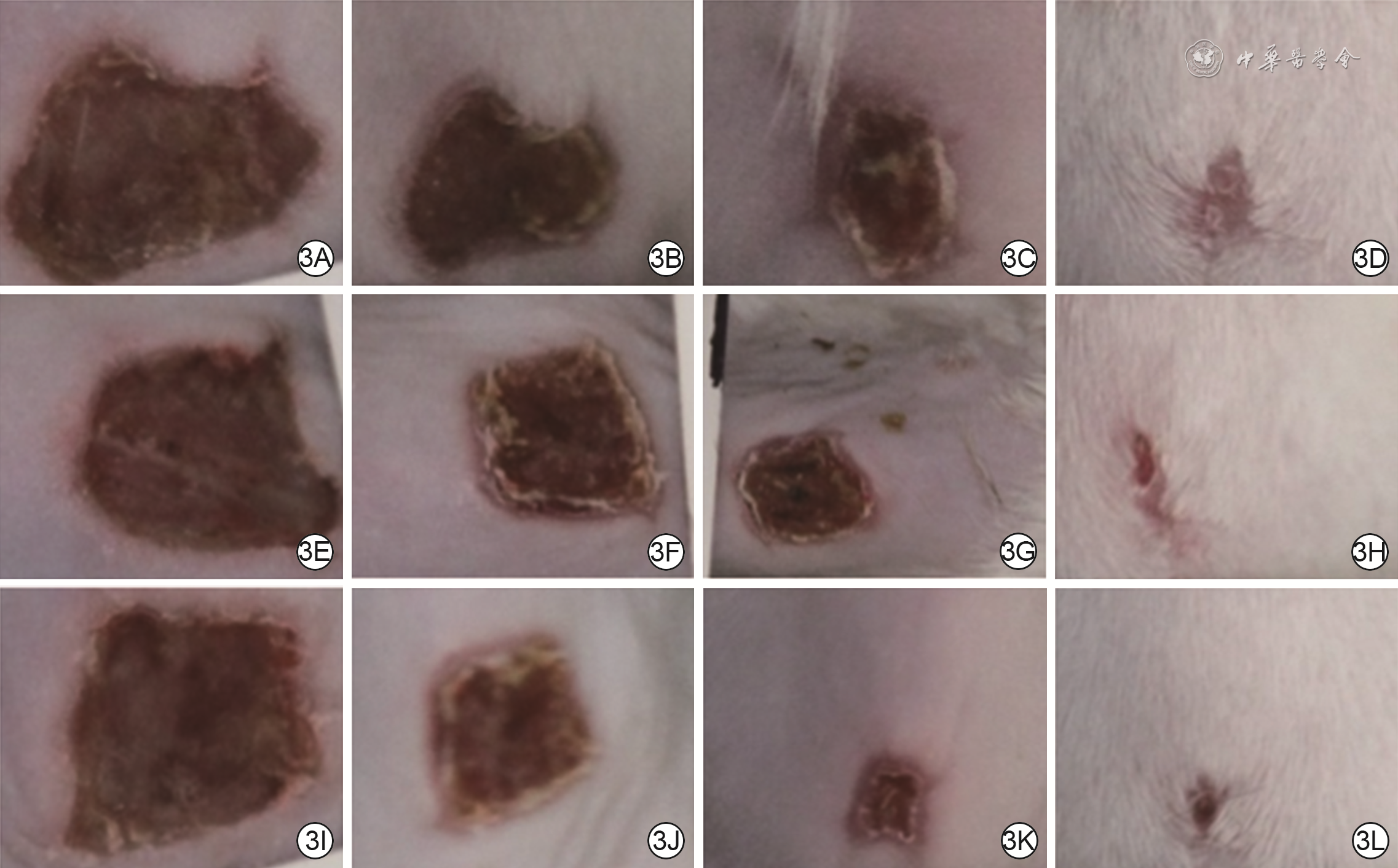
3 3组糖尿病全层皮肤缺损大鼠伤后各时间点创面愈合情况。3A、3B、3C、3D.分别为磷酸盐缓冲液组伤后3、7、10、14 d创面情况,创面逐渐缩小;3E、3F、3G、3H.分别为单纯脂肪间充质干细胞(ADSC)组伤后3、7、10、14 d创面情况,图3E、3F、3G、3H 中创面分别较图3A、3B、3C、3D有所缩小;3I、3J、3K、3L.分别为肝细胞生长因子修饰ADSC组伤后3、7、10、14 d创面情况,图3J、3K、3L中创面分别较图3F、3G、3H有所缩小,较图3B、3C、3D明显缩小

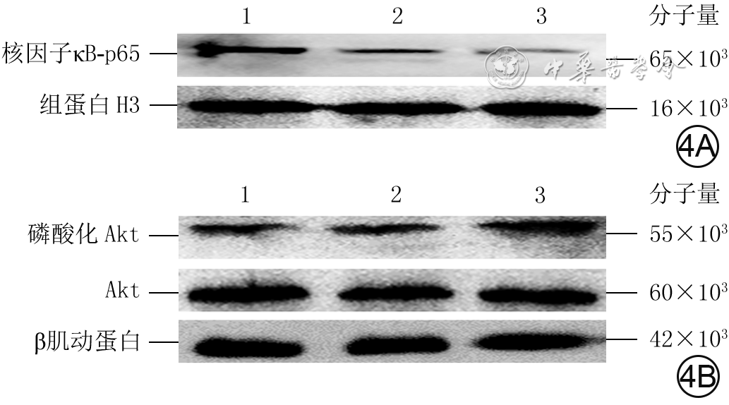
4 蛋白质印迹法检测3组糖尿病全层皮肤缺损大鼠创周皮肤组织中伤后3 d核因子κB-p65蛋白表达与伤后7 d Akt磷酸化水平。4A.核因子κB-p65;4B.Akt磷酸化
注:1、2、3分别为磷酸盐缓冲液组、单纯脂肪间充质干细胞(ADSC)组、肝细胞生长因子修饰ADSC组;Akt为蛋白激酶B
表1 实时荧光定量反转录PCR法检测大鼠创周皮肤组织炎症因子mRNA表达的引物序列及产物大小
指标名称 引物序列(5′→3′) 产物大小(bp) 肿瘤坏死因子α 上游:AGAACTCCAGGCGGTGTCTGTG 225 下游:GTGGCAAATCGGCTGACGGTGT 白细胞介素1β 上游:CACTTCACAAGTCGGAGGCT 114 下游:TCTGACAGTGCATCATCGCT 白细胞介素10 上游:TCCCAGTCAGCCAGACCCACA 144 下游:GGCAACCCAAGTAACCCTTAA 3-磷酸甘油醛脱氢酶 上游:GGCACAGTCAAGGCTGAGAATG 143 下游:ATGGTGGTGAAGACGCCAGTA  下载: 导出CSV
表2 3组糖尿病全层皮肤缺损大鼠伤后各时间点创面愈合率比较(%,
组别 鼠数(只) 3 d 7 d 10 d 14 d PBS组 9 21.4±1.3 61.4±1.5 80.1±2.1 92.4±1.8 单纯ADSC组 9 25.1±2.1 67.2±1.3 89.3±1.4 95.1±2.1 HGF修饰ADSC组 9 31.5±1.0 75.2±2.0 92.2±1.3 99.1±1.8 t1值 1.452 0.393 0.436 0.211 P1值 0.005 0.028 0.008 0.015 t2值 4.982 3.011 4.211 7.503 P2值 0.010 0.032 0.016 0.021 注:PBS为磷酸盐缓冲液,ADSC为脂肪间充质干细胞,HGF为肝细胞生长因子;时间因素主效应,F=1 968.321,P<0.001;处理因素主效应,F=592.785,P<0.001;两者交互作用,F=13.353,P<0.001;t1值、P1值,t2值、P2值分别为HGF修饰ADSC组与PBS组、单纯ADSC组各时间点比较所得
下载: 导出CSV
表3 3组糖尿病全层皮肤缺损大鼠伤后3 d创周皮肤组织中3种炎症因子mRNA表达比较(
组别 鼠数(只) TNF-α IL-1β IL-10 PBS组 9 1.00±0.16 1.00±0.08 1.00±0.11 单纯ADSC组 9 0.60±0.04 0.47±0.05 1.57±0.23 HGF修饰ADSC组 9 0.35±0.06 0.21±0.03 2.32±0.28 F值 97.003 484.640 85.088 P值 <0.001 <0.001 <0.001 t1值 7.281 17.700 -6.644 P1值 <0.001 <0.001 <0.001 t2值 6.231 13.083 -6.381 P2值 <0.001 <0.001 <0.001 注:PBS为磷酸盐缓冲液,ADSC为脂肪间充质干细胞,HGF为肝细胞生长因子,TNF-α为肿瘤坏死因子α,IL为白细胞介素;F值、P值为3组间各指标总体比较所得;t1值、P1值,t2值、P2值分别为HGF修饰ADSC组与PBS组、单纯ADSC组各指标比较所得
下载: 导出CSV
表4 3组糖尿病全层皮肤缺损大鼠伤后7 d创周皮肤组织中2种胶原的mRNA表达比较(
组别 鼠数(只) Ⅰ型胶原 Ⅲ型胶原 PBS组 9 1.00±0.06 1.00±0.03 单纯ADSC组 9 1.28±0.17 1.31±0.21 HGF修饰ADSC组 9 2.16±0.18 2.88±0.16 F值 158.373 388.256 P值 <0.001 <0.001 t1值 -5.126 -4.347 P1值 <0.001 <0.001 t2值 -10.694 -19.876 P2值 <0.001 <0.001 注:PBS为磷酸盐缓冲液,ADSC为脂肪间充质干细胞,HGF为肝细胞生长因子;F值、P值为3组间各指标总体比较所得;t1值、P1值,t2值、P2值分别为HGF修饰ADSC组与PBS组、单纯ADSC组各指标比较所得
下载: 导出CSV
表5 3组糖尿病全层皮肤缺损大鼠创周皮肤组织中伤后3 d核因子κB-p65蛋白表达与伤后7 d Akt磷酸化水平比较(
组别 鼠数(只) 核因子κB-p65 Akt磷酸化 PBS组 9 0.86±0.14 0.23±0.08 单纯ADSC组 9 0.38±0.07 0.38±0.11 HGF修饰ADSC组 9 0.17±0.04 0.74±0.12 F值 132.627 59.502 P值 <0.001 <0.001 t1值 9.447 -3.367 P1值 <0.001 0.004 t2值 7.783 -6.819 P2值 <0.001 <0.001 注:PBS为磷酸盐缓冲液,ADSC为脂肪间充质干细胞,HGF为肝细胞生长因子,Akt为蛋白激酶B;F值、P值为3组间各指标总体比较所得;t1值、P1值,t2值、P2值分别为HGF修饰ADSC组与PBS组、单纯ADSC组各指标比较所得
下载: 导出CSV
-

 下载:
下载:


计量
- 文章访问数: 3201
- HTML全文浏览量: 162
- PDF下载量: 61
- 被引次数: 0
