


Effects and mechanism of estrogen receptor β agonist on the migration and oxidative stress of human umbilical vein endothelial cell under high glucose condition
-
摘要:
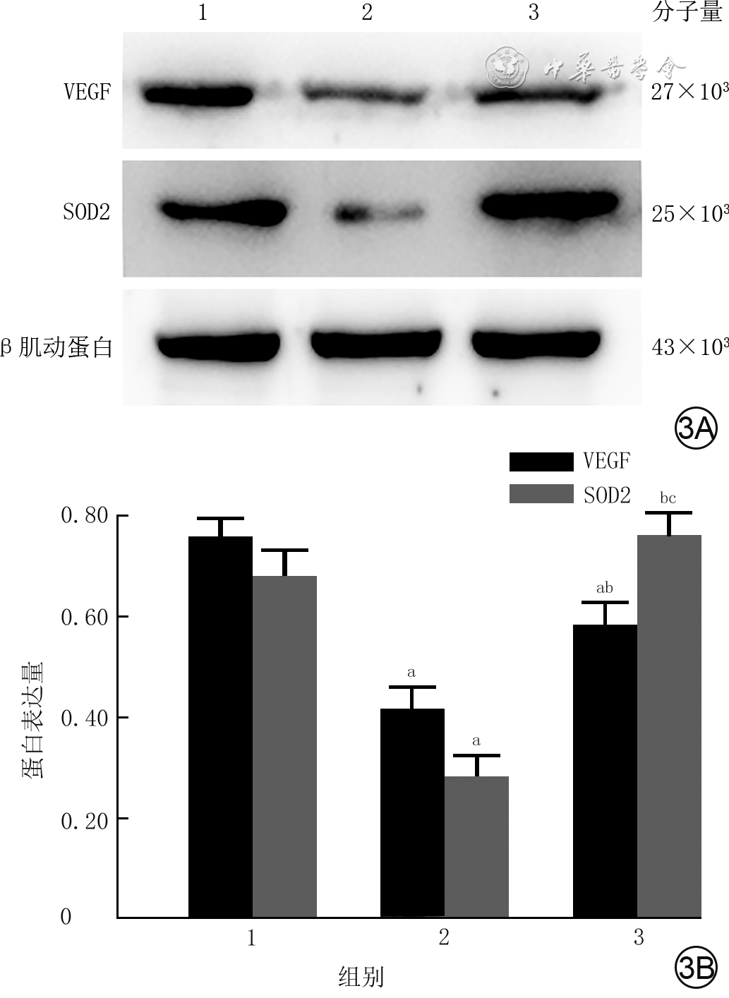
目的 探讨雌激素受体β(ERβ)激动剂对高糖作用下人脐静脉内皮细胞(HUVEC)迁移和氧化应激的影响及相关机制。 方法 采用实验研究方法。将HUVEC常规培养传代,取对数生长期细胞进行后续实验。将细胞按随机数字表法分为3组,正常对照组采用含5.5 mmol/L D-葡萄糖的RPMI 1640细胞培养基(下同)培养,单纯高糖组采用仅含25.0 mmol/L D-葡萄糖的细胞培养基培养,高糖+二芳基丙腈(DPN)组采用含25.0 mmol/L D-葡萄糖+10 μmol/L DPN的细胞培养基培养。采用划痕试验检测划痕后24 h 3组细胞迁移率,荧光探针法检测培养5 d 3组细胞内活性氧水平(以红色荧光强度表示),蛋白质印迹法检测培养5 d 3组细胞内血管内皮生长因子(VEGF)及超氧化物歧化酶2(SOD2)的蛋白表达。以上每个检测均取细胞同前行相同分组及相应培养,每个指标检测每组细胞样本数均为5。对数据行单因素方差分析、LSD-t检验。 结果 划痕后24 h,单纯高糖组细胞迁移率[(36±5)%]明显低于正常对照组和高糖+DPN组[(76±4)%、(65±5)%,t=14.511、9.603,P<0.01],高糖+DPN组细胞迁移率明显低于正常对照组(t=3.943,P<0.01)。培养5 d,单纯高糖组细胞内活性氧水平(1.81±0.12)明显高于正常对照组、高糖+DPN组(1.00±0.14、0.91±0.15,t=9.679、10.549,P<0.01),高糖+DPN组细胞内活性氧水平与正常对照组相近(t=1.031,P>0.05)。培养5 d,单纯高糖组细胞内VEGF和SOD2蛋白表达均明显低于正常对照组(t=14.175、13.787,P<0.01)和高糖+DPN组(t=6.321、17.750,P<0.01);高糖+DPN组VEGF蛋白表达明显低于正常对照组(t=7.206,P<0.01),SOD2蛋白表达明显高于正常对照组(t=2.890,P<0.05)。 结论 激活ERβ可通过促进VEGF和SOD2的表达,明显改善高糖对HUVEC迁移的抑制,缓解高糖诱导的氧化应激损伤。 Abstract:Objective To investigate the effects and related mechanism of estrogen receptor β (ERβ) agonist on the migration and oxidative stress of human umbilial vein endothelial cells (HUVECs) under high glucose condition. Methods The experimental research method was adopted. HUVECs were routinely cultured and passaged, and then cells of the logarithmic growth phase were collected for the subsequent experiments. The cells were divided into three groups according to the random number table, including normal control group (cultured with Roswell Park Memorial Institute 1640 cell culture medium (the same cell culture medium below) containing 5.5 mmol/L D-glucose), high glucose alone group (cultured with cell culture medium containing 25.0 mmol/L D-glucose alone), and high glucose+ERβ agonist diarylpropionitrile (DPN) group (cultured with cell culture medium containing 25.0 mmol/L D-glucose and 10 μmol/L DPN). Scratch test was conducted to detect the cell migration rate in the 3 groups at 24 h post scratching. Fluorescent probe method was used to detect the reactive oxygen species (ROS, denoted by red fluorescence intensity) of cells in the 3 groups on 5 d post culture. Western blotting was used to detect the protein expression levels of vascular endothelial growth factor (VEGF) and superoxide dismutase 2 (SOD2) of cells in the 3 groups on 5 d post culture. In the above-mentioned experiments, cells were grouped and cultured correspondingly as before, the number of samples in each group was 5. Data were statistically analyzed with one-way analysis of variance and least significant difference t test. Results At 24 h post scratching, the cell migration rate in high glucose alone group was (36±5)%, which was significantly lower than (76±4)% of normal control group and (65±5)% of high glucose+DPN group (t=14.511, 9.603, P<0.01), and the cell migration rate in high glucose+DPN group was significantly lower than that in normal control group (t=3.943, P<0.01). On 5 d post culture, the level of ROS of cells in high glucose alone group (1.81±0.12) was significantly increased compared with normal control group and high glucose+DPN group (1.00±0.14, 0.91±0.15, t=9.679, 10.549, P<0.01), while the level of ROS of cells in normal control group and high glucose+DPN group were close (t=1.031, P>0.05). On 5 d post culture, the protein expression levels of VEGF and SOD2 of cells in high glucose alone group were significantly lower than the levels of normal control group (t=14.175, 13.787, P<0.01) and high glucose+DPN group (t=6.321, 17.750, P<0.01). The protein expression level of VEGF of cells in high glucose+DPN group was significantly lower than the level of normal control group (t=7.206, P<0.05), while the protein expression level of SOD2 of cells in high glucose+DPN group was significantly higher than the level of normal control group (t=2.890, P<0.05). Conclusions The activation of ERβ can improve the inhibition of HUVECs migration induced by high glucose and alleviate oxidative stress injury induced by high glucose, which may be achieved by promoting the expression of VEGF and SOD2. -
Key words:
- Estrogen receptor beta /
- Endothelial cells /
- Cell migration assays /
- Oxidative stress /
- High glucose
-
参考文献
(20) [1] ZouboulisCC, ChenWC, ThorntonMJ, et al. Sexual hormones in human skin[J]. Horm Metab Res, 2007, 39(2):85-95. DOI: 10.1055/s-2007-961807. [2] CampbellL, EmmersonE, DaviesF, et al. Estrogen promotes cutaneous wound healing via estrogen receptor beta independent of its antiinflammatory activities[J]. J Exp Med, 2010, 207(9): 1825-1833. DOI: 10.1084/jem.20100500. [3] WarnerM, HuangB, GustafssonJA. Estrogen receptor β as a pharmaceutical target[J]. Trends Pharmacol Sci, 2017, 38(1):92-99. DOI: 10.1016/j.tips.2016.10.006. [4] SuthprasertpornN, SuwannaN, ThangniponW. Protective effects of diarylpropionitrile against hydrogen peroxide-induced damage in human neuroblastoma SH-SY5Y cells[J]. Drug Chem Toxicol, 2019:1-8. DOI: 10.1080/01480545.2019.1658768. [5] PeržeľováV, SabolF, VasilenkoT, et al. Pharmacological activation of estrogen receptors-α and -β differentially modulates keratinocyte differentiation with functional impact on wound healing[J]. Int J Mol Med, 2016, 37(1):21-28. DOI: 10.3892/ijmm.2015.2351. [6] HermanWH. The economic costs of diabetes: is it time for a new treatment paradigm?[J]. Diabetes Care, 2013, 36(4): 775-776. DOI: 10.2337/dc13-0270. [7] Cano SanchezM, LancelS, BoulangerE, et al. Targeting oxidative stress and mitochondrial dysfunction in the treatment of impaired wound healing: a systematic review[J]. Antioxidants (Basel), 2018, 7(8): 98. DOI: 10.3390/antiox7080098. [8] MilanPB, LotfibakhshaieshN, JoghataieMT, et al. Accelerated wound healing in a diabetic rat model using decellularized dermal matrix and human umbilical cord perivascular cells[J]. Acta Biomaterialia, 2016, 45: 234-246. DOI: 10.1016/j.actbio.2016.08.053. [9] TepperOM, CaplaJM, GalianoRD, et al. Adult vasculogenesis occurs through in situ recruitment, proliferation and tubulization of circulating bone marrow-derived cells[J]. Blood, 2005, 105(3): 1068-1077. DOI: 10.1182/blood-2004-03-1051. [10] TsaiHW, WangPH, TsuiKH. Mesenchymal stem cell in wound healing and regeneration[J]. J Chin Med Assoc, 2018, 81(3):223-224. DOI: 10.1016/j.jcma.2017.06.011. [11] OkonkwoUA, DiPietroLA. Diabetes and wound angiogenesis[J]. Int J Mol Sci, 2017, 18(7): 1419. DOI: 10.3390/ijms18071419. [12] 胡建军 血管相关迁移细胞蛋白调控血管新生及血管平滑肌细胞迁移 重庆 重庆大学 2016 胡建军. 血管相关迁移细胞蛋白调控血管新生及血管平滑肌细胞迁移[D].重庆:重庆大学,2016.
[13] QuattriniC, JeziorskaM, BoultonAJM, et al. Reduced vascular endothelial growth factor expression and intra-epidermal nerve fiber loss in human diabetic neuropathy[J]. Diabetes Care, 2008, 31(1): 140-145. DOI: 10.2337/dc07-1556. [14] SchlingemannRO, Van NoordenCJF, DiekmanMJM, et al. VEGF levels in plasma in relation to platelet activation, glycemic control, and microvascular complications in type 1 diabetes[J]. Diabetes Care, 2013, 36(6): 1629-1634. DOI: 10.2337/dc12-1951. [15] LiXL. The association between MCP-1, VEGF polymorphisms and their serum levels in patients with diabetic foot ulcer[J]. Medicine (Baltimore), 2018, 97(24): e10959. DOI: 10.1097/MD.0000000000010959. [16] 吴东蕾, 刘静. 氧化应激损伤与口腔疾病相关性的研究进展[J]. 国际口腔医学杂志, 2019, 46(1): 62-67. DOI: 10.7518/gjkq.2019.01.011. [17] NewsholmeP, CruzatVF, KeaneKN, et al. Molecular mechanisms of ROS production and oxidative stress in diabetes[J]. Biochem J, 2016, 473(24):4527-4550. DOI: 10.1042/BCJ20160503C. [18] ZhouXQ, LiM, XiaoMF, et al. ERβ accelerates diabetic wound healing by ameliorating hyperglycemia-induced persistent oxidative stress[J]. Front Endocrinol (Lausanne), 2019, 10:499. DOI: 10.3389/fendo.2019.00499. [19] ZhangT, LiangXY, ShiLY, et al. Estrogen receptor and PI3K/Akt signaling pathway involvement in S-(-)equol-induced activation of Nrf2/ARE in endothelial cells[J]. PLoS One, 2013, 8(11): e79075. DOI: 10.1371/journal.pone.0079075. [20] ZhanY, LiuZY, LiM, et al. ERβ expression in the endothelium ameliorates ischemia/reperfusion-mediated oxidative burst and vascular injury[J]. Free Radic Biol Med, 2016, 96: 223-233. DOI: 10.1016/j.freeradbiomed.2016.04.029. -

1 划痕试验观察3组人脐静脉内皮细胞划痕后各时间点迁移情况 光学显微镜×40。1A、1B、1C.分别为正常对照组、单纯高糖组、高糖+二芳基丙腈(DPN)组划痕后即刻;1D、1E、1F.分别为正常对照组、单纯高糖组、高糖+DPN组划痕后24 h,图1D、1F剩余划痕面积较小,图1E剩余划痕面积较大

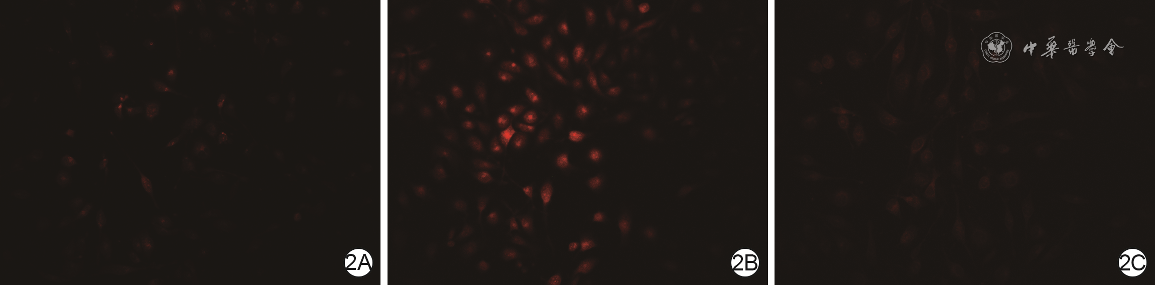
2 3组人脐静脉内皮细胞培养5 d活性氧水平(红色荧光)观察 DCFH-DA×400。2A.正常对照组活性氧水平较低;2B.单纯高糖组活性氧水平高;2C.高糖+二芳基丙腈组活性氧水平较低,且与图2A相近
注:2',7'-二氯二氢荧光素二乙酸酯(DCFH-DA)荧光探针阳性为红色荧光
-

 下载:
下载:


图(3)
计量
- 文章访问数: 443
- HTML全文浏览量: 192
- PDF下载量: 12
- 被引次数: 0
