


Clinical effects of flaps with cervical cutaneous branch of transverse cervical artery in repairing neck radiation ulcers
-
摘要:
目的 探讨应用颈横动脉颈段皮支皮瓣修复颈部放射性溃疡的临床效果。 方法 采用回顾性观察性研究方法。2016年1月—2019年12月,暨南大学附属广州红十字会医院采用颈横动脉颈段皮支皮瓣修复颈部放射性溃疡患者8例,其中男6例、女2例,年龄52~75岁。溃疡发生距放射治疗平均14.5年,入院时溃疡面积为5.0 cm×3.0 cm~7.0 cm×6.0 cm。切除溃疡及周围纤维化组织后创面面积为6.0 cm×5.0 cm~13.0 cm×6.5 cm。7例患者Ⅰ期行皮瓣切取、创面修复术;另1例患者Ⅰ期行皮瓣供区预扩张,Ⅱ期行皮瓣切取、创面修复术。皮瓣面积为8.0 cm×7.0 cm~15.0 cm×8.5 cm。7例患者皮瓣供区创面直接缝合;1例患者皮瓣供区创面部分缝合后,取自体大腿薄中厚皮修复。术前取溃疡组织行病理学检查,观察术后皮瓣成活情况和皮瓣供区愈合情况,随访时观察皮瓣情况、颈部溃疡复发及功能情况、皮瓣供区瘢痕增生情况。 结果 术前溃疡组织病理学检查显示,溃疡全层皮肤坏死,溃疡基底及周围有明显纤维组织增生、胶原化、小灶钙化;间质有大量慢性炎症细胞及少量急性炎症细胞浸润,排除肿瘤复发。8例患者术后皮瓣均成活,创面得到有效修复,皮瓣供区愈合良好。术后随访6~24个月,皮瓣外观良好,溃疡无复发,颈部活动功能得到明显改善,皮瓣供区未见明显瘢痕增生。 结论 颈部放射性溃疡是颈部放射治疗后较严重的远期并发症,保守治疗难以愈合。颈横动脉颈段皮支皮瓣靠近颈部,血运丰富、解剖恒定、易于切取,治疗颈部放射性溃疡后创面愈合良好,无明显瘢痕增生,功能明显改善。 -
关键词:
- 伤口愈合 /
- 外科皮瓣 /
- 放射性溃疡 /
- 颈横动脉颈段皮支皮瓣
Abstract:Objective To explore the clinical effects of flaps with cervical cutaneous branch of transverse cervical artery in repairing neck radiation ulcer. Methods The retrospective observational research was conducted. From January 2016 to December 2019, 8 cases with neck radiation ulcer were admitted to Guangzhou Red Cross Hospital of Jinan University and repaired with flaps based on cervical cutaneous branch of transverse cervical artery. There were 6 males and 2 females, aged 52-75 years. The ulcers occurred 14.5 years after radiotherapy on average, with ulcer areas of 5.0 cm×3.0 cm-7.0 cm×6.0 cm on admission. The wound areas were ranged from 6.0 cm×5.0 cm to 13.0 cm×6.5 cm after ulcers and fibrotic tissue around were resected. Seven cases underwent resection of flaps and wound repair operation on the first stage, and the other 1 case underwent pre-expansion of flap donor area on the first stage and resection of flap and wound repair operation on the second stage, with flap sizes of 8.0 cm×7.0 cm-15.0 cm×8.5 cm. The wounds in the donor areas of flaps in 7 patients were sutured directly, and the wound in the donor area of flap in the other 1 patient was repaired with thin split-thickness skin graft from thigh after being sutured partially. The preoperative ulcer tissue was collected for pathological examination, and the postoperative survival of the flaps and healing of the flap donor areas were observed. The flaps, the recurrence of the neck ulcers and neck function, and the scar hyperplasia in the donor areas of flaps were observed during follow-up. Results Preoperative pathological examination of ulcer tissue showed that full-thickness necrosis occurred in ulcer skin, obvious fibrotic tissue hyperplasia, collagenization, and small-scale calcification in the base and surrounding tissue of the ulcers, and a large amount of chronic inflammatory cells and a small amount of acute inflammatory cells infiltration were observed in intercellular substance, which excluded the recurrence of tumor. All the flaps in 8 cases survived, the wounds were repaired effectively, and the postoperative donor areas of flaps healed well. During postoperative follow-up of 6-24 months, the flaps had good appearances without recurrence of ulcer, the movement function of neck was significantly improved, and no obvious scar hyperplasia was observed in the donor areas of flaps. Conclusions Radiation ulcer in the neck is a serious long-term complication of neck after radiotherapy, which is difficult to heal with conservative treatment. The flap with cervical cutaneous branch of transverse cervical artery is close to the neck, with rich blood supply, constant anatomy, and is easy to cut. Neck radiation ulcers treated with the flaps showed good wound healing and improved functions, with no obvious scar hyperplasia. -
所有作者均声明不存在利益冲突糖尿病创面是糖尿病患者常见的严重并发症之一。海军军医大学第一附属医院夏照帆院士团队联合福建医科大学附属协和医院陈昭宏教授团队在《Burns & Trauma》杂志发文《Exosomes derived from human amniotic epithelial cells accelerate diabetic wound healing via PI3K-Akt-mTOR-mediated promotion in angiogenesis and fibroblast function》,探讨人羊膜上皮细胞外泌体(hAEC-Exo)对糖尿病创面愈合的作用,并初步阐明其机制。作者首先将经超速离心法分离获得的hAEC-Exo通过透射电镜、动态光散射、流式细胞术进行鉴定,观察到hAEC-Exo直径为(105.89±10.36)nm,呈杯状或球状形态,且外泌体表面标志物CD63和TSG101阳性。hAEC-Exo可显著促进人Fb(HFb)和人脐静脉内皮细胞(HUVEC)的增殖,促进HFb迁移,提升HUVEC的血管形成能力。微阵列芯片和RT-PCR检测观察到,hAEC-Exo中前15位富集的微小RNA(miRNA)表达增多,与创面愈合密切相关。进一步生物信息学分析和蛋白质印迹法检测显示,hAEC-Exo可活化HFb和HUVEC的磷脂酰肌醇3-激酶-蛋白激酶B-哺乳动物雷帕霉素靶蛋白(PI3K-Akt-mTOR)通路,该通路选择性抑制剂LY294002可部分抵消hAEC-Exo对HFb和HUVEC的促增殖、迁移和血管形成作用。体内创面实验证实,hAEC-Exo可提高db/db小鼠创面愈合速度,改善真皮胶原合成、沉积和排列,增加真皮毛细血管密度,但hAEC-Exo对db/db小鼠创面愈合的促进作用可被LY294002阻断。该研究从miRNA的角度探讨了hAEC-Exo通过活化PI3K-Akt-mTOR通路调控HFb和HUVEC活性,促进糖尿病创面愈合,为糖尿病创面的治疗提供了新的角度和方法。孙佳辰,编译自《Burns Trauma》,2020,8:tkaa020;申传安,审校
-
参考文献
(21) [1] WaghmareCM.Radiation burn--from mechanism to management[J].Burns,2013,39(2):212-219.DOI: 10.1016/j.burns.2012.09.012. [2] 陈伟雄,王跃建,张剑利,等.鼻咽癌放疗后颈部慢性放射性溃疡的皮瓣修复[J].临床耳鼻咽喉头颈外科杂志,2013,27(9):465-467.DOI: 10.13201/j.issn.1001-1781.2013.09.007. [3] ZhouY,ZhangY.Single- versus 2-stage reconstruction for chronic post-radiation chest wall ulcer: a 10-year retrospective study of chronic radiation-induced ulcers[J].Medicine (Baltimore),2019,98(8):e14567.DOI: 10.1097/MD.0000000000014567. [4] LiX,ZhangF,LiuX,et al.Staged treatment of chest wall radiation-induced ulcer with negative pressure wound therapy and latissimus dorsi myocutaneous flap transplantation[J].J Craniofac Surg,2019,30(5):e450-e453.DOI: 10.1097/SCS.0000000000005514. [5] WeiKC, YangKC, ChenLW, et al. Management of fluoroscopy-induced radiation ulcer: one-stage radical excision and immediate reconstruction[J]. Sci Rep, 2016,6:35875. DOI: 10.1038/srep35875. [6] YarnoldJ,BrotonsMC.Pathogenetic mechanisms in radiation fibrosis[J].Radiother Oncol,2010,97(1):149-161.DOI: 10.1016/j.radonc.2010.09.002. [7] AkitaS,HayashidaK,TakakiS,et al.The neck burn scar contracture: a concept of effective treatment[J/OL].Burns Trauma,2017,5:22[2020-08-07].https://pubmed.ncbi.nlm.nih.gov/28717655/. DOI: 10.1186/s41038-017-0086-8. [8] 沈余明,沈祖尧,王乃佐,等.严重放射性溃疡的修复[J].中国修复重建外科杂志,2000,14(4):208-210. [9] 马显杰,鲁开化,艾玉峰, 等.颈横动脉颈段皮支皮瓣的临床应用[J].中国美容整形外科杂志,2006,17(4):265-267. DOI: 10.3969/j.issn.1673-7040.2006.04.008. [10] ReissisM,ReissisD,BottiniGB,et al.A morphometric analysis of the suitability of the transverse cervical artery as a recipient artery in head and neck free flap microvascular reconstruction[J].Surg Radiol Anat,2018,40(8):891-897. DOI: 10.1007/s00276-018-2019-z. [11] 陈峥,鄂占森,杨世梅,等.颈横动脉主干起始位置及背段分支的超声研究[J].生物医学工程与临床,2014,18(5):454-457. [12] 马显杰.颈横动脉颈段皮支轴型皮瓣的临床应用[J].中华整形烧伤外科杂志,1993,9(1):22-24.DOI: 10.3760/j.issn:1009-4598.1993.01.014. [13] WangX,WangH.Nonexpanded prefabricated anterior perforator of transverse cervical artery flap for full facial reconstruction[J].J Craniofac Surg,2019,30(4):1206-1207. DOI: 10.1097/SCS.0000000000005061. [14] ChenS,LiY,YangZ,et al.Surgical treatment for facial port wine stain by prefabricated expanded cervical flap carried by superficial temporal artery[J].J Craniofac Surg,2019,30(7):2124-2127.DOI: 10.1097/SCS.0000000000005612. [15] 陈建武,马显杰,郭树忠.以颈横动脉分支为蒂的锁骨上皮瓣和颈段皮支皮瓣的区别与联系[J].中华整形外科杂志,2018,34(3):239-242. DOI: 10.3760/cma.j.issn.1009-4598.2018.03.019. [16] ChenB,SongH,XuM,et al.Reconstruction of cica-contracture on the face and neck with skin flap and expanded skin flap pedicled by anterior branch of transverse cervical artery[J].J Craniomaxillofac Surg,2016,44(9):1280-1286. DOI: 10.1016/j.jcms.2016.04.020. [17] SongB,ChenJ,MaX,et al.The pre-expanded subclavicular island flap: a new tool for facial reconstruction[J].J Plast Reconstr Aesthet Surg,2016,69(12):1653-1661. DOI: 10.1016/j.bjps.2016.09.001. [18] 马显杰,董立维,李杨,等.扩张后颈横动脉颈段皮支皮瓣的临床应用[J].中华整形外科杂志,2015,31(3):165-167. DOI: 10.3760/cma.j.issn.1009-4598.2015.03.002. [19] SongH,ChaiJ.Pre-expanded transverse cervical artery perforator flap[J].Clin Plast Surg,2017,44(1):41-47.DOI: 10.1016/j.cps.2016.08.002. [20] 许澎,王淑琴,燕辛,等.颈横动脉颈段皮支扩张皮瓣整复面部烧伤后瘢痕挛缩畸形[J].中华烧伤杂志,2016,32(8):458-462.DOI: 10.3760/cma.j.issn.1009-2587.2016.08.004. [21] 周莉萍,张文浩,王玉龙.远位埋没缝合切口减张技术联合电子线照射治疗瘢痕疙瘩九例[J].中华整形外科杂志,2019,35(3):278-281. DOI: 10.3760/cma.j.issn.1009-4598.2019.03.013. -
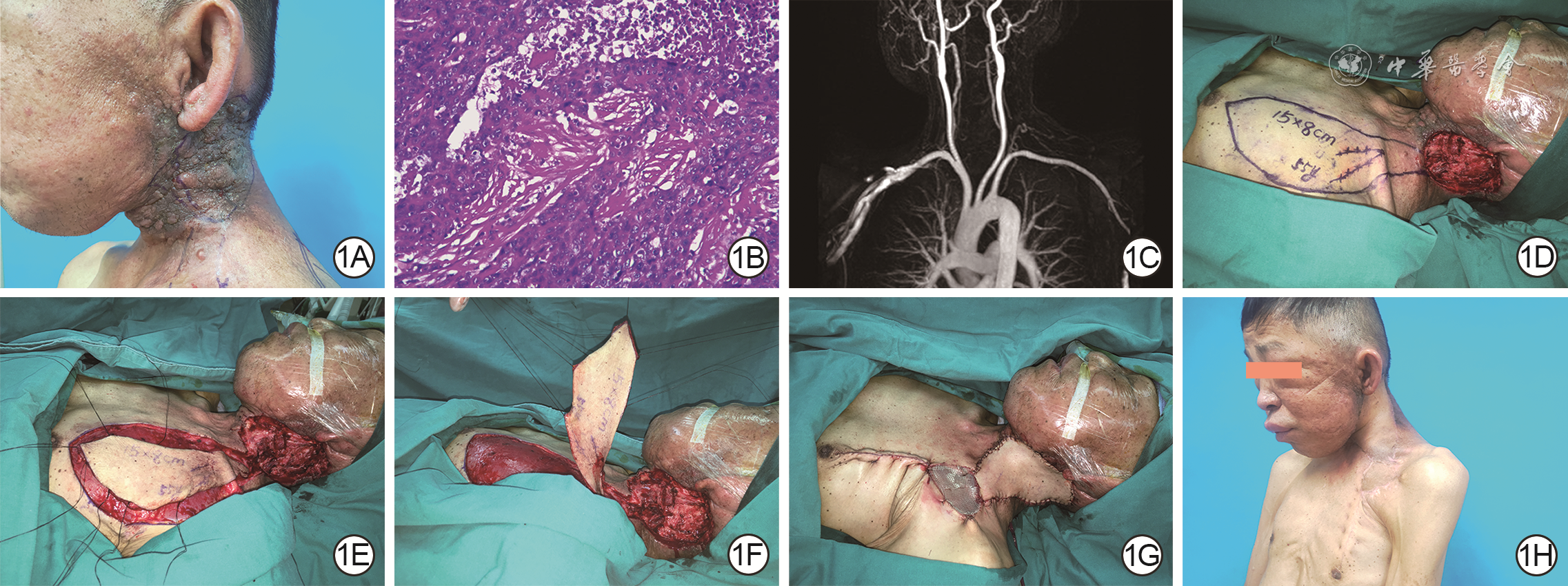
1 左侧颈横动脉颈段皮支皮瓣修复例1鼻咽癌患者颈部淋巴结放射治疗12年后左侧颈部放射性溃疡。1A.术前溃疡周围皮肤变硬,面部明显水肿,颈部活动受限;1B.术前溃疡组织病理学检查提示全层皮肤坏死,溃疡周围严重纤维化,伴较多淋巴细胞浸润 苏木精-伊红×100;1C.术前磁共振血管成像提示患者颈横动脉颈段皮支存在,充盈良好;1D.术中切除溃疡及周围纤维化组织并设计皮瓣;1E.切取皮瓣;1F.皮瓣分离到颈横动脉颈段皮支穿出点;1G.皮瓣与创面边缘间断缝合,皮瓣供区减张缝合缩小后用自体大腿薄中厚皮片修复;1H.术后1年颈部活动度得到改善,溃疡无复发,皮瓣供区未见明显瘢痕增生
-

 下载:
下载:

图(2)
计量
- 文章访问数: 251
- HTML全文浏览量: 88
- PDF下载量: 41
- 被引次数: 0
