


Regulatory effects of bio-intensity electric field on transformation of human skin fibroblasts
-
摘要:
目的 探讨生物强度电场对人皮肤成纤维细胞(HSF)转化的调节作用。 方法 采用实验研究方法。取HSF,分为经200 mV/mm电场处理6 h的200 mV/mm电场组和置于电场装置中不通电处理6 h的模拟电场组,在活细胞工作站中观察细胞形态和排列变化;记录处理0、6 h细胞数,并计算细胞数变化率;观察并计算3 h内细胞运动方向、位移速度、轨迹速度(以上实验模拟电场组样本数为34、200 mV/mm电场组样本数为30);采用免疫荧光法检测处理3 h细胞α平滑肌肌动蛋白(α-SMA)的蛋白表达(样本数为3)。取HSF分为置于电场装置中不通电处理3 h的模拟电场组和经相应强度电场处理3 h的100 mV/mm电场组、200 mV/mm电场组、400 mV/mm电场组,另取HSF分为置于电场装置中不通电处理6 h的模拟电场组和经200 mV/mm电场处理相应时间的电场处理1 h组、电场处理3 h组、电场处理6 h组,采用蛋白质印迹法检测α-SMA、增殖细胞核抗原(PCNA)的蛋白表达(样本数为3)。对数据行Mann-Whitney U检验、单因素方差分析、独立样本t检验及LSD检验。 结果 处理6 h,与模拟电场组相比,200 mV/mm电场组细胞形态拉长,并产生局部粘连;模拟电场组细胞任意排列,200 mV/mm电场组细胞呈有规律的纵向排列;2组细胞数变化率相近(P>0.05)。处理3 h内,200 mV/mm电场组细胞有明显的向正极运动趋势,模拟电场组细胞绕原点运动;与模拟电场组比较,200 mV/mm电场组细胞位移速度和轨迹速度均明显加快(Z值分别为-5.33、-5.41,P<0.01),方向性显著增强(Z=-4.39,P<0.01)。处理3 h,200 mV/mm电场组细胞α-SMA蛋白表达较模拟电场组明显增加(t=-9.81,P<0.01)。处理3 h,100 mV/mm电场组、200 mV/mm电场组、400 mV/mm电场组细胞α-SMA蛋白表达分别为1.195±0.057、1.606±0.041、1.616±0.039,均明显多于模拟电场组的0.649±0.028(P<0.01)。与100 mV/mm电场组比较,200 mV/mm电场组、400 mV/mm电场组细胞α-SMA蛋白表达均明显增加(P<0.01)。电场处理1 h组、电场处理3 h组、电场处理6 h组细胞α-SMA蛋白表达分别为0.730±0.032、1.561±0.031、1.553±0.045,均明显多于模拟电场组的0.464±0.020(P<0.01);与电场处理1 h组比较,电场处理3 h组、电场处理6 h组细胞α-SMA蛋白表达均明显增加(P<0.01)。处理3 h,与模拟电场组比较,100 mV/mm电场组、200 mV/mm电场组、400 mV/mm电场组细胞PCNA蛋白表达均明显减少(P<0.05或P<0.01);与100 mV/mm电场组比较,200 mV/mm电场组、400 mV/mm电场组细胞PCNA蛋白表达均明显减少(P<0.05或P<0.01);与200 mV/mm电场组比较,400 mV/mm电场组细胞PCNA蛋白表达明显减少(P<0.01)。与模拟电场组比较,电场处理1 h组、电场处理3 h组、电场处理6 h组细胞PCNA蛋白表达均明显减少(P<0.01);与电场处理1 h组比较,电场处理3 h组、电场处理6 h组细胞PCNA蛋白表达均明显减少(P<0.05或P<0.01);与电场处理3 h组比较,电场处理6 h组细胞PCNA蛋白表达明显减少(P<0.01)。 结论 生物强度电场可诱导HSF迁移、促进Fb向肌Fb转化,且转化有一定的时间及电场强度依赖性。 Abstract:Objective To investigate the regulatory effects of bio-intensity electric field on the transformation of human skin fibroblasts (HSFs). Methods The experimental research methods were used. HSFs were collected and divided into 200 mV/mm electric field group treated with 200 mV/mm electric field for 6 h and simulated electric field group placed in the electric field device without electricity for 6 h. Changes in morphology and arrangement of cells were observed in the living cell workstation; the number of cells at 0 and 6 h of treatment was recorded, and the rate of change in cell number was calculated; the direction of cell movement, movement velocity, and trajectory velocity within 3 h were observed and calculated (the number of samples was 34 in the simulated electric field group and 30 in 200 mV/mm electric field group in the aforementioned experiments); the protein expression of α-smooth muscle actin (α-SMA) in cells after 3 h of treatment was detected by immunofluorescence method (the number of sample was 3). HSFs were collected and divided into simulated electric field group placed in the electric field device without electricity for 3 h, and 100 mV/mm electric field group, 200 mV/mm electric field group, and 400 mV/mm electric field group which were treated with electric fields of corresponding intensities for 3 h. Besides, HSFs were divided into simulated electric field group placed in the electric field device without electricity for 6 h, and electric field treatment 1 h group, electric field treatment 3 h group, and electric field treatment 6 h group treated with 200 mV/mm electric field for corresponding time. The protein expressions of α-SMA and proliferating cell nuclear antigen (PCNA) were detected by Western blotting (the number of sample was 3). Data were statistically analyzed with Mann-Whitney U test, one-way analysis of variance, independent sample t test, and least significant difference test. Results After 6 h of treatment, compared with that in simulated electric field group, the cells in 200 mV/mm electric field group were elongated in shape and locally adhered; the cells in simulated electric field group were randomly arranged, while the cells in 200 mV/mm electric field group were arranged in a regular longitudinal direction; the change rates in the number of cells in the two groups were similar (P>0.05). Within 3 h of treatment, the cells in 200 mV/mm electric field group had an obvious tendency to move toward the positive electrode, and the cells in simulated electric field group moved around the origin; compared with those in simulated electric field group, the movement velocity and trajectory velocity of the cells in 200 mV/mm electric field group were increased significantly (with Z values of -5.33 and -5.41, respectively, P<0.01), and the directionality was significantly enhanced (Z=-4.39, P<0.01). After 3 h of treatment, the protein expression of α-SMA of cells in 200 mV/mm electric field group was significantly higher than that in simulated electric field group (t=-9.81, P<0.01). After 3 h of treatment, the protein expressions of α-SMA of cells in 100 mV/mm electric field group, 200 mV/mm electric field group, and 400 mV/mm electric field group were 1.195±0.057, 1.606±0.041, and 1.616±0.039, respectively, which were significantly more than 0.649±0.028 in simulated electric field group (P<0.01). Compared with that in 100 mV/mm electric field group, the protein expressions of α-SMA of cells in 200 mV/mm electric field group and 400 mV/mm electric field group were significantly increased (P<0.01). The protein expressions of α-SMA of cells in electric field treatment 1 h group, electric field treatment 3 h group, and electric field treatment 6 h group were 0.730±0.032, 1.561±0.031, and 1.553±0.045, respectively, significantly more than 0.464±0.020 in simulated electric field group (P<0.01). Compared with that in electric field treatment 1 h group, the protein expressions of α-SMA in electric field treatment 3 h group and electric field treatment 6 h group were significantly increased (P<0.01). After 3 h of treatment, compared with that in simulated electric field group, the protein expressions of PCNA of cells in 100 mV/mm electric field group, 200 mV/mm electric field group, and 400 mV/mm electric field group were significantly decreased (P<0.05 or P<0.01); compared with that in 100 mV/mm electric field group, the protein expressions of PCNA of cells in 200 mV/mm electric field group and 400 mV/mm electric field group were significantly decreased (P<0.05 or P<0.01); compared with that in 200 mV/mm electric field group, the protein expression of PCNA of cells in 400 mV/mm electric field group was significantly decreased (P<0.01). Compared with that in simulated electric field group, the protein expressions of PCNA of cells in electric field treatment 1 h group, electric field treatment 3 h group, and electric field treatment 6 h group were significantly decreased (P<0.01); compared with that in electric field treatment 1 h group, the protein expressions of PCNA of cells in electric field treatment 3 h group and electric field treatment 6 h group were significantly decreased (P<0.05 or P<0.01); compared with that in electric field treatment 3 h group, the protein expression of PCNA of cells in electric field treatment 6 h group was significantly decreased (P<0.01). Conclusions The bio-intensity electric field can induce the migration of HSFs and promote the transformation of fibroblasts to myofibroblasts, and the transformation displays certain dependence on the time and intensity of electric field. -
Key words:
- Skin /
- Fibroblasts /
- Myofibroblasts /
- Cell proliferation /
- Cell transformation /
- Bio-intensity electric fields
-
(1)较早地在烧伤领域提出持续炎症-免疫抑制-分解代谢综合征(PICS)这一概念。
持续炎症-免疫抑制-分解代谢综合征(persistent inflammation-immunosuppression-catabolism syndrome,PICS)是在全身性感染或非感染如烧创伤等进入慢性危重症(chronic critical illness,CCI)阶段时,出现的以持续性炎症、免疫抑制及蛋白质高分解代谢为特征的临床综合征。PICS防治困难、发病率高,是导致重症患者长期生活质量低下及远期死亡的重要原因,已成为重症患者治疗的新挑战 [ 1, 2] 。
大面积烧伤是一个累及全身的严重创伤事件,其病理生理学过程及临床表现与PICS类似,但目前关于大面积烧伤患者并发PICS的临床研究甚少。本研究总结大面积烧伤患者继发PICS的临床特征,对大面积烧伤患者继发PICS的影响因素进行多元回归分析,以期有助于临床早期识别、阻止或逆转可能的高危因素,进而改善大面积烧伤患者的预后。
1. 对象与方法
本回顾性病例系列研究符合《赫尔辛基宣言》的基本原则,根据暨南大学附属广州红十字会医院(以下简称本单位)伦理委员会政策,可以在不泄露患者身份信息的前提下对其临床资料进行分析、使用。
1.1 入选标准
纳入标准:(1)烧伤总面积≥30%TBSA;(2)年龄≥18岁,性别不限;(3)住院天数>14 d。排除标准:病历资料不完整者。
1.2 PICS诊断标准
(1)住院天数>14 d;(2)持续性的炎症反应:C反应蛋白>150 mg/L;(3)免疫抑制:淋巴细胞计数<0.8×10 9/L;(4)分解代谢综合征:血清白蛋白<30 g/L。
1.3 临床资料与分组
2017年1月—2021年12月,本单位烧伤ICU(BICU)收治220例符合入选标准的大面积成年烧伤患者,其中男168例、女52例,年龄18~84(43±14)岁。通过查询本单位的电子病历系统和重症护理系统,按照PICS发生情况将患者分为PICS组(84例)和非PICS组(136例)。
1.4 统计指标
1.4.1 一般资料
性别、年龄(分层:<65岁、≥65岁)、入院时合并基础疾病(糖尿病、高血压)情况和急性生理学和慢性健康状况评价Ⅱ(APACHEⅡ)评分、入院时和入院14 d脓毒症相关性器官功能衰竭评价(SOFA)评分、治疗期间行机械通气超过48 h比例。
1.4.2 专科情况
烧伤总面积、Ⅲ度烧伤面积、伤后48 h内入院比例、伤后30 d深度创面暴露面积(包括未行手术治疗的深Ⅱ度和Ⅲ度烧伤创面、暴露的肉芽组织创面、植皮区/供皮区感染创面面积)。
1.4.3 结局指标
住院天数、住院总费用、手术次数以及死亡情况。
1.5 统计学处理
采用SPSS 22.0统计软件进行数据分析。符合正态分布的计量资料数据以
2. 结果
2.1 一般资料
PICS组患者入院时APACHEⅡ评分和SOFA评分、治疗期间行机械通气超过48 h比例均明显高于非PICS组( P<0.05),2组患者其余一般资料均相近( P>0.05)。见 表1。
表1 2组大面积烧伤患者一般资料比较组别 例数 性别[例(%)] 年龄[例(%)] 入院时合并基础疾病[例(%)] 入院时APACHEⅡ评分(分, 入院时SOFA评分[分, M( Q 1, Q 3)] 入院14 d SOFA评分[分, M( Q 1, Q 3)] 治疗期间行机械通气超过48 h[例(%)] 男 女 <65岁 ≥65岁 糖尿病 高血压 PICS组 84 65(77.4) 19(22.6) 80(95.2) 4(4.8) 3(3.6) 10(11.9) 11±5 4(3,5) 3(2,4) 46(54.8) 非PICS组 136 103(75.7) 33(24.3) 128(94.1) 8(5.9) 4(2.9) 8(5.9) 7±4 2(1,3) 2(1,3) 54(39.7) 统计量值 χ 2=0.08 χ 2<0.01 χ 2<0.01 χ 2=2.50 t=6.78 Z=-4.75 Z=-1.90 χ 2=4.74 P值 0.780 0.960 1.000 0.113 <0.001 <0.001 0.057 0.029 注:PICS为持续炎症-免疫抑制-分解代谢综合征,APACHEⅡ为急性生理学和慢性健康状况评价Ⅱ,SOFA为脓毒症相关性器官功能衰竭评价 2.2 专科情况
PICS组患者烧伤总面积、Ⅲ度烧伤面积、伤后30 d深度创面暴露面积均明显大于非PICS组( P<0.05),但伤后48 h内入院比例明显低于非PICS组( P<0.05)。见 表2。
表2 2组大面积烧伤患者专科情况比较组别 例数 烧伤总面积(%TBSA, Ⅲ度烧伤面积[%TBSA, M( Q 1, Q 3)] 伤后48 h内入院[例(%)] 伤后30 d深度创面暴露面积[%TBSA, M( Q 1, Q 3)] PICS组 84 73±20 56(36,80) 57(67.9) 25(15,35) 非PICS组 136 55±20 29(6,43) 112(82.4) 8(0,13) 统计量值 t=6.29 Z=-7.25 χ 2=6.13 Z=-8.73 P值 <0.001 <0.001 0.013 <0.001 注:PICS为持续炎症-免疫抑制-分解代谢综合征,TBSA为体表总面积;深度创面暴露面积包括未行手术治疗的深Ⅱ度和Ⅲ度烧伤创面、暴露的肉芽组织创面、植皮区/供皮区感染创面面积 2.3 结局指标
PICS组患者住院天数、住院总费用、手术次数均明显多于非PICS组( P<0.05),但2组患者死亡情况相近( P>0.05)。见 表3。
表3 2组大面积烧伤患者结局指标比较组别 例数 住院天数[d, M( Q 1, Q 3)] 住院总费用[万元, M( Q 1, Q 3)] 手术次数[次, M( Q 1, Q 3)] 死亡[例(%)] PICS组 84 66(42,86) 85.3(48.1,110.6) 5(3,7) 2(2.4) 非PICS组 136 40(26,48) 35.7(13.0,47.8) 2(1,4) 3(2.2) 统计量值 Z=-7.12 Z=-8.48 Z=-6.87 χ 2<0.01 P值 <0.001 <0.001 <0.001 0.633 注:PICS为持续炎症-免疫抑制-分解代谢综合征 2.4 多因素logistic回归分析结果
以继发PICS的情况(PICS=1,非PICS=0)为因变量,将单因素分析中差异具有统计学意义的指标作为自变量,将烧伤总面积、Ⅲ度烧伤面积、伤后30 d深度创面暴露面积、入院时APACHEⅡ评分、入院时SOFA评分以原始值代入,伤后48 h内是否入院赋值(是=1,否=2)、治疗期间行机械通气是否超过48 h赋值(是=1,否=2),进行多因素logistic回归分析。结果显示,入院时APACHEⅡ评分、伤后30 d深度创面暴露面积均为大面积烧伤患者继发PICS的独立危险因素( P<0.05)。见 表4。
表4 影响220例大面积烧伤患者继发PICS的多因素logistic回归分析阳性结果因素 回归系数 标准误 比值比 95%置信区间 P值 入院时APACHEⅡ评分(分) 0.14 12.13 1.15 1.06~1.25 <0.001 伤后30 d深度创面暴露面积(%TBSA) 0.07 30.07 1.07 1.05~1.10 <0.001 注:PICS为持续炎症-免疫抑制-分解代谢综合征,APACHEⅡ为急性生理学和慢性健康状况评价Ⅱ,TBSA为体表总面积;深度创面暴露面积包括未行手术治疗的深Ⅱ度和Ⅲ度烧伤创面、暴露的肉芽组织创面、植皮区/供皮区感染创面面积 3. 讨论
近年来,随着重症器官支持治疗理念和技术的发展,越来越多的重症患者从MODS早期死亡高峰阶段幸存下来,成为CCI患者。2012年,Gentile等 [ 3] 提出PICS的新概念。PICS提供了一个理解长期住院CCI患者病理生理状态的新视角。该概念被提出后,其合理性逐渐被接受。在脓毒症、多发伤、严重创伤等患者中的研究显示,PICS患者住院天数多、医疗资源消耗巨大、长期生活质量低下、中远期病死率高,值得临床医师高度重视。
大面积烧伤患者的病程中,存在典型的持续炎症反应、免疫抑制以及高分解代谢状态。大面积烧伤患者是否存在PICS,其发生、发展的特点如何,是本研究团队开展这一回顾性病例系列研究的目的。
本单位收治的220例符合入选标准的大面积成年烧伤患者中有84例符合PICS诊断标准,PICS的发病率为38.18%,高于在多发伤患者中的11.7% [ 4] ,与老年脓毒症患者中的37.1%相近 [ 5] 。一般情况下,年龄≥65岁、合并基础疾病是重症患者继发PICS的危险因素。本研究显示,PICS组与非PICS组大面积烧伤患者在年龄、入院时合并基础疾病(糖尿病、高血压)方面的差异均无统计学意义( P>0.05),可能与本单位BICU收治的烧伤患者普遍较年轻,大部分为从事体力劳动的青壮年,基础疾病较少有关。
本研究中,PICS组患者烧伤总面积、Ⅲ度烧伤面积均明显大于非PICS组。烧伤面积与深度基本决定了烧伤的严重程度,大面积烧伤将导致大量促炎性细胞因子释放,进而触发和增强炎症反应与高分解代谢,推测其可能促成了PICS的发生。
有文献指出,烧伤后最初阶段的复苏不全往往是导致SIRS持续或全身状态恶化的重要因素 [ 6, 7] 。与PICS组相比,非PICS组伤后48 h内入院的患者占比较高。一般情况下,伤后48 h内入院的患者通常能接受较规范的液体复苏以及后续治疗。本研究中,伤后48 h后入院的患者,一部分是受伤地点离本单位较远,长途转运或多方辗转后才到达本单位接受治疗;一部分是在外院治疗了一段时间,创面处理不恰当,患者出现了一系列并发症后转入本单位的,因此PICS的发病率也高。不合适的创面处理通常会造成创面的感染、暴露,影响后续创面处理的效果,增加了创面封闭的难度,因此手术次数也会相应增多。推测此为本研究中PICS组患者的手术次数较非PICS组多的主要原因。
本研究中PICS组治疗期间行机械通气超过48 h的患者比例明显高于非PICS组,这与许多关于PICS的研究结果 [ 8, 9] 一致。有研究显示,SOFA评分对烧伤患者的预后有较好的预测价值 [ 10] 。在本研究中,2组患者仅入院时这个时间点的SOFA评分比较差异有统计学意义( P<0.05),且SOFA评分非大面积烧伤患者继发PICS的独立危险因素。SOFA评分由氧合指数、血小板计数、胆红素水平、血管活性药物使用情况、格拉斯哥昏迷量表(GCS)评分等组成,其中的胆红素水平、GCS评分对于烧伤患者早期的评估,特异度并不高 [ 10] 。有学者指出,由于烧伤患者脓毒症休克出现迅速,但胆红素指标对于肝脏功能变化反应较慢,并且胆红素水平非烧伤患者的常规检测项目,因此认为,应将其从对烧伤及烧伤脓毒症休克患者的评分中剔除 [ 11] 。而包含血糖水平、将镇静与非镇静患者分别评估(镇静患者评估肠内营养耐受情况,非镇静患者评估意识状态)的“烧伤SOFA”评分,可能更适合重症烧伤患者。
进一步的多因素logistic回归分析结果显示,入院时APACHEⅡ评分、伤后30 d深度创面暴露面积是大面积烧伤患者继发PICS的独立危险因素。APACHEⅡ评分是目前临床危重症患者病情评估的主要评分系统,由急性生理学评分、年龄评分、慢性健康状况评分三部分组成,得分越高表示病情越重。本研究中,PICS组患者病情更严重、伤后48 h内入院的患者比例更低,可能是导致入院时APACHEⅡ评分更高的原因。而此评分包括了本研究纳入的如年龄、合并基础疾病等诸多影响因素,较全面地反映了患者的病情,可能是其成为大面积烧伤患者继发PICS独立危险因素的重要原因。同样,在对脓毒症、创伤等的研究中观察到,APACHEⅡ评分高的患者更易出现PICS [ 4, 8] ,与本研究结果一致。然而入院时病情严重程度是否与后期出现的PICS直接相关?伤后48 h后入院的已经出现并发症的患者,入院时的APACHEⅡ评分是否能反映其病情的严重程度?这需要大样本、更细的分层研究来证实。
本研究中2组患者烧伤总面积和Ⅲ度烧伤面积有明显差异,但这2个指标不是大面积烧伤患者继发PICS的独立危险因素,而伤后30 d深度创面暴露面积是大面积烧伤患者继发PICS的独立危险因素。大面积深度创面的长时间暴露对烧伤患者内环境的稳态、代谢、免疫等带来深远的影响。而创面坏死组织的去除,自体皮或皮肤替代物覆盖创面不仅可以减少炎症因子的释放以及其带来的全身炎症反应、免疫抑制,亦可以通过恢复体温调节、减少热量损失和水分蒸发等减轻高代谢反应,是打断烧伤后持续炎症反应-免疫抑制-高分解代谢这一循环的有效手段 [ 12, 13] 。
为了缩短大面积烧伤患者创面暴露时间,减少并发症的发生。本单位自2007年开始,对重症烧伤患者实行统一管理,伤后5 d左右为患者行第1次切削痂手术,对同一深度烧伤面积区间的患者实施统一手术方案;同时,结合深Ⅱ度创面愈合后作为供皮区,头部、阴囊反复供皮,以及控制手术出血及损伤等方法,分次分批手术覆盖创面。对于外院转入、创面处理不及时或不恰当且已经出现并发症的患者,亦在维护脏器功能的同时,积极进行手术干预,尽量减少创面暴露的时间和面积。既往研究显示,本单位BICU治疗的烧伤总面积<50%TBSA、51%~80%TBSA、>80%TBSA的患者,平均创面愈合时间分别为36、43、79 d [ 14] 。
持续炎症-免疫抑制-高分解代谢多层面与多环节的恶性循环,最终影响了PICS患者的结局。目前,针对PICS的治疗,包括抗感染治疗、免疫治疗、物理治疗、营养治疗等,但PICS是由一系列的介质引起的,介质相互关联和依存,存在多层面、多环节的恶性循环,目前许多关键节点或环路尚不清楚,因此治疗棘手。大面积烧伤的患者病死率与感染以及全身炎症反应等导致的脏器功能损伤有关,其中PICS一般与创面暴露导致的全身炎症反应相关。本研究结果显示,虽然PICS在大面积烧伤患者中发病率不低,但经过积极的手术治疗,结合重症患者脏器功能支持技术,总体预后良好,2组患者病死率相近。这也提示,无论是对于烧伤本身的治疗还是对于烧伤后并发症的治疗,都不能忽略对创面本身的处理。
另外,本研究也显示,PICS组患者手术次数、住院天数、住院总费用均明显多于非PICS组,提示继发PICS的患者,需要更多的手术干预和住院天数以及更高的住院费用。因此,尽管PICS预后良好,但其仍会给患者以及医院带来巨大负担。
综上所述,本研究中,大面积烧伤患者继发PICS的发病率较高,入院时APACHEⅡ评分以及伤后30 d深度创面暴露面积为大面积烧伤患者继发PICS的独立危险因素,说明入院时病情严重的患者更易出现PICS,提示深度创面处理对于阻断大面积烧伤患者持续炎症-免疫抑制-高分解代谢这一恶性循环的重要性。然而本研究属于单中心回顾性研究,样本量小,可能造成选择性偏倚,需要大样本多中心的研究完善其结果。同时,PICS诊断标准仍有争议,涉及PICS诊断的指标,例如血清白蛋白水平、C反应蛋白等是否能较好地体现烧伤患者的代谢、炎症反应情况,仍值得商榷;烧伤患者继发PICS是否对其瘢痕的形成、远期生活质量以及生存产生影响,也仍需要进一步研究。
王文平:设计实验、实施实验、采集数据、分析数据、撰写文章;冀然、邬亚婷:实施实验、采集数据;张泽:实施实验、采集数据、分析数据;张恒术:撰写并审阅文章,获取研究经费,行政、技术和材料支持;张琼:分析数据、技术和材料支持;江旭品、滕苗:设计实验,撰写并审阅文章,获取研究经费,行政、技术和材料支持所有作者均声明不存在利益冲突 -
参考文献
(40) [1] JiR,TengM,ZhangZ,et al.Electric field down-regulates CD9 to promote keratinocytes migration through AMPK pathway[J].Int J Med Sci,2020,17(7):865-873.DOI: 10.7150/ijms.42840. [2] YanT,JiangX,GuoX,et al.Electric field-induced suppression of PTEN drives epithelial-to-mesenchymal transition via mTORC1 activation[J].J Dermatol Sci,2017,85(2):96-105.DOI: 10.1016/j.jdermsci.2016.11.007. [3] BostanLE,AlmqvistS,PullarCE.A pulsed current electric field alters protein expression creating a wound healing phenotype in human skin cells[J].Regen Med,2020,15(5):1611-1623.DOI: 10.2217/rme-2019-0087. [4] NuccitelliR.A role for endogenous electric fields in wound healing[J].Curr Top Dev Biol,2003,58:1-26.DOI: 10.1016/s0070-2153(03)58001-2. [5] AbeR,DonnellySC,PengT,et al.Peripheral blood fibrocytes: differentiation pathway and migration to wound sites[J].J Immunol,2001,166(12):7556-7562.DOI: 10.4049/jimmunol.166.12.7556. [6] DeesC,ChakrabortyD,DistlerJHW.Cellular and molecular mechanisms in fibrosis[J].Exp Dermatol,2021,30(1):121-131.DOI: 10.1111/exd.14193. [7] RemstDF,Blaney DavidsonEN,van der KraanPM.Unravelling osteoarthritis-related synovial fibrosis: a step closer to solving joint stiffness[J].Rheumatology (Oxford),2015,54(11):1954-1963.DOI: 10.1093/rheumatology/kev228. [8] KamilS,MohanRR.Corneal stromal wound healing: major regulators and therapeutic targets[J].Ocul Surf,2021,19:290-306.DOI: 10.1016/j.jtos.2020.10.006. [9] 刘杰,任淅,郭小伟,等.直流电场对BALB/c小鼠乳鼠真皮成纤维细胞定向迁移与排列的作用及其机制[J].中华烧伤杂志,2016,32(4):224-231.DOI: 10.3760/cma.j.issn.1009-2587.2016.04.007. [10] 冀然,张泽,王文平,等.生物强度电场对人表皮细胞株HaCaT和小鼠表皮细胞运动性及CD9表达的影响[J].中华烧伤杂志,2021,37(1):34-41.DOI: 10.3760/cma.j.cn501120-20200115-00023. [11] TredgetEE,LeviB,DonelanMB.Biology and principles of scar management and burn reconstruction[J].Surg Clin North Am,2014,94(4):793-815.DOI: 10.1016/j.suc.2014.05.005. [12] BarrettLW,FearVS,WaithmanJC,et al.Understanding acute burn injury as a chronic disease[J/OL].Burns Trauma,2019,7:23[2022-03-16].https://pubmed.ncbi.nlm.nih.gov/31534977/. DOI: 10.1186/s41038-019-0163-2. [13] DesmoulièreA,DarbyIA,GabbianiG.Normal and pathologic soft tissue remodeling: role of the myofibroblast, with special emphasis on liver and kidney fibrosis[J].Lab Invest,2003,83(12):1689-1707.DOI: 10.1097/01.lab.0000101911.53973.90. [14] CulleyOJ,LouisB,PhilippeosC,et al.Differential expression of insulin-like growth factor 1 and Wnt family member 4 correlates with functional heterogeneity of human dermal fibroblasts[J].Front Cell Dev Biol,2021,9:628039.DOI: 10.3389/fcell.2021.628039. [15] TomasekJJ,GabbianiG,HinzB,et al.Myofibroblasts and mechano-regulation of connective tissue remodelling[J].Nat Rev Mol Cell Biol,2002,3(5):349-363.DOI: 10.1038/nrm809. [16] XinY,MinP,XuH,et al.CD26 upregulates proliferation and invasion in keloid fibroblasts through an IGF-1-induced PI3K/AKT/mTOR pathway[J/OL].Burns Trauma,2020,8:tkaa025[2022-03-16].https://pubmed.ncbi.nlm.nih.gov/33150188/. DOI: 10.1093/burnst/tkaa025. [17] ModarressiA,PietramaggioriG,GodboutC,et al.Hypoxia impairs skin myofibroblast differentiation and function[J].J Invest Dermatol,2010,130(12):2818-2827.DOI: 10.1038/jid.2010.224. [18] Demidova-RiceTN,HamblinMR,HermanIM.Acute and impaired wound healing: pathophysiology and current methods for drug delivery, part 2: role of growth factors in normal and pathological wound healing: therapeutic potential and methods of delivery[J].Adv Skin Wound Care,2012,25(8):349-370.DOI: 10.1097/01.ASW.0000418541.31366.a3. [19] PutnikP,KresojaŽ,BosiljkovT,et al.Comparing the effects of thermal and non-thermal technologies on pomegranate juice quality: a review[J].Food Chem,2019,279:150-161.DOI: 10.1016/j.foodchem.2018.11.131. [20] SnyderS,DeJuliusC,WillitsRK.Electrical stimulation increases random migration of human dermal fibroblasts[J].Ann Biomed Eng,2017,45(9):2049-2060.DOI: 10.1007/s10439-017-1849-x. [21] SuessPM,SmithSA,MorrisseyJH.Platelet polyphosphate induces fibroblast chemotaxis and myofibroblast differentiation[J].J Thromb Haemost,2020,18(11):3043-3052.DOI: 10.1111/jth.15066. [22] NguyenEB,WishnerJ,SlowinskaK.The effect of pulsed electric field on expression of ECM proteins: collagen, elastin, and MMP1 in human dermal fibroblasts[J].J Electroanal Chem (Lausanne),2018,812:265-272.DOI: 10.1016/j.jelechem.2018.01.050. [23] ChaponnierC,GabbianiG.Pathological situations characterized by altered actin isoform expression[J].J Pathol,2004,204(4):386-395.DOI: 10.1002/path.1635. [24] FroidureA,Marchal-DuvalE,Homps-LegrandM,et al.Chaotic activation of developmental signalling pathways drives idiopathic pulmonary fibrosis[J].Eur Respir Rev,2020,29(158):190140. DOI: 10.1183/16000617.0140-2019. [25] Vaamonde-GarciaC,MalaiseO,CharlierE,et al.15-Deoxy-Δ-12, 14-prostaglandin J2 acts cooperatively with prednisolone to reduce TGF-β-induced pro-fibrotic pathways in human osteoarthritis fibroblasts[J].Biochem Pharmacol,2019,165:66-78.DOI: 10.1016/j.bcp.2019.03.039. [26] SidgwickGP,BayatA.Extracellular matrix molecules implicated in hypertrophic and keloid scarring[J].J Eur Acad Dermatol Venereol,2012,26(2):141-152.DOI: 10.1111/j.1468-3083.2011.04200.x. [27] TanJ,WuJ.Current progress in understanding the molecular pathogenesis of burn scar contracture[J/OL].Burns Trauma,2017,5:14[2022-03-16]. https://pubmed.ncbi.nlm.nih.gov/28546987/. DOI: 10.1186/s41038-017-0080-1. [28] WangL,KongW,LiuB,et al.Proliferating cell nuclear antigen promotes cell proliferation and tumorigenesis by up-regulating STAT3 in non-small cell lung cancer[J].Biomed Pharmacother,2018,104:595-602.DOI: 10.1016/j.biopha.2018.05.071. [29] ZhengW,XuS.Analysis of differential expression proteins of paclitaxel-treated lung adenocarcinoma cell A549 using tandem mass tag-based quantitative proteomics[J].Onco Targets Ther,2020,13:10297-10313.DOI: 10.2147/OTT.S259895. [30] ChiangCP,LangMJ,LiuBY,et al.Expression of proliferating cell nuclear antigen (PCNA) in oral submucous fibrosis, oral epithelial hyperkeratosis and oral epithelial dysplasia in Taiwan[J].Oral Oncol,2000,36(4):353-359.DOI: 10.1016/s1368-8375(00)00014-2. [31] ChenW,WuC,ChenY,et al.Downregulation of ceramide synthase 1 promotes oral cancer through endoplasmic reticulum stress[J].Int J Oral Sci,2021,13(1):10.DOI: 10.1038/s41368-021-00118-4. [32] KisK,LiuX,HagoodJS.Myofibroblast differentiation and survival in fibrotic disease[J].Expert Rev Mol Med,2011,13:e27.DOI: 10.1017/S1462399411001967. [33] ShenM,YoungA,C.PCNAAutexier, a focus on replication stress and the alternative lengthening of telomeres pathway[J].DNA Repair (Amst),2021,100:103055.DOI: 10.1016/j.dnarep.2021.103055. [34] DharadharS,van DijkWJ,ScheffersS,et al.Insert L1 is a central hub for allosteric regulation of USP1 activity[J].EMBO Rep,2021,22(4):e51749.DOI: 10.15252/embr.202051749. [35] LiB,WangR,WangY,et al.Regulation of smooth muscle contraction by monomeric non-RhoA GTPases[J].Br J Pharmacol,2020,177(17):3865-3877.DOI: 10.1111/bph.15172. [36] PfitzerG,WirthA,LuciusC,et al.Regulation of smooth muscle contraction by calcium, monomeric GTPases of the Rho subfamily and their effector kinases[J].Adv Exp Med Biol,2003,538:89-99; discussion 99.DOI: 10.1007/978-1-4419-9029-7_8. [37] LiX,WangF,LanY,et al.GDF-5 induces epidermal stem cell migration via RhoA-MMP9 signalling[J].J Cell Mol Med,2021,25(4):1939-1948.DOI: 10.1111/jcmm.15925. [38] QiY,LiangX,DaiF,et al.RhoA/ROCK pathway activation is regulated by AT1 receptor and participates in smooth muscle migration and dedifferentiation via promoting actin cytoskeleton polymerization[J].Int J Mol Sci,2020,21(15):5398. DOI: 10.3390/ijms21155398. [39] LeinhosL,PetersJ,KrullS,et al.Hypoxia suppresses myofibroblast differentiation by changing RhoA activity[J].J Cell Sci,2019,132(5):jcs223230. DOI: 10.1242/jcs.223230. [40] TsaiCH, LinBJ, ChaoPH. α2β1 integrin and RhoA mediates electric field-induced ligament fibroblast migration directionality[J].J Orthop Res, 2013,31(2):322-327. DOI: 10.1002/jor.22215. -
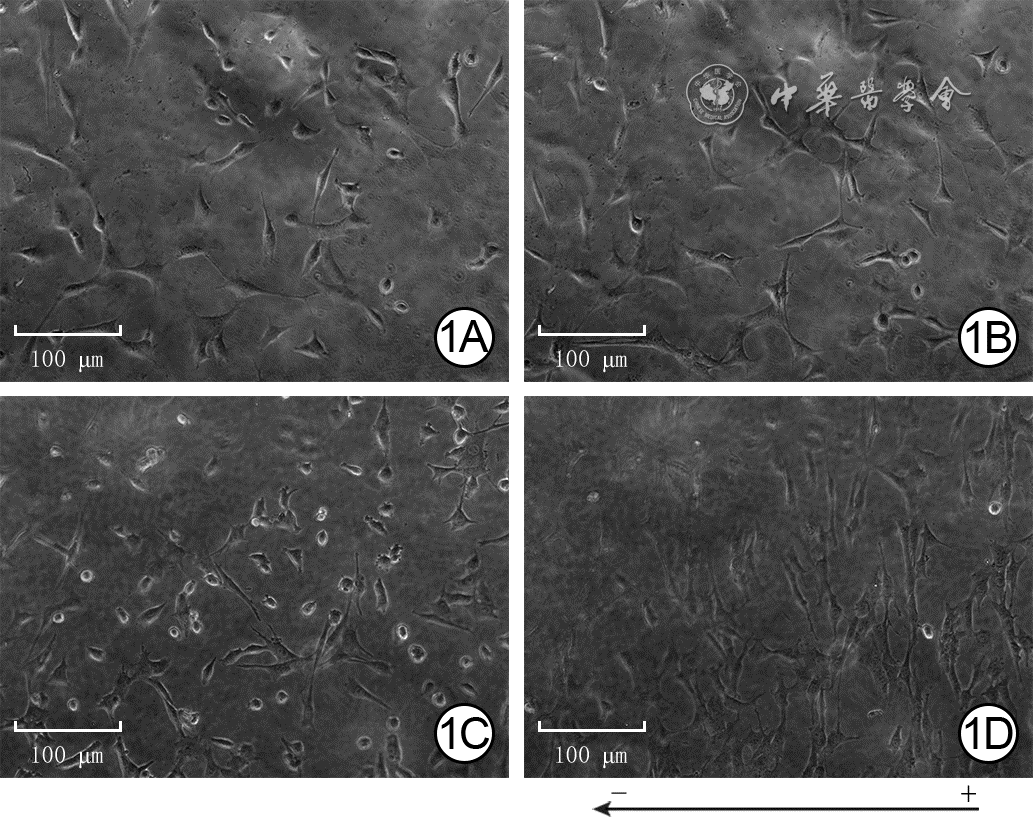
1 采用活细胞工作站观察电场刺激各时间点2组人皮肤成纤维细胞的形态和排列 倒置相差显微镜×100,图中标尺为100 μm。 1A、1B.分别为模拟电场组处理0(即刻)、6 h,细胞均呈任意方向排列;1C、1D.分别为200 mV/mm电场组处理0、6 h,图1C细胞呈任意方向排列,图1D细胞形态拉长,呈纵向排列,细胞长轴与电场方向垂直
注:图1D下方箭头为电场方向,“+” 为正极、“-” 为负极
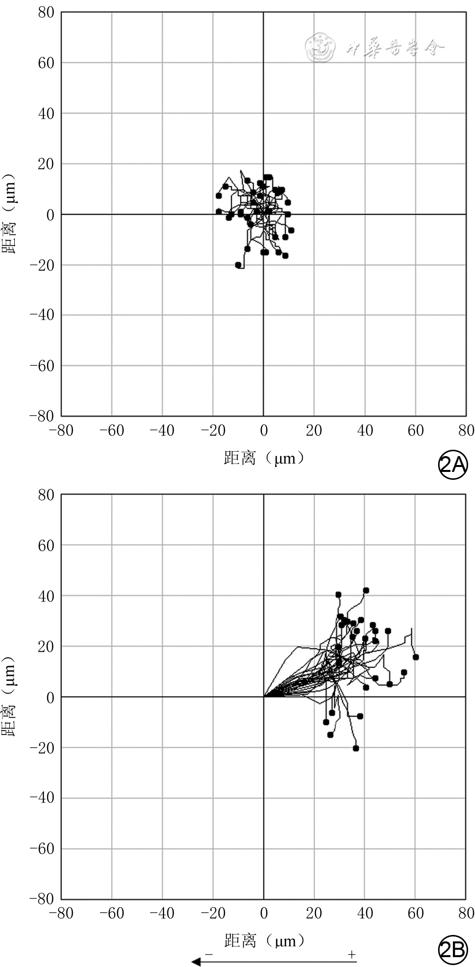
2 采用活细胞工作站观察2组人皮肤成纤维细胞经电场处理3 h内运动轨迹 倒置相差显微镜×100。2A.模拟电场组细胞绕原点运动;2B.200 mV/mm电场组细胞趋向于正极运动
注:细胞运动起点为坐标(0,0),运动终点为4个象限中的圆点,连接前述2个点之间的曲线为细胞运动轨迹,圆点位于左上、左下象限代表细胞向负极方向迁移,圆点位于右上、右下象限代表细胞向正极方向迁移;图2B下方箭头为电场方向,“ + ” 为正极、“-”为负极
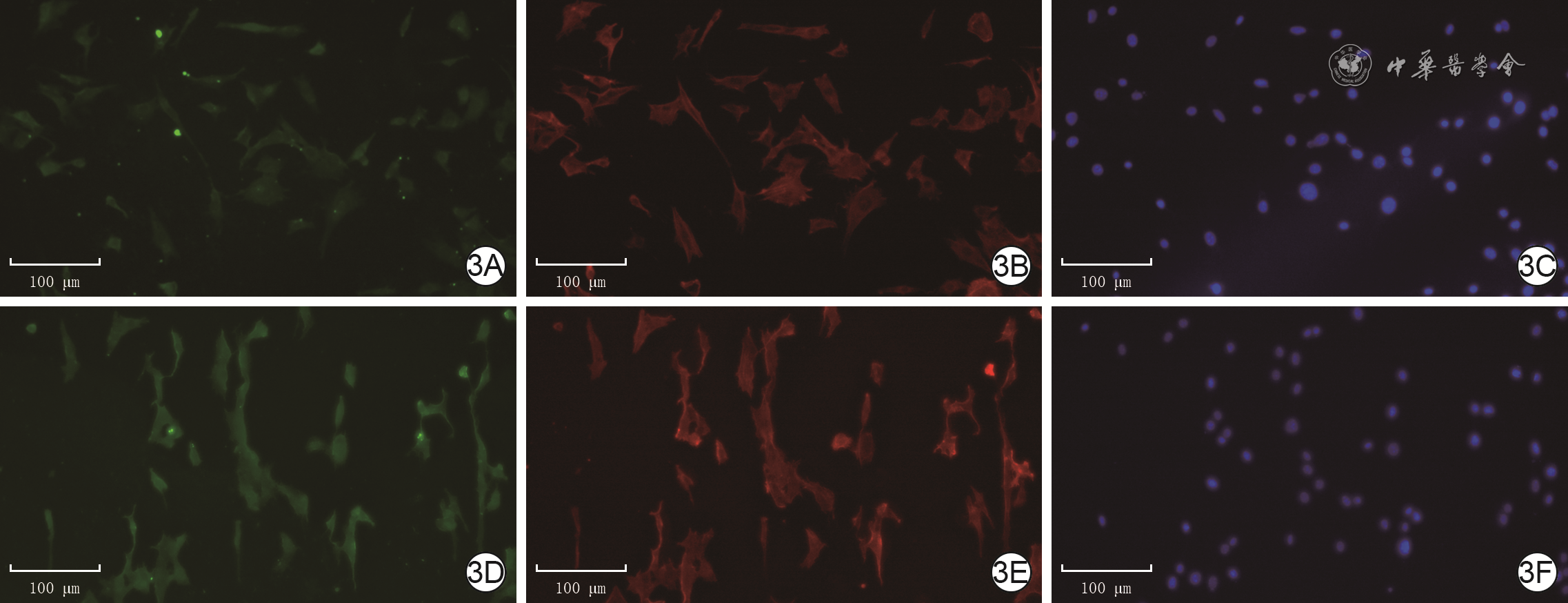
3 免疫荧光法检测电场处理3 h后2组人皮肤成纤维细胞α-SMA的表达 Alexa Fluor 488-4′,6-二脒基-2-苯基吲哚-罗丹明标记的鬼笔环肽×100,图中标尺为100 μm。3A、3B、3C.分别为模拟电场组细胞α-SMA蛋白表达、纤维状肌动蛋白排列、细胞核形态,α-SMA蛋白表达较低,纤维状肌动蛋白无序排列,细胞核完整;3D、3E、3F.分别为200 mV/mm电场组细胞α-SMA蛋白表达、纤维肌动蛋白排列、细胞核形态,图3D中α-SMA蛋白表达较图3A增加,纤维状肌动蛋白纵向排列,细胞核完整
注:绿色荧光标记α平滑肌肌动蛋白(α-SMA),红色荧光标记纤维状肌动蛋白,蓝色荧光标记细胞核
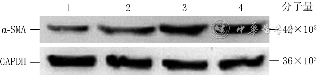
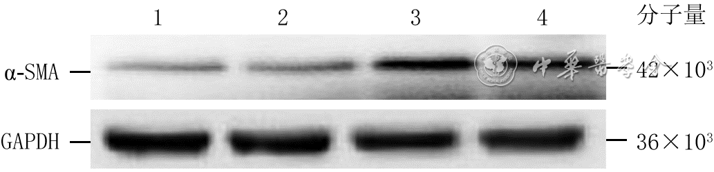
4 蛋白质印迹法检测不同强度电场处理3 h后4组人皮肤成纤维细胞α-SMA蛋白表达
注:α-SMA为α平滑肌肌动蛋白,GAPDH为3-磷酸甘油醛脱氢酶;条带上方1、2、3、4分别指模拟电场组、100 mV/mm电场组、200 mV/mm电场组、400 mV/mm电场组
5 蛋白质印迹法检测电场处理不同时间4组人皮肤成纤维细胞α-SMA蛋白表达
注:α-SMA为α平滑肌肌动蛋白,GAPDH为3-磷酸甘油醛脱氢酶;条带上方1、2、3、4分别指模拟电场组(处理6 h)、电场处理1 h组、电场处理3 h组、电场处理6 h组
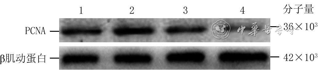
6 蛋白质印迹法检测不同强度电场处理3 h后4组人皮肤成纤维细胞增殖细胞核抗原(PCNA)蛋白表达
注:条带上方1、2、3、4分别指模拟电场组、100 mV/mm电场组、200 mV/mm电场组、400 mV/mm电场组
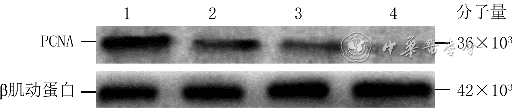
7 蛋白质印迹法检测电场处理不同时间4组人皮肤成纤维细胞增殖细胞核抗原(PCNA)蛋白表达
注:条带上方1、2、3、4分别指模拟电场组(处理6 h)、电场处理1 h组、电场处理3 h组、电场处理6 h组
表1 2组人皮肤成纤维细胞经电场处理3 h内cosθ及位移速度和轨迹速度比较[M(Q1,Q3)]
组别 样本数 cosθ 位移速度(μm/min) 轨迹速度(μm/min) 模拟电场组 34 -0.184(-0.336,0.371) 0.207(0.160,0.261) 0.165(0.126,0.204) 200 mV/mm电场组 30 0.833(0.694,0.925) 0.470(0.419,0.523) 0.383(0.347,0.438) Z值 -4.39 -5.33 -5.41 P值 <0.001 <0.001 <0.001  下载: 导出CSV
《中华烧伤与创面修复杂志》第六届编辑委员会通讯编委名单按姓氏拼音排序
贲道锋 卞徽宁 曹永倩 晁生武 陈辉 陈婧 陈朗 陈铭锐 陈鹏 陈晓东 陈忠勇 程君涛 迟云飞 储国平 党永明 邓呈亮 狄海萍 丁国兵 丁若虹 董茂龙 段红杰 段鹏 樊东力 房贺 冯光 付忠华 郭毅斌 韩兆峰 侯春胜 胡德林 胡炯宇 胡骁骅 胡晓燕 黄红军 纪世召 江华 姜丽萍 姜玉峰 雷娜 黎宁 李东杰 李峰 李靖 李晓东 李晓鲁 梁钢 梁鹏飞 林才 林国安 林源 刘德伍 刘健 刘军 刘淑华 龙奕 卢长虹 鲁峰 吕开阳 吕强 马思远 牛轶雯 欧阳军 乔亮 覃凤均 邱学文 曲滨 任超 沈江涌 石继红 宋慧锋 苏海涛 苏永涛 孙勇 孙瑜 谭江琳 唐修俊 滕苗 田社民 涂家金 汪虹 汪洋 王爱萍 王德怀 王洪涛 王会军 王良喜 王爽 王献珍 王志永 温冰 邬佳敏 吴红 吴继炎 吴巍巍 吴祖煌 向飞 向军 谢举临 谢松涛 辛海明 许喜生 许学文 薛斌 杨建民 杨敏烈 杨薛康 姚明 姚兴伟 叶祥柏 易成刚 易南 于东宁 岳丽青 翟红军 詹日兴 张博 张东霞 张红艳 张菊芳 张玲娟 张庆红 张彦琦 张寅 张元海 张志 赵全 赵冉 赵雄 郑德义 郑东风 郑军 周国富 周俊峄 周琴 周万芳 朱峰 朱宇刚 祝筱梅 邹立津 邹晓防
下载: 导出CSV
-


 下载:
下载:
计量
- 文章访问数: 422
- HTML全文浏览量: 89
- PDF下载量: 30
- 被引次数: 0
