


-
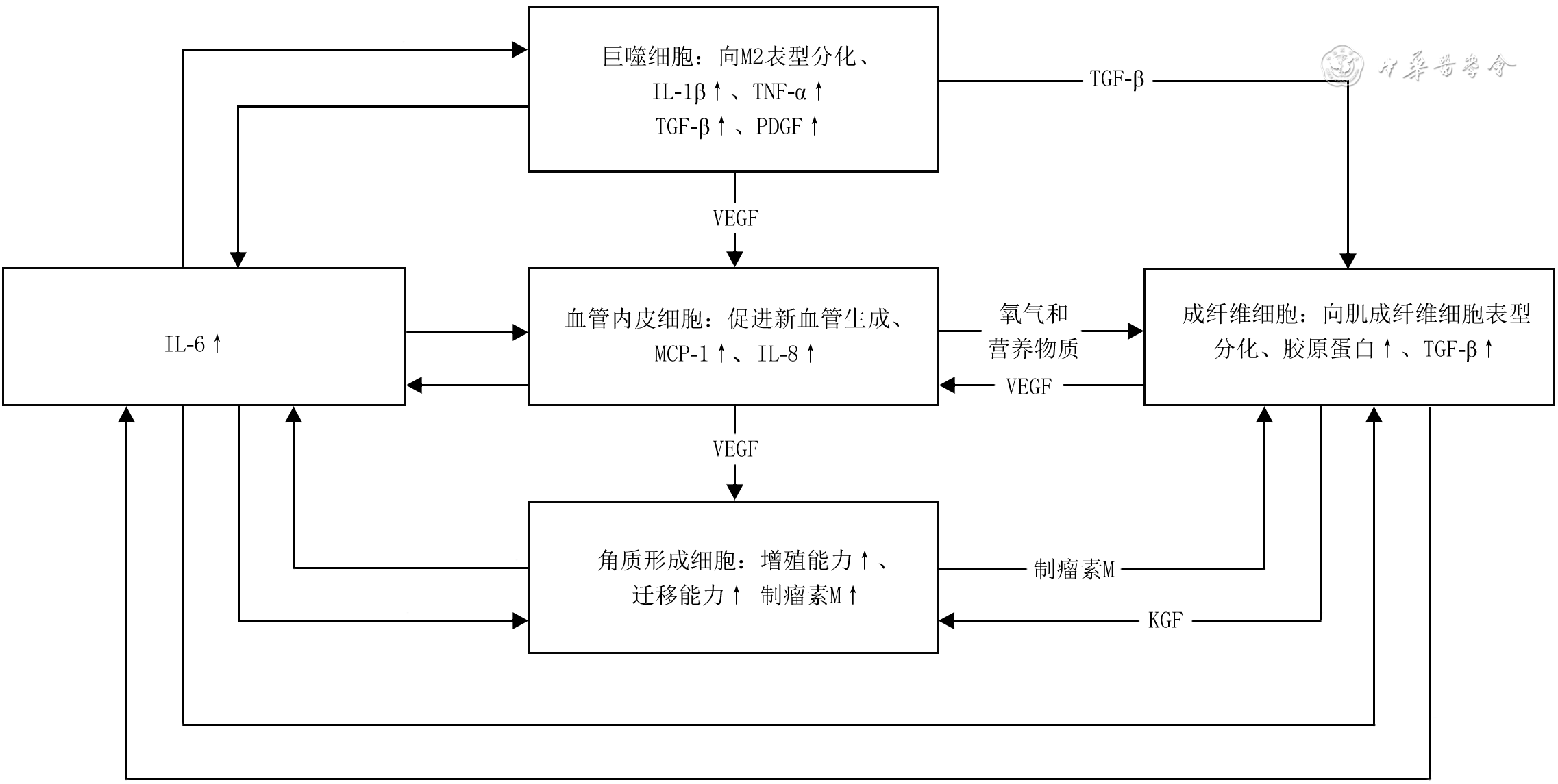
摘要: 增生性瘢痕是受损皮肤的细胞外基质过度堆积导致的病理性修复结果,不同程度地影响患者的外观和功能。瘢痕形成的程度与创面愈合过程中炎症反应的强弱直接相关,过度或延长的炎症反应会增加增生性瘢痕的发生率。白细胞介素6(IL-6)是一种多效性细胞因子,可参与调控由成纤维细胞、巨噬细胞、角质形成细胞和血管内皮细胞组成的促纤维化网络,与增生性瘢痕的形成密切相关。该文就IL-6及其信号通路在增生性瘢痕形成中的作用进行综述。Abstract: Hypertrophic scar is a pathological repair result of excessive accumulation of extracellular matrix after skin damage, which affects the appearance and function of patients with varying degrees. The degree of scar formation is directly related to the strength of inflammatory reaction during wound healing, and excessive or prolonged inflammatory response increases the incidence of hypertrophic scars. Interleukin-6 (IL-6) is a pleiotropic cytokine that is involved in regulating the fibrotic network composed of fibroblasts, macrophages, keratinocytes, and vascular endothelial cells, and is closely related to the formation of hypertrophic scars. This article reviews the role of IL-6 and its signaling pathway in hypertrophic scar formation.
-
Key words:
- Interleukin-6 /
- Inflammation /
- Fibro-sis /
- Skin /
- Hypertrophic scar
-
参考文献
(37) [1] 岑瑛, 刘睿奇. 瘢痕的研究新进展及临床治疗[J].中华烧伤杂志,2019,35(12):839-841. DOI: 10.3760/cma.j.issn.1009-2587.2019.12.002. [2] WilgusTA. Inflammation as an orchestrator of cutaneous scar formation: a review of the literature[J]. Plast Aesthet Res, 2020,7:54.DOI: 10.20517/2347-9264.2020.150. [3] TanJ, WuJ. Current progress in understanding the molecular pathogenesis of burn scar contracture[J/OL]. Burns Trauma, 2017,5:14[2022-04-12]. https://pubmed.ncbi.nlm.nih.gov/28546987/.DOI: 10.1186/s41038-017-0080-1. [4] OgawaR, AkitaS, AkaishiS, et al. Diagnosis and treatment of keloids and hypertrophic scars-Japan Scar Workshop Consensus Document 2018[J/OL]. Burns Trauma, 2019,7:39[2022-04-12]. https://pubmed.ncbi.nlm.nih.gov/31890718/. DOI: 10.1186/s41038-019-0175-y. [5] ChippE, CharlesL, ThomasC, et al. A prospective study of time to healing and hypertrophic scarring in paediatric burns: every day counts[J/OL]. Burns Trauma, 2017,5:3[2022-04-12]. https://pubmed.ncbi.nlm.nih.gov/28116323/. DOI: 10.1186/s41038-016-0068-2. [6] van ZuijlenPP, RuurdaJJ, van VeenHA, et al. Collagen morphology in human skin and scar tissue: no adaptations in response to mechanical loading at joints[J]. Burns, 2003,29(5):423-431. DOI: 10.1016/s0305-4179(03)00052-4. [7] WongVW, PaternoJ, SorkinM, et al. Mechanical force prolongs acute inflammation via T-cell-dependent pathways during scar formation[J]. FASEB J, 2011,25(12):4498-4510. DOI: 10.1096/fj.10-178087. [8] ShiJ, ShiS, XieW, et al. IL-10 alleviates lipopolysaccharide- induced skin scarring via IL-10R/STAT3 axis regulating TLR4/NF-κB pathway in dermal fibroblasts[J]. J Cell Mol Med, 2021,25(3):1554-1567. DOI: 10.1111/jcmm.16250. [9] ShiJ, ShiS, XieW, et al. IL-10 alleviates lipopolysaccharide- induced skin scarring via IL-10R/STAT3 axis regulating TLR4/NF-κB pathway in dermal fibroblasts[J]. J Cell Mol Med, 2021, 25(3):1554-1567. DOI: 10.1111/jcmm.16250. [10] QianLW, FourcaudotAB, YamaneK, et al. Exacerbated and prolonged inflammation impairs wound healing and increases scarring[J]. Wound Repair Regen, 2016,24(1):26-34. DOI: 10.1111/wrr.12381. [11] DongX, MaoS, WenH. Upregulation of proinflammatory genes in skin lesions may be the cause of keloid formation (Review)[J]. Biomed Rep, 2013,1(6):833-836. DOI: 10.3892/br.2013.169. [12] BermanB, MaderalA, RaphaelB. Keloids and hypertrophic scars: pathophysiology, classification, and treatment[J]. Dermatol Surg, 2017,43 Suppl 1:S3-18. DOI: 10.1097/DSS.0000000000000819. [13] 李丽, 王林. 正常皮肤与病理性瘢痕组织中HIF-1α和促炎细胞因子水平比较及两者相关性研究[J].中国美容医学,2019,28(4):71-73. [14] LiechtyKW, AdzickNS, CrombleholmeTM. Diminished interleukin 6 (IL-6) production during scarless human fetal wound repair[J]. Cytokine, 2000,12(6):671-676. DOI: 10.1006/cyto.1999.0598. [15] 刘娜, 张川, 杨磊, 等. 白细胞介素-6单克隆抗体抑制机械张力诱发小鼠增生性瘢痕[J].中华实验外科杂志,2017,34(12):2090-2092. DOI: 10.3760/cma.j.issn.1001-9030.2017.12.024. [16] HuangJ, ChenJ, WoY, et al. CO2 fractional laser combined with 5-fluorouracil ethosomal gel treatment of hypertrophic scar macro-, microscopic, and molecular mechanism of action in a rabbit animal model[J]. Rejuvenation Res, 2021,24(2):131-138. DOI: 10.1089/rej.2019.2204. [17] MurakamiM, KamimuraD, HiranoT. Pleiotropy and specificity: insights from the interleukin 6 family of cytokines[J]. Immunity, 2019,50(4):812-831. DOI: 10.1016/j.immuni.2019.03.027. [18] NaseemS, HussainT, ManzoorS. Interleukin-6: a promising cytokine to support liver regeneration and adaptive immunity in liver pathologies[J]. Cytokine Growth Factor Rev, 2018,39:36-45. DOI: 10.1016/j.cytogfr.2018.01.002. [19] SchumacherN, Rose-JohnS. ADAM17 activity and IL-6 trans-signaling in inflammation and cancer[J]. Cancers (Basel), 2019, 11(11):1736.DOI: 10.3390/cancers11111736. [20] LokauJ, AgtheM, FlynnCM, et al. Proteolytic control of interleukin-11 and interleukin-6 biology[J]. Biochim Biophys Acta Mol Cell Res, 2017,1864(11 Pt B):2105-2117. DOI: 10.1016/j.bbamcr.2017.06.008. [21] KaurS, BansalY, KumarR, et al. A panoramic review of IL-6: structure, pathophysiological roles and inhibitors[J]. Bioorg Med Chem, 2020,28(5):115327. DOI: 10.1016/j.bmc.2020.115327. [22] ChenW, YuanH, CaoW, et al. Blocking interleukin-6 trans-signaling protects against renal fibrosis by suppressing STAT3 activation[J]. Theranostics, 2019,9(14):3980-3991. DOI: 10.7150/thno.32352. [23] Epstein ShochetG, BrookE, Bardenstein-WaldB, et al. TGF-β pathway activation by idiopathic pulmonary fibrosis (IPF) fibroblast derived soluble factors is mediated by IL-6 trans- signaling[J]. Respir Res, 2020,21(1):56. DOI: 10.1186/s12931-020-1319-0. [24] HeinkS, YogevN, GarbersC, et al. Trans-presentation of IL-6 by dendritic cells is required for the priming of pathogenic TH17 cells[J]. Nat Immunol, 2017,18(1):74-85. DOI: 10.1038/ni.3632. [25] JonesSA, JenkinsBJ. Recent insights into targeting the IL-6 cytokine family in inflammatory diseases and cancer[J]. Nat Rev Immunol, 2018,18(12):773-789. DOI: 10.1038/s41577-018-0066-7. [26] JohnsonBZ, StevensonAW, PrêleCM, et al. The role of IL-6 in skin fibrosis and cutaneous wound healing[J]. Biomedicines, 2020, 8(5):101.DOI: 10.3390/biomedicines8050101. [27] ČomaM, FröhlichováL, UrbanL, et al. Molecular changes underlying hypertrophic scarring following burns involve specific deregulations at all wound healing stages (inflammation, proliferation and maturation)[J]. Int J Mol Sci, 2021,22(2):897.DOI: 10.3390/ijms22020897. [28] HasegawaM, SatoS, IhnH, et al. Enhanced production of interleukin-6 (IL-6), oncostatin M and soluble IL-6 receptor by cultured peripheral blood mononuclear cells from patients with systemic sclerosis[J]. Rheumatology (Oxford), 1999,38(7):612-617. DOI: 10.1093/rheumatology/38.7.612. [29] RayS, JuX, SunH, et al. The IL-6 trans-signaling-STAT3 pathway mediates ECM and cellular proliferation in fibroblasts from hypertrophic scar[J]. J Invest Dermatol, 2013,133(5):1212-1220. DOI: 10.1038/jid.2012.499. [30] DufourAM, AlvarezM, RussoB, et al. Interleukin-6 and type-I collagen production by systemic sclerosis fibroblasts are differentially regulated by interleukin-17A in the presence of transforming growth factor-beta 1[J]. Front Immunol, 2018,9:1865. DOI: 10.3389/fimmu.2018.01865. [31] Shapouri-MoghaddamA, MohammadianS, VaziniH, et al. Macrophage plasticity, polarization, and function in health and disease[J]. J Cell Physiol, 2018,233(9):6425-6440. DOI: 10.1002/jcp.26429. [32] XuX, GuS, HuangX, et al. The role of macrophages in the formation of hypertrophic scars and keloids[J/OL]. Burns Trauma, 2020,8:tkaa006[2022-05-07]. https://pubmed.ncbi.nlm.nih.gov/32341919/.DOI: 10.1093/burnst/taa006. [33] BrauneJ, WeyerU, HobuschC, et al. IL-6 regulates M2 polarization and local proliferation of adipose tissue macrophages in obesity[J]. J Immunol, 2017,198(7):2927-2934. DOI: 10.4049/jimmunol.1600476. [34] WernerS, KriegT, SmolaH. Keratinocyte-fibroblast interactions in wound healing[J]. J Invest Dermatol, 2007,127(5):998-1008. DOI: 10.1038/sj.jid.5700786. [35] PengY, WuS, TangQ, et al. KGF-1 accelerates wound contraction through the TGF-β1/Smad signaling pathway in a double-paracrine manner[J]. J Biol Chem, 2019,294(21):8361-8370. DOI: 10.1074/jbc.RA118.006189. [36] ZhuM, YangM, YangQ, et al. Chronic hypoxia-induced microvessel proliferation and basal membrane degradation in the bone marrow of rats regulated through the IL-6/JAK2/STAT3/MMP-9 pathway[J]. Biomed Res Int, 2020,2020:9204708. DOI: 10.1155/2020/9204708. [37] LinZQ, KondoT, IshidaY, et al. Essential involvement of IL-6 in the skin wound-healing process as evidenced by delayed wound healing in IL-6-deficient mice[J]. J Leukoc Biol, 2003,73(6):713-721. DOI: 10.1189/jlb.0802397. -

 下载:
下载:
图(1)
计量
- 文章访问数: 270
- HTML全文浏览量: 48
- PDF下载量: 28
- 被引次数: 0
