


Expression of microRNA-296 in rabbit hypertrophic scars and its role to human fibroblasts
-
摘要:
目的 探讨微小RNA-296(miR-296)在兔增生性瘢痕中的表达及对人成纤维细胞(HFb)的作用。 方法 采用实验研究方法。将12只雌雄不限成年新西兰长耳兔按完全随机法分为正常对照组和瘢痕组,每组6只。瘢痕组根据文献制作兔耳增生性瘢痕模型,正常对照组兔不行任何处理。于瘢痕组建模后60 d,采用苏木精-伊红染色法观察2组兔耳瘢痕/皮肤组织中成纤维细胞(Fb)生长与排列,采用实时荧光定量反转录PCR法检测2组兔耳瘢痕/皮肤组织中miR-296和转化生长因子β1(TGF-β1)的mRNA表达量,另对miR-296与TGF-β1mRNA表达量的相关性进行Pearson回归分析。取2批HFb,第1批分为TGF-β1野生型+miR-296阴性对照组和TGF-β1野生型+miR-296模拟物组,第2批分为TGF-β1突变型+miR-296阴性对照组和TGF-β1突变型+miR-296模拟物组,分别转染对应的序列。转染后48 h,采用荧光素酶报告基因检测试剂盒检测每组细胞TGF-β1的荧光素酶和肾荧光素酶的表达,以其比值反映基因表达水平。取2批HFb,每批细胞均分为miR-296阴性对照组和miR-296模拟物组,分别转染对应序列。第1批细胞转染后0(即刻)、12、24、36、48 h,采用噻唑蓝法检测细胞增殖情况。第2批细胞转染后24 h,采用蛋白质印迹法检测细胞中TGF-β1和Ⅰ型胶原蛋白表达情况。细胞实验中样本数均为3。对数据行析因设计方差分析、独立样本t检验。 结果 瘢痕组建模后60 d,瘢痕组兔耳瘢痕组织中Fb过度增生且排列紊乱,正常对照组兔耳皮肤组织中Fb生长与排列无异常;瘢痕组兔耳瘢痕组织中miR-296的mRNA表达量(0.65±0.11)显著低于正常对照组兔耳皮肤组织(1.19±0.12,t=5.175,P<0.01),瘢痕组兔耳瘢痕组织中TGF-β1的mRNA表达量(1.47±0.06)显著高于正常对照组兔耳皮肤组织(1.10±0.03,t=12.410,P<0.01)。Pearson回归分析显示,12只兔耳瘢痕/皮肤组织中miR-296和TGF-β1的mRNA表达量呈明显负相关(F=7.278,P<0.05)。转染后48 h,TGF-β1野生型+miR-296模拟物组细胞TGF-β1的基因表达明显低于TGF-β1野生型+miR-296阴性对照组(t=35.190,P<0.01),2个TGF-β1突变型组TGF-β1基因表达相近(P>0.05)。miR-296模拟物组细胞增殖能力在转染后12、24、36、48 h明显低于miR-296阴性对照组(t=3.275、11.980、10.460、17.260,P<0.05或P<0.01)。转染后24 h,miR-296阴性对照组细胞TGF-β1和Ⅰ型胶原的蛋白表达量均明显高于miR-296模拟物组(t=3.758、29.390,P<0.05或P<0.01)。 结论 兔增生性瘢痕中miR-296表达下调,miR-296能够通过下调TGF-β1的表达抑制HFb的增殖和Ⅰ型胶原蛋白的表达。 Abstract:Objective To investigate the expression of microRNA-296 (miR-296) in rabbit hypertrophic scars and its role in human fibroblasts (HFbs). Methods The experimental method was used. Twelve healthy adult New Zealand long-eared rabbits regardless gender were randomly divided into normal control group and scar group, with 6 rabbits in each group. The rabbit ear hypertrophic scar model was created in scar group according to the literature, and the rabbits in normal control group did not receive any treatment. On 60 days after setting up the models in scar group, hematoxylin-eosin staining was performed to observe the growth and arrangement of fibroblasts (Fbs) in the ear scars and skin tissue of rabbits in the two groups. The mRNA expressions of miR-296 and transforming growth factor-β1 (TGF-β1) in ear scars and skin tissue of rabbits in the two groups were detected by real-time fluorescent quantitative reverse transcription polymerase chain reaction, and the correlation of mRNA between miR-296 and TGF-β1 was performed with Pearson regression analysis. Two batches of HFbs were used and transfected respectively with corresponding sequences, with the 1st batch being divided into TGF-β1 wild type+miR-296 negative control group and TGF-β1 wild type+miR-296 mimic group and the 2nd batch being divided into TGF-β1 mutant type+miR-296 negative control group and TGF-β1 mutant type+miR-296 mimic group. At 48 h after transfection, luciferase reporter gene detection kit was used to detect the luciferase and renal luciferase expression of TGF-β1 in the cells of each group, with their ratio being used to reflect the gene expression level. Two batches of HFbs were used, and each batch of cells were divided into miR-296 negative control group and miR-296 mimic group, being transfected with the corresponding sequences. At 0 (immediately), 12, 24, 36, and 48 h after transfecting the first batch of cells, the cell proliferation was detected by thiazolyl blue method. At 24 h after transfecting the second batch of cells, the expression of TGF-β1 and collagen type Ⅰ was detected by Western blotting. The number of samples in cell experiments was 3. Data were statistically analyzed with analysis of variance for factorial design, independent sample t test. Results On 60 days after setting up the models in scar group, the Fbs of rabbit ear scar tissue in scar group proliferated and arranged disorderly, while the growth and arrangement of Fbs in rabbit ear skin tissue in normal control group were normal. The mRNA expression of miR-296 of rabbit scar tissue in scar group (0.65±0.11) was significantly lower than 1.19±0.12 of rabbit ear skin tissue in normal control group (t=5.175, P<0.01). The mRNA expression of TGF-β1 of rabbit ear scar tissue in scar group (1.47±0.06) was significantly higher than 1.10±0.03 of rabbit ear skin tissue in normal control group (t=12.410, P<0.01). Pearson regression analysis showed that there was a negative correlation between the mRNA expression of miR-296 and TGF-β1 in the ear scars and skin tissue of 12 rabbits (F=7.278, P<0.05). At 48 h after transfection, the gene expression of TGF-β1 of cells in TGF-β1 wild type+miR-296 mimic group was significantly lower than that in TGF-β1 wild type+miR-296 negative control group (t=35.190, P<0.01), while the gene expression of TGF-β1 of cells in the two TGF-β1 mutant type groups were close (P>0.05). The HFbs proliferation ability in miR-296 mimic group was significantly lower than that in miR-296 negative control group at 12, 24, 36, and 48 h after transfection(t=3.275, 11.980, 10.460, 17.260, P<0.05 or P<0.01). At 24 h after transfection, the protein expressions of TGF-β1 and type Ⅰ collagen of cells in miR-296 negative control group were significantly higher than those in miR-296 mimic group (t=3.758, 29.390, P<0.05 or P<0.01). Conclusions The miR-296 expression in rabbit hypertrophic scars is down-regulated; miR-296 can inhibit the proliferation of HFbs and the expression of type Ⅰ collagen by down regulating the expression of TGF-β1. -
Key words:
- Cicatrix /
- Fibroblasts /
- Transforming growth factor beta 1 /
- microRNA
-
参考文献
(30) [1] KirkpatrickLD,ShuppJW,SmithRD,et al.Galectin-1 production is elevated in hypertrophic scar[J].Wound Repair Regen,2021,29(1):117-128.DOI: 10.1111/wrr.12869. [2] LeX,WuWW.The therapeutic effect of interleukin-18 on hypertrophic scar through inducing Fas ligand expression[J].Burns,2021,47(2):430-438.DOI: 10.1016/j.burns.2020.07.008. [3] TanJ,ZhouJ,HuangL,et al.Hypertrophic scar improvement by early intervention with ablative fractional carbon dioxide laser treatment[J].Lasers Surg Med,2021,53(4):450-457.DOI: 10.1002/lsm.23301. [4] LiQ,ChenX,ChenL,et al.LINC00173 promotes the apoptosis of hypertrophic scar fibroblasts through increasing β-catenin expression[J].Mol Cell Biochem,2021,476(2):1005-1014.DOI: 10.1007/s11010-020-03966-6. [5] LabergeA,MerjanehM,ArifS,et al.Shedding of proangiogenic microvesicles from hypertrophic scar myofibroblasts[J].Exp Dermatol,2021,30(1):112-120.DOI: 10.1111/exd.14178. [6] ZhangT,WangXF,WangZC,et al.Current potential therapeutic strategies targeting the TGF-β/Smad signaling pathway to attenuate keloid and hypertrophic scar formation[J].Biomed Pharmacother,2020,129:110287.DOI: 10.1016/j.biopha.2020.110287. [7] SongJ,LiX,LiJ.Emerging evidence for the roles of peptide in hypertrophic scar[J].Life Sci,2020,241:117174.DOI: 10.1016/j.lfs.2019.117174. [8] NongX,RajbanshiG,ChenL,et al.Effect of artesunate and relation with TGF-β1 and SMAD3 signaling on experimental hypertrophic scar model in rabbit ear[J].Arch Dermatol Res,2019,311(10):761-772.DOI: 10.1007/s00403-019-01960-7. [9] WeiG,XuQ,LiuL,et al.LY2109761 reduces TGF-β1-induced collagen production and contraction in hypertrophic scar fibroblasts[J].Arch Dermatol Res,2018,310(8):615-623.DOI: 10.1007/s00403-018-1849-1. [10] HuangY,WangY,WangX,et al.The effects of the transforming growth factor-β1 (TGF-β1) signaling pathway on cell proliferation and cell migration are mediated by ubiquitin specific protease 4 (USP4) in hypertrophic scar tissue and primary fibroblast cultures[J].Med Sci Monit,2020,26:e920736.DOI: 10.12659/MSM.920736. [11] LiYH,YangJ,ZhengZ,et al.Botulinum toxin type A attenuates hypertrophic scar formation via the inhibition of TGF-β1/Smad and ERK pathways[J].J Cosmet Dermatol,2021,20(5):1374-1380.DOI: 10.1111/jocd.13842. [12] LiL,HanW,ChenY,et al.MiR-3613-3p inhibits hypertrophic scar formation by down-regulating arginine and glutamate-rich 1[J].Mol Cell Biochem,2021,476(2):1025-1036.DOI: 10.1007/s11010-020-03968-4. [13] ZhangQ,GuoB,HuiQ,et al.miR-137 inhibits proliferation and metastasis of hypertrophic scar fibroblasts via targeting pleiotrophin[J].Cell Physiol Biochem,2018,49(3):985-995.DOI: 10.1159/000493236. [14] LiuB,GuoZ,GaoW.miR-181b-5p promotes proliferation and inhibits apoptosis of hypertrophic scar fibroblasts through regulating the MEK/ERK/p21 pathway[J].Exp Ther Med,2019,17(3):1537-1544.DOI: 10.3892/etm.2019.7159. [15] FangL,EllimsAH,MooreXL,et al.Circulating microRNAs as biomarkers for diffuse myocardial fibrosis in patients with hypertrophic cardiomyopathy[J].J Transl Med,2015,13:314.DOI: 10.1186/s12967-015-0672-0. [16] MotawiTK,ShakerOG,El-MaraghySA,et al.Serum microRNAs as potential biomarkers for early diagnosis of hepatitis C virus-related hepatocellular carcinoma in Egyptian patients[J].PLoS One,2015,10(9):e0137706.DOI: 10.1371/journal.pone.0137706. [17] CantaluppiV,GattiS,MedicaD,et al.Microvesicles derived from endothelial progenitor cells protect the kidney from ischemia-reperfusion injury by microRNA-dependent reprogramming of resident renal cells[J].Kidney Int,2012,82(4):412-427.DOI: 10.1038/ki.2012.105. [18] ZhangT,ShenZ,ZhengJ,et al.Effect of UVA1 on hypertrophic scarring in the rabbit ear model[J].Biosci Rep,2020,40(1):BSR20190007. DOI: 10.1042/BSR20190007. [19] de BakkerE,van der PuttenM,HeymansMW,et al.Prognostic tools for hypertrophic scar formation based on fundamental differences in systemic immunity[J].Exp Dermatol,2021,30(1):169-178.DOI: 10.1111/exd.14139. [20] YangJH,YoonJY,MoonJ,et al.Expression of inflammatory and fibrogenetic markers in acne hypertrophic scar formation: focusing on role of TGF-β and IGF-1R[J].Arch Dermatol Res,2018,310(8):665-673.DOI: 10.1007/s00403-018-1856-2. [21] LiuJ,ZhaoB,ZhuH,et al.Wnt4 negatively regulates the TGF-β1-induced human dermal fibroblast-to-myofibroblast transition via targeting Smad3 and ERK[J].Cell Tissue Res,2020,379(3):537-548.DOI: 10.1007/s00441-019-03110-x. [22] DobaczewskiM,ChenW,FrangogiannisNG.Transforming growth factor (TGF)-β signaling in cardiac remodeling[J].J Mol Cell Cardiol,2011,51(4):600-606.DOI: 10.1016/j.yjmcc.2010.10.033. [23] ChenH,XuY,YangG,et al.Mast cell chymase promotes hypertrophic scar fibroblast proliferation and collagen synthesis by activating TGF-β1/Smads signaling pathway[J].Exp Ther Med,2017,14(5):4438-4442.DOI: 10.3892/etm.2017.5082. [24] ShiDM,LiLX,BianXY,et al.miR-296-5p suppresses EMT of hepatocellular carcinoma via attenuating NRG1/ERBB2/ERBB3 signaling[J].J Exp Clin Cancer Res,2018,37(1):294.DOI: 10.1186/s13046-018-0957-2. [25] FuQ,SongX,LiuZ,et al.miRomics and proteomics reveal a miR-296-3p/PRKCA/FAK/Ras/c-Myc feedback loop modulated by HDGF/DDX5/β-catenin complex in lung adenocarcinoma[J].Clin Cancer Res,2017,23(20):6336-6350.DOI: 10.1158/1078-0432.CCR-16-2813. [26] ZhuL,DengH,HuJ,et al.The promising role of miR-296 in human cancer[J].Pathol Res Pract,2018,214(12):1915-1922.DOI: 10.1016/j.prp.2018.09.026. [27] ZhangXW,WangL,DingH.Long noncoding RNA AK089579 inhibits epithelial-to-mesenchymal transition of peritoneal mesothelial cells by competitively binding to microRNA-296-3p via DOK2 in peritoneal fibrosis[J].FASEB J,2019,33(4):5112-5125.DOI: 10.1096/fj.201801111RR. [28] ChenY,GaoH,LiY.Inhibition of LncRNA FOXD3-AS1 suppresses the aggressive biological behaviors of thyroid cancer via elevating miR-296-5p and inactivating TGF-β1/Smads signaling pathway[J].Mol Cell Endocrinol,2020,500:110634.DOI: 10.1016/j.mce.2019.110634. [29] ChenM,ChenC,LuoH,et al.MicroRNA-296-5p inhibits cell metastasis and invasion in nasopharyngeal carcinoma by reversing transforming growth factor-β-induced epithelial-mesenchymal transition[J].Cell Mol Biol Lett,2020,25(1):49.DOI: 10.1186/s11658-020-00240-x. [30] CaoZ,LiuW,QuX,et al.miR-296-5p inhibits IL-1β-induced apoptosis and cartilage degradation in human chondrocytes by directly targeting TGF-β1/CTGF/p38MAPK pathway[J].Cell Cycle,2020,19(12):1443-1453.DOI: 10.1080/15384101.2020.1750813. -

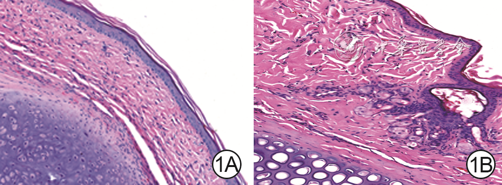
1 瘢痕组建模后60 d,2组兔耳瘢痕/皮肤组织中成纤维细胞(Fb)排列 苏木精-伊红×200。1A.正常对照组兔耳皮肤组织中Fb生长正常且排列均匀;1B. 瘢痕组兔耳瘢痕组织中Fb增生且排列异常

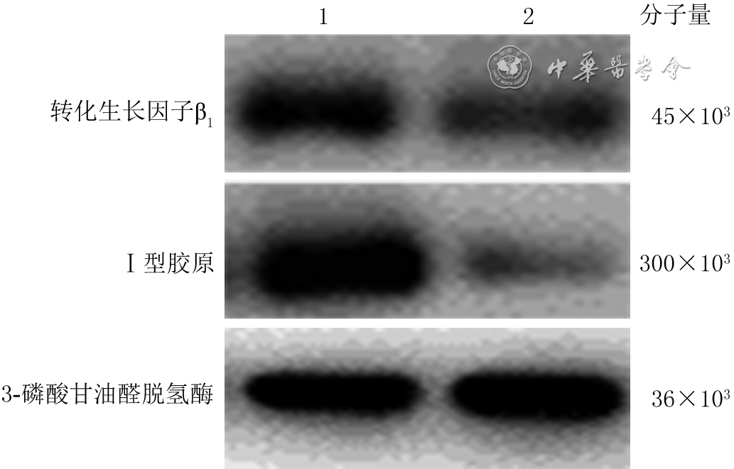
2 蛋白质印迹法检测2组人成纤维细胞转染后24 h转化生长因子β1和Ⅰ型胶原蛋白表达
注:1、2分别为微小RNA-296(miR-296)阴性对照组和miR-296模拟物组
表1 2组人成纤维细胞转染后各时间点细胞增殖情况比较(
组别 样本数 0 h 12 h 24 h 36 h 48 h miR-296阴性对照组 15 0.140±0.034 0.350±0.041 0.548±0.030 0.764±0.044 0.815±0.021 miR-296模拟物组 15 0.142±0.022 0.258±0.025 0.330±0.010 0.488±0.006 0.570±0.013 t值 0.065 3.275 11.980 10.460 17.260 P值 0.952 0.031 0.001 0.001 0.001 注:miR为微小RNA;各组各时间点样本数均为3;处理因素主效应,F=26.760,P<0.001;时间因素主效应,F=269.400,P<0.001;两者交互作用,F=404.200,P<0.001  下载: 导出CSV
-

 下载:
下载:

图(2) / 表(1)
计量
- 文章访问数: 282
- HTML全文浏览量: 97
- PDF下载量: 14
- 被引次数: 0
