


The effect and mechanism of exosomes derived from human amniotic epithelial cells on the proliferation and migration of HaCaT in high glucose environment
-
摘要:
目的 探讨人羊膜上皮细胞外泌体(hAEC-Exo)对高糖环境下HaCaT增殖和迁移的作用及其相关机制。 方法 采用实验研究方法。取2019年1—6月于福建医科大学附属协和医院妇产科10名足月分娩健康孕妇羊膜组织,分离原代人羊膜上皮细胞(hAEC)。观察培养第2、4、7天原代hAEC生长状态和形态改变,采用流式细胞术检测细胞表面标志物CD73、CD90、CD29、CD34及人白细胞抗原DR(HLA-DR)的表达,取第2~4代细胞用于后续实验。超速离心法分离hAEC-Exo。将HaCaT与hAEC-Exo共培养3 h,采用倒置荧光显微镜观察HaCaT对hAEC-Exo的摄取情况。取HaCaT,分为磷酸盐缓冲液(PBS)组、hAEC-Exo组和二甲基亚砜(DMSO)+PBS组、DMSO+hAEC-Exo组、LY294002+hAEC-Exo组,每组3孔,并进行相应处理,采用细胞计数试剂盒8(CCK-8)法检测培养0(即刻)、12、24、36、48、60 h细胞增殖活力;划痕试验检测划痕后0、24、48、72 h划痕愈合情况,并计算划痕愈合率;Transwell实验检测培养48 h穿膜细胞数;蛋白质印迹法检测培养24 h后磷脂酰肌醇3-激酶-蛋白激酶B-哺乳动物雷帕霉素靶蛋白(PI3K-Akt-mTOR)通路相关的哺乳动物雷帕霉素靶蛋白(mTOR)、磷酸化mTOR(p-mTOR)、蛋白激酶B(Akt)、磷酸化Akt(p-Akt)的蛋白表达。对数据行重复测量方差分析、单因素方差分析及独立样本t检验。 结果 原代hAEC培养第2天多数呈卵圆形,大小均一;培养第4、7天,细胞形态呈典型的鹅卵石样单层排列。原代hAEC高表达间充质干细胞表面标志物CD73、CD90及CD29,不表达或低表达造血干细胞相关表面标志物CD34及HLA-DR。培养3 h,hAEC-Exo成功被HaCaT内吞入细胞质,并聚集于细胞核周围。培养12、24、36、48、60 h,hAEC-Exo组HaCaT增殖活力明显高于PBS组(t=3.691、10.861、12.121、10.531、14.931,P<0.01)。划痕后24、48、72 h,PBS组HaCaT划痕愈合率明显低于hAEC-Exo组(t=3.342、6.427、5.485,P<0.05或P<0.01)。培养48 h,hAEC-Exo组HaCaT穿膜数显著多于PBS组(t=5.385,P<0.01)。培养24 h,hAEC-Exo组HaCaT中p-mTOR和p-Akt蛋白表达量明显高于PBS组(t=4.240、5.586,P<0.01),2组HaCaT中mTOR和Akt蛋白表达量相近(P>0.05)。培养24 h,DMSO+hAEC-Exo组HaCaT中p-mTOR和p-Akt的蛋白表达量明显高于DMSO+PBS组(t=6.155、8.338,P<0.01)和LY294002+hAEC-Exo组(t=5.030、3.960,P<0.01),3组HaCaT中mTOR和Akt蛋白表达量相近(P>0.05)。DMSO+hAEC-Exo组HaCaT培养12、24、36、48、60 h增殖活力为0.78±0.05、1.23±0.07、1.60±0.09、1.86±0.09、2.03±0.08,明显高于DMSO+PBS组的0.46±0.04、0.69±0.07、0.98±0.08、1.16±0.08、1.26±0.11(t=4.376、7.398、8.488、9.766、10.730,P<0.01);DMSO+hAEC-Exo组HaCaT培养24、36、48、60 h增殖活力明显高于LY294002+hAEC-Exo组的0.96±0.09、1.20±0.08、1.39±0.08、1.55±0.10(t=3.639、5.447、6.605、6.693,P<0.05或P<0.01)。DMSO+hAEC-Exo组HaCaT划痕后24、48、72 h划痕愈合率明显高于DMSO+PBS组(t=4.003、6.349、7.714,P<0.01)和LY294002+hAEC-Exo组(t=3.805、4.676、4.067,P<0.05或P<0.01)。培养48 h,DMSO+hAEC-Exo组HaCaT穿膜数明显多于DMSO+PBS组和LY294002+hAEC-Exo组(t=7.464、1.232,P<0.01)。 结论 PI3K-Akt-mTOR通路介导hAEC-Exo促进高糖环境下HaCaT增殖和迁移。 Abstract:Objective To investigate the effect and mechanism of exosomes derived from human amniotic epithelial cells (hAEC-Exos) on the proliferation and migration of HaCaT in high glucose environment. Methods The experimental research method was adopted. The amniotic membrane tissue was collected from 10 healthy pregnant women at full term delivery in the Department of Obstetrics and Gynecology of Fujian Medical University Union Hospital from January to June 2019, and the primary human amniotic epithelial cells (hAECs) were isolated. The growth status and morphological changes of the primary hAECs on the 2nd, 4th, and 7th day of culture were observed, and the expressions of the cells surface markers of CD73, CD90, CD29, CD34, and human leukocyte antigen DR (HLA-DR). The 2nd to 4th passages of hAECs were used in the following experiments. The hAEC-Exos were separated by ultracentrifugation method. The HaCaT and hAEC-Exos were co-cultured for 3 h, and the uptake of hAEC-Exos by HaCaT was observed by inverted fluorescence microscopy. The HaCaT were divided into phosphate buffer solution (PBS) group and hAEC-Exos group or dimethyl sulfoxide (DMSO)+PBS group, DMSO+hAEC-Exos group, and LY294002+hAEC-Exos group, which were dealt correspondingly, with 3 wells in each group. Cell counting kit 8 (CCK-8) method was used to detect cell proliferation activity after 0 (immediately), 12, 24, 36, 48, and 60 h of culture. The scratch test was conducted to detect the scratch healing at 0, 24, 48, and 72 h after the scratch, and the scratch healing rate was calculated, respectively. The Transwell experiment was conducted to detect the number of transmembrane cells after 48 h of culture. The Western blotting was used to detect the protein expressions of mammalian target of rapamycin (mTOR), phosphorylated mTOR (p-mTOR), protein kinase B (Akt), and phosphorylated Akt (p-Akt) related to phosphatidylinositol 3-kinase-Akt-mTOR (PI3K-Akt-mTOR) pathway after 24 h of culture. Data were statistically analyzed with analysis of variance for repeated measurement, one-way analysis of variance, and independent sample t test. Results Most of the primary hAECs were oval and uniform in size on the 2nd day of culture. The hAECs were arranged in a typical cobblestone-like monolayer on the 4th and 7th day of culture. The primary hAECs highly expressed CD73, CD90, and CD29 of mesenchymal stem cell related surface markers, and were with no or low expressions of CD34 and HLA-DR of hematopoietic stem cell related surface markers. After 3 h of culture, hAEC-Exos were successfully endocytosed by HaCaT into the cytoplasm and gathered around the nucleus. After 12, 24, 36, 48, and 60 h of culture, the proliferation activity of HaCaT in hAEC-Exos group was significantly higher than that in PBS group (t=3.691, 10.861, 12.121, 10.531, 14.931, P<0.01). At 24, 48, and 72 h after scratch, the scratch healing rates of HaCaT in PBS group were significantly lower than those in hAEC-Exos group (t=3.342, 6.427, 5.485, P<0.05 or P<0.01). After 48 h of culture, the number of transmembrane HaCaT in hAEC-Exos group was significantly more than that in PBS group (t=5.385, P<0.01). After 24 h of culture, the protein expressions of p-mTOR and p-Akt in HaCaT of hAEC-Exos group were significantly higher than those in PBS group (t=4.240, 5.586, P<0.01), while the protein expressions of mTOR and Akt in HaCaT of the two groups were similar (P>0.05). After 24 h of culture, the protein expressions of p-mTOR and p-Akt in HaCaT of DMSO+hAEC-Exos group were significantly higher than those in DMSO+PBS group (t=6.155, 8.338, P<0.01) and LY294002+hAEC-Exos group (t=5.030, 3.960, P<0.01), while the protein expressions of mTOR and Akt in HaCaT of the three groups were similar (P>0.05). The proliferation activity of HaCaT in DMSO+hAEC-Exos group at 12, 24, 36, 48, and 60 h of culture was 0.78±0.05, 1.23±0.07, 1.60±0.09, 1.86±0.09, and 2.03±0.08, which was significantly higher than 0.46±0.04, 0.69±0.07, 0.98±0.08, 1.16±0.08, and 1.26±0.11 in DMSO+PBS group (t=4.376, 7.398, 8.488, 9.766, 10.730, P<0.01). The proliferation activity of HaCaT in DMSO+hAEC-Exos group at 24, 36, 48, and 60 h was significantly higher than 0.96±0.09, 1.20±0.08, 1.39±0.08, and 1.55±0.10 in LY294002+hAEC-Exos group (t=3.639, 5.447, 6.605, 6.693, P<0.05 or P<0.01). The scratch healing rates of HaCaT in DMSO+hAEC-Exos group at 24, 48, and 72 h after scratch were significantly higher than those in DMSO+PBS group (t=4.003, 6.349, 7.714, P<0.01) and LY294002+hAEC-Exos group (t=3.805, 4.676, 4.067, P<0.05 or P<0.01). After 48 h of culture, the number of transmembrane HaCaT in DMSO+hAEC-Exos group was significantly more than that in DMSO+PBS group and LY294002+hAEC-Exos group, respectively (t=7.464, 1.232, P<0.01). Conclusions PI3K-Akt-mTOR pathway can promote the proliferation and migration of HaCaT in high glucose environment by mediating hAEC-Exos. -
(1)较早地在烧伤领域提出持续炎症-免疫抑制-分解代谢综合征(PICS)这一概念。
持续炎症-免疫抑制-分解代谢综合征(persistent inflammation-immunosuppression-catabolism syndrome,PICS)是在全身性感染或非感染如烧创伤等进入慢性危重症(chronic critical illness,CCI)阶段时,出现的以持续性炎症、免疫抑制及蛋白质高分解代谢为特征的临床综合征。PICS防治困难、发病率高,是导致重症患者长期生活质量低下及远期死亡的重要原因,已成为重症患者治疗的新挑战 [ 1, 2] 。
大面积烧伤是一个累及全身的严重创伤事件,其病理生理学过程及临床表现与PICS类似,但目前关于大面积烧伤患者并发PICS的临床研究甚少。本研究总结大面积烧伤患者继发PICS的临床特征,对大面积烧伤患者继发PICS的影响因素进行多元回归分析,以期有助于临床早期识别、阻止或逆转可能的高危因素,进而改善大面积烧伤患者的预后。
1. 对象与方法
本回顾性病例系列研究符合《赫尔辛基宣言》的基本原则,根据暨南大学附属广州红十字会医院(以下简称本单位)伦理委员会政策,可以在不泄露患者身份信息的前提下对其临床资料进行分析、使用。
1.1 入选标准
纳入标准:(1)烧伤总面积≥30%TBSA;(2)年龄≥18岁,性别不限;(3)住院天数>14 d。排除标准:病历资料不完整者。
1.2 PICS诊断标准
(1)住院天数>14 d;(2)持续性的炎症反应:C反应蛋白>150 mg/L;(3)免疫抑制:淋巴细胞计数<0.8×10 9/L;(4)分解代谢综合征:血清白蛋白<30 g/L。
1.3 临床资料与分组
2017年1月—2021年12月,本单位烧伤ICU(BICU)收治220例符合入选标准的大面积成年烧伤患者,其中男168例、女52例,年龄18~84(43±14)岁。通过查询本单位的电子病历系统和重症护理系统,按照PICS发生情况将患者分为PICS组(84例)和非PICS组(136例)。
1.4 统计指标
1.4.1 一般资料
性别、年龄(分层:<65岁、≥65岁)、入院时合并基础疾病(糖尿病、高血压)情况和急性生理学和慢性健康状况评价Ⅱ(APACHEⅡ)评分、入院时和入院14 d脓毒症相关性器官功能衰竭评价(SOFA)评分、治疗期间行机械通气超过48 h比例。
1.4.2 专科情况
烧伤总面积、Ⅲ度烧伤面积、伤后48 h内入院比例、伤后30 d深度创面暴露面积(包括未行手术治疗的深Ⅱ度和Ⅲ度烧伤创面、暴露的肉芽组织创面、植皮区/供皮区感染创面面积)。
1.4.3 结局指标
住院天数、住院总费用、手术次数以及死亡情况。
1.5 统计学处理
采用SPSS 22.0统计软件进行数据分析。符合正态分布的计量资料数据以
2. 结果
2.1 一般资料
PICS组患者入院时APACHEⅡ评分和SOFA评分、治疗期间行机械通气超过48 h比例均明显高于非PICS组( P<0.05),2组患者其余一般资料均相近( P>0.05)。见 表1。
表1 2组大面积烧伤患者一般资料比较组别 例数 性别[例(%)] 年龄[例(%)] 入院时合并基础疾病[例(%)] 入院时APACHEⅡ评分(分, 入院时SOFA评分[分, M( Q 1, Q 3)] 入院14 d SOFA评分[分, M( Q 1, Q 3)] 治疗期间行机械通气超过48 h[例(%)] 男 女 <65岁 ≥65岁 糖尿病 高血压 PICS组 84 65(77.4) 19(22.6) 80(95.2) 4(4.8) 3(3.6) 10(11.9) 11±5 4(3,5) 3(2,4) 46(54.8) 非PICS组 136 103(75.7) 33(24.3) 128(94.1) 8(5.9) 4(2.9) 8(5.9) 7±4 2(1,3) 2(1,3) 54(39.7) 统计量值 χ 2=0.08 χ 2<0.01 χ 2<0.01 χ 2=2.50 t=6.78 Z=-4.75 Z=-1.90 χ 2=4.74 P值 0.780 0.960 1.000 0.113 <0.001 <0.001 0.057 0.029 注:PICS为持续炎症-免疫抑制-分解代谢综合征,APACHEⅡ为急性生理学和慢性健康状况评价Ⅱ,SOFA为脓毒症相关性器官功能衰竭评价 2.2 专科情况
PICS组患者烧伤总面积、Ⅲ度烧伤面积、伤后30 d深度创面暴露面积均明显大于非PICS组( P<0.05),但伤后48 h内入院比例明显低于非PICS组( P<0.05)。见 表2。
表2 2组大面积烧伤患者专科情况比较组别 例数 烧伤总面积(%TBSA, Ⅲ度烧伤面积[%TBSA, M( Q 1, Q 3)] 伤后48 h内入院[例(%)] 伤后30 d深度创面暴露面积[%TBSA, M( Q 1, Q 3)] PICS组 84 73±20 56(36,80) 57(67.9) 25(15,35) 非PICS组 136 55±20 29(6,43) 112(82.4) 8(0,13) 统计量值 t=6.29 Z=-7.25 χ 2=6.13 Z=-8.73 P值 <0.001 <0.001 0.013 <0.001 注:PICS为持续炎症-免疫抑制-分解代谢综合征,TBSA为体表总面积;深度创面暴露面积包括未行手术治疗的深Ⅱ度和Ⅲ度烧伤创面、暴露的肉芽组织创面、植皮区/供皮区感染创面面积 2.3 结局指标
PICS组患者住院天数、住院总费用、手术次数均明显多于非PICS组( P<0.05),但2组患者死亡情况相近( P>0.05)。见 表3。
表3 2组大面积烧伤患者结局指标比较组别 例数 住院天数[d, M( Q 1, Q 3)] 住院总费用[万元, M( Q 1, Q 3)] 手术次数[次, M( Q 1, Q 3)] 死亡[例(%)] PICS组 84 66(42,86) 85.3(48.1,110.6) 5(3,7) 2(2.4) 非PICS组 136 40(26,48) 35.7(13.0,47.8) 2(1,4) 3(2.2) 统计量值 Z=-7.12 Z=-8.48 Z=-6.87 χ 2<0.01 P值 <0.001 <0.001 <0.001 0.633 注:PICS为持续炎症-免疫抑制-分解代谢综合征 2.4 多因素logistic回归分析结果
以继发PICS的情况(PICS=1,非PICS=0)为因变量,将单因素分析中差异具有统计学意义的指标作为自变量,将烧伤总面积、Ⅲ度烧伤面积、伤后30 d深度创面暴露面积、入院时APACHEⅡ评分、入院时SOFA评分以原始值代入,伤后48 h内是否入院赋值(是=1,否=2)、治疗期间行机械通气是否超过48 h赋值(是=1,否=2),进行多因素logistic回归分析。结果显示,入院时APACHEⅡ评分、伤后30 d深度创面暴露面积均为大面积烧伤患者继发PICS的独立危险因素( P<0.05)。见 表4。
表4 影响220例大面积烧伤患者继发PICS的多因素logistic回归分析阳性结果因素 回归系数 标准误 比值比 95%置信区间 P值 入院时APACHEⅡ评分(分) 0.14 12.13 1.15 1.06~1.25 <0.001 伤后30 d深度创面暴露面积(%TBSA) 0.07 30.07 1.07 1.05~1.10 <0.001 注:PICS为持续炎症-免疫抑制-分解代谢综合征,APACHEⅡ为急性生理学和慢性健康状况评价Ⅱ,TBSA为体表总面积;深度创面暴露面积包括未行手术治疗的深Ⅱ度和Ⅲ度烧伤创面、暴露的肉芽组织创面、植皮区/供皮区感染创面面积 3. 讨论
近年来,随着重症器官支持治疗理念和技术的发展,越来越多的重症患者从MODS早期死亡高峰阶段幸存下来,成为CCI患者。2012年,Gentile等 [ 3] 提出PICS的新概念。PICS提供了一个理解长期住院CCI患者病理生理状态的新视角。该概念被提出后,其合理性逐渐被接受。在脓毒症、多发伤、严重创伤等患者中的研究显示,PICS患者住院天数多、医疗资源消耗巨大、长期生活质量低下、中远期病死率高,值得临床医师高度重视。
大面积烧伤患者的病程中,存在典型的持续炎症反应、免疫抑制以及高分解代谢状态。大面积烧伤患者是否存在PICS,其发生、发展的特点如何,是本研究团队开展这一回顾性病例系列研究的目的。
本单位收治的220例符合入选标准的大面积成年烧伤患者中有84例符合PICS诊断标准,PICS的发病率为38.18%,高于在多发伤患者中的11.7% [ 4] ,与老年脓毒症患者中的37.1%相近 [ 5] 。一般情况下,年龄≥65岁、合并基础疾病是重症患者继发PICS的危险因素。本研究显示,PICS组与非PICS组大面积烧伤患者在年龄、入院时合并基础疾病(糖尿病、高血压)方面的差异均无统计学意义( P>0.05),可能与本单位BICU收治的烧伤患者普遍较年轻,大部分为从事体力劳动的青壮年,基础疾病较少有关。
本研究中,PICS组患者烧伤总面积、Ⅲ度烧伤面积均明显大于非PICS组。烧伤面积与深度基本决定了烧伤的严重程度,大面积烧伤将导致大量促炎性细胞因子释放,进而触发和增强炎症反应与高分解代谢,推测其可能促成了PICS的发生。
有文献指出,烧伤后最初阶段的复苏不全往往是导致SIRS持续或全身状态恶化的重要因素 [ 6, 7] 。与PICS组相比,非PICS组伤后48 h内入院的患者占比较高。一般情况下,伤后48 h内入院的患者通常能接受较规范的液体复苏以及后续治疗。本研究中,伤后48 h后入院的患者,一部分是受伤地点离本单位较远,长途转运或多方辗转后才到达本单位接受治疗;一部分是在外院治疗了一段时间,创面处理不恰当,患者出现了一系列并发症后转入本单位的,因此PICS的发病率也高。不合适的创面处理通常会造成创面的感染、暴露,影响后续创面处理的效果,增加了创面封闭的难度,因此手术次数也会相应增多。推测此为本研究中PICS组患者的手术次数较非PICS组多的主要原因。
本研究中PICS组治疗期间行机械通气超过48 h的患者比例明显高于非PICS组,这与许多关于PICS的研究结果 [ 8, 9] 一致。有研究显示,SOFA评分对烧伤患者的预后有较好的预测价值 [ 10] 。在本研究中,2组患者仅入院时这个时间点的SOFA评分比较差异有统计学意义( P<0.05),且SOFA评分非大面积烧伤患者继发PICS的独立危险因素。SOFA评分由氧合指数、血小板计数、胆红素水平、血管活性药物使用情况、格拉斯哥昏迷量表(GCS)评分等组成,其中的胆红素水平、GCS评分对于烧伤患者早期的评估,特异度并不高 [ 10] 。有学者指出,由于烧伤患者脓毒症休克出现迅速,但胆红素指标对于肝脏功能变化反应较慢,并且胆红素水平非烧伤患者的常规检测项目,因此认为,应将其从对烧伤及烧伤脓毒症休克患者的评分中剔除 [ 11] 。而包含血糖水平、将镇静与非镇静患者分别评估(镇静患者评估肠内营养耐受情况,非镇静患者评估意识状态)的“烧伤SOFA”评分,可能更适合重症烧伤患者。
进一步的多因素logistic回归分析结果显示,入院时APACHEⅡ评分、伤后30 d深度创面暴露面积是大面积烧伤患者继发PICS的独立危险因素。APACHEⅡ评分是目前临床危重症患者病情评估的主要评分系统,由急性生理学评分、年龄评分、慢性健康状况评分三部分组成,得分越高表示病情越重。本研究中,PICS组患者病情更严重、伤后48 h内入院的患者比例更低,可能是导致入院时APACHEⅡ评分更高的原因。而此评分包括了本研究纳入的如年龄、合并基础疾病等诸多影响因素,较全面地反映了患者的病情,可能是其成为大面积烧伤患者继发PICS独立危险因素的重要原因。同样,在对脓毒症、创伤等的研究中观察到,APACHEⅡ评分高的患者更易出现PICS [ 4, 8] ,与本研究结果一致。然而入院时病情严重程度是否与后期出现的PICS直接相关?伤后48 h后入院的已经出现并发症的患者,入院时的APACHEⅡ评分是否能反映其病情的严重程度?这需要大样本、更细的分层研究来证实。
本研究中2组患者烧伤总面积和Ⅲ度烧伤面积有明显差异,但这2个指标不是大面积烧伤患者继发PICS的独立危险因素,而伤后30 d深度创面暴露面积是大面积烧伤患者继发PICS的独立危险因素。大面积深度创面的长时间暴露对烧伤患者内环境的稳态、代谢、免疫等带来深远的影响。而创面坏死组织的去除,自体皮或皮肤替代物覆盖创面不仅可以减少炎症因子的释放以及其带来的全身炎症反应、免疫抑制,亦可以通过恢复体温调节、减少热量损失和水分蒸发等减轻高代谢反应,是打断烧伤后持续炎症反应-免疫抑制-高分解代谢这一循环的有效手段 [ 12, 13] 。
为了缩短大面积烧伤患者创面暴露时间,减少并发症的发生。本单位自2007年开始,对重症烧伤患者实行统一管理,伤后5 d左右为患者行第1次切削痂手术,对同一深度烧伤面积区间的患者实施统一手术方案;同时,结合深Ⅱ度创面愈合后作为供皮区,头部、阴囊反复供皮,以及控制手术出血及损伤等方法,分次分批手术覆盖创面。对于外院转入、创面处理不及时或不恰当且已经出现并发症的患者,亦在维护脏器功能的同时,积极进行手术干预,尽量减少创面暴露的时间和面积。既往研究显示,本单位BICU治疗的烧伤总面积<50%TBSA、51%~80%TBSA、>80%TBSA的患者,平均创面愈合时间分别为36、43、79 d [ 14] 。
持续炎症-免疫抑制-高分解代谢多层面与多环节的恶性循环,最终影响了PICS患者的结局。目前,针对PICS的治疗,包括抗感染治疗、免疫治疗、物理治疗、营养治疗等,但PICS是由一系列的介质引起的,介质相互关联和依存,存在多层面、多环节的恶性循环,目前许多关键节点或环路尚不清楚,因此治疗棘手。大面积烧伤的患者病死率与感染以及全身炎症反应等导致的脏器功能损伤有关,其中PICS一般与创面暴露导致的全身炎症反应相关。本研究结果显示,虽然PICS在大面积烧伤患者中发病率不低,但经过积极的手术治疗,结合重症患者脏器功能支持技术,总体预后良好,2组患者病死率相近。这也提示,无论是对于烧伤本身的治疗还是对于烧伤后并发症的治疗,都不能忽略对创面本身的处理。
另外,本研究也显示,PICS组患者手术次数、住院天数、住院总费用均明显多于非PICS组,提示继发PICS的患者,需要更多的手术干预和住院天数以及更高的住院费用。因此,尽管PICS预后良好,但其仍会给患者以及医院带来巨大负担。
综上所述,本研究中,大面积烧伤患者继发PICS的发病率较高,入院时APACHEⅡ评分以及伤后30 d深度创面暴露面积为大面积烧伤患者继发PICS的独立危险因素,说明入院时病情严重的患者更易出现PICS,提示深度创面处理对于阻断大面积烧伤患者持续炎症-免疫抑制-高分解代谢这一恶性循环的重要性。然而本研究属于单中心回顾性研究,样本量小,可能造成选择性偏倚,需要大样本多中心的研究完善其结果。同时,PICS诊断标准仍有争议,涉及PICS诊断的指标,例如血清白蛋白水平、C反应蛋白等是否能较好地体现烧伤患者的代谢、炎症反应情况,仍值得商榷;烧伤患者继发PICS是否对其瘢痕的形成、远期生活质量以及生存产生影响,也仍需要进一步研究。
所有作者均声明不存在利益冲突广告目次深圳市源兴医药股份有限公司 ………………………………………………………………………………………… 插页3美纳里尼(中国)投资有限公司 ………………………………………………………………………………………… 插页5上海铠唏尔医疗器械贸易有限公司 …………………………………………………………………………………… 插页6南海朗肽制药有限公司 ………………………………………………………………………………………… 对中文目次1江西省科星生物工程有限公司 ………………………………………………………………………………… 对中文目次2上海腾瑞制药股份有限公司 …………………………………………………………………………………… 对英文目次1保赫曼(上海)贸易有限公司 …………………………………………………………………………………… 对英文目次2浙江医学科技开发有限公司 …………………………………………………………………………………………… 插页7苏州汇涵医用科技发展有限公司 ……………………………………………………………………………………… 插页8苏州爱得科技发展股份有限公司 ……………………………………………………………………………………… 对正文珠海亿胜生物制药有限公司 ……………………………………………………………………………………………… 封三武汉维斯第医用科技股份有限公司 ……………………………………………………………………………………… 封底 -
参考文献
(27) [1] BurgessJL,WyantWA,Abdo AbujamraB,et al.Diabetic wound-healing science[J].Medicina (Kaunas),2021,57(10):1072. DOI: 10.3390/medicina57101072. [2] RatajczakJ,WysoczynskiM,HayekF,et al.Membrane-derived microvesicles: important and underappreciated mediators of cell-to-cell communication[J].Leukemia,2006,20(9):1487-1495.DOI: 10.1038/sj.leu.2404296. [3] FerreiraADF, GomesDA. Stem cell extracellular vesicles in skin repair[J].Bioengineering (Basel),2018,6(1):4. DOI: 10.3390/bioengineering6010004. [4] van NielG, D'AngeloG, RaposoG.Shedding light on the cell biology of extracellular vesicles[J].Nat Rev Mol Cell Biol,2018,19(4):213-228. DOI: 10.1038/nrm.2017.125. [5] ZhangW,BaiX,ZhaoB,et al.Cell-free therapy based on adipose tissue stem cell-derived exosomes promotes wound healing via the PI3K/Akt signaling pathway[J].Exp Cell Res,2018,370(2):333-342. DOI: 10.1016/j.yexcr.2018.06.035. [6] ZhangJ,ChenC,HuB,et al.Exosomes derived from human endothelial progenitor cells accelerate cutaneous wound healing by promoting angiogenesis through Erk1/2 signaling[J].Int J Biol Sci,2016,12(12):1472-1487.DOI: 10.7150/ijbs.15514. [7] LiX, LiuL, YangJ, et al. Exosome derived from human umbilical cord mesenchymal stem cell mediates miR-181c attenuating burn-induced excessive inflammation[J]. EBioMedicine, 2016,8:72-82. DOI: 10.1016/j.ebiom.2016.04.030. [8] ZhaoB,LiX,ShiX,et al.Exosomal microRNAs derived from human amniotic epithelial cells accelerate wound healing by promoting the proliferation and migration of fibroblasts[J].Stem Cells Int,2018,2018:5420463.DOI: 10.1155/2018/5420463. [9] ZhengY,ZhengS,FanX,et al.Amniotic epithelial cells accelerate diabetic wound healing by modulating inflammation and promoting neovascularization[J].Stem Cells Int,2018,2018:1082076.DOI: 10.1155/2018/1082076. [10] ZhengY,JiS,WuH,et al.Topical administration of cryopreserved living micronized amnion accelerates wound healing in diabetic mice by modulating local microenvironment[J].Biomaterials,2017,113:56-67.DOI: 10.1016/j.biomaterials.2016.10.031. [11] WeiP,ZhongC,YangX,et al.Exosomes derived from human amniotic epithelial cells accelerate diabetic wound healing via PI3K-AKT-mTOR-mediated promotion in angiogenesis and fibroblast function[J/OL].Burns Trauma,2020,8:tkaa020[2021-04-24].https://pubmed.ncbi.nlm.nih.gov/32923490/.DOI: 10.1093/burnst/tkaa020. [12] XiaoGY,ChengCC,ChiangYS,et al.Exosomal miR-10a derived from amniotic fluid stem cells preserves ovarian follicles after chemotherapy[J].Sci Rep,2016,6:23120.DOI: 10.1038/srep23120. [13] FortunatoO,GaspariniP,BoeriM,et al.Exo-miRNAs as a new tool for liquid biopsy in lung cancer[J].Cancers (Basel),2019,11(6):888. DOI: 10.3390/cancers11060888. [14] HuY,RaoSS,WangZX,et al.Exosomes from human umbilical cord blood accelerate cutaneous wound healing through miR-21-3p-mediated promotion of angiogenesis and fibroblast function[J].Theranostics,2018,8(1):169-184.DOI: 10.7150/thno.21234. [15] HuangH,CuiW,QiuW,et al.Impaired wound healing results from the dysfunction of the Akt/mTOR pathway in diabetic rats[J].J Dermatol Sci,2015,79(3):241-251.DOI: 10.1016/j.jdermsci.2015.06.002. [16] CastilhoRM,SquarizeCH,GutkindJS.Exploiting PI3K/mTOR signaling to accelerate epithelial wound healing[J].Oral Dis,2013,19(6):551-558.DOI: 10.1111/odi.12070. [17] ShawTJ,MartinP.Wound repair: a showcase for cell plasticity and migration[J].Curr Opin Cell Biol,2016,42:29-37.DOI: 10.1016/j.ceb.2016.04.001. [18] HuSC,LanCE.High-glucose environment disturbs the physiologic functions of keratinocytes: focusing on diabetic wound healing[J].J Dermatol Sci,2016,84(2):121-127.DOI: 10.1016/j.jdermsci.2016.07.008. [19] ZhaoB,LiuJQ,YangC,et al.Human amniotic epithelial cells attenuate TGF-β1-induced human dermal fibroblast transformation to myofibroblasts via TGF-β1/Smad3 pathway[J].Cytotherapy,2016,18(8):1012-1024.DOI: 10.1016/j.jcyt.2016.04.009. [20] ZhaoB,LiuJQ,ZhengZ,et al.Human amniotic epithelial stem cells promote wound healing by facilitating migration and proliferation of keratinocytes via ERK, JNK and AKT signaling pathways[J].Cell Tissue Res,2016,365(1):85-99.DOI: 10.1007/s00441-016-2366-1. [21] CamussiG,DeregibusMC,BrunoS,et al.Exosomes/microvesicles as a mechanism of cell-to-cell communication[J].Kidney Int,2010,78(9):838-848.DOI: 10.1038/ki.2010.278. [22] FriedmanRC,FarhKK,BurgeCB,et al.Most mammalian mRNAs are conserved targets of microRNAs[J].Genome Res,2009,19(1):92-105.DOI: 10.1101/gr.082701.108. [23] MengZ,ZhouD,GaoY,et al.miRNA delivery for skin wound healing[J].Adv Drug Deliv Rev,2018,129:308-318.DOI: 10.1016/j.addr.2017.12.011. [24] MulhollandEJ,DunneN,McCarthyHO.MicroRNA as therapeutic targets for chronic wound healing[J].Mol Ther Nucleic Acids,2017,8:46-55.DOI: 10.1016/j.omtn.2017.06.003. [25] FahsF, BiX, YuFS, et al. New insights into microRNAs in skin wound healing[J]. IUBMB Life, 2015,67(12):889-896. DOI: 10.1002/iub.1449. [26] LiD,LiXI,WangA,et al.MicroRNA-31 promotes skin wound healing by enhancing keratinocyte proliferation and migration[J].J Invest Dermatol,2015,135(6):1676-1685.DOI: 10.1038/jid.2015.48. [27] DeppeJ,SteinritzD,SantovitoD,et al.Upregulation of miR-203 and miR-210 affect growth and differentiation of keratinocytes after exposure to sulfur mustard in normoxia and hypoxia[J].Toxicol Lett,2016,244:81-87.DOI: 10.1016/j.toxlet.2015.09.012. -
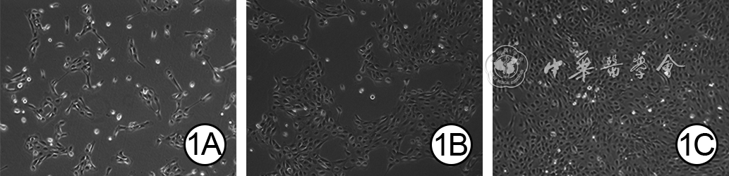
1 原代人羊膜上皮细胞(hAEC)培养第2、4、7天形态观察 倒置荧光显微镜×100。1A.培养第2天,hAEC贴壁生长,形态、大小均一;1B、1C.分别为培养第4、7天,hAEC增殖活跃,细胞密度增加,呈典型的鹅卵石样单层排列
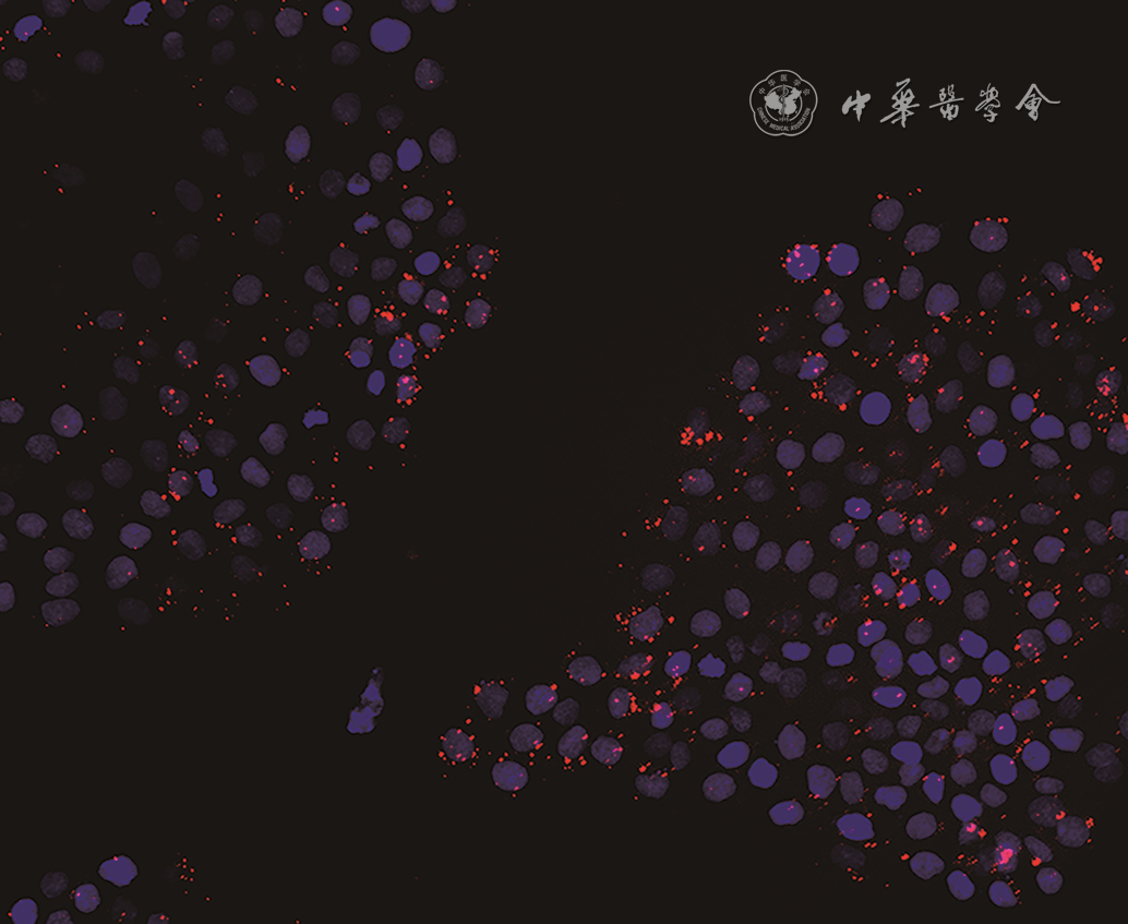
2 人羊膜上皮细胞外泌体(hAEC-Exo)与HaCaT共培养3 h,hAEC-Exo被HaCaT内吞入细胞质,并聚集于细胞核周围 PKH26-4',6-二脒基-2-苯基吲哚×200
注:红色指示外泌体,蓝色指示细胞核
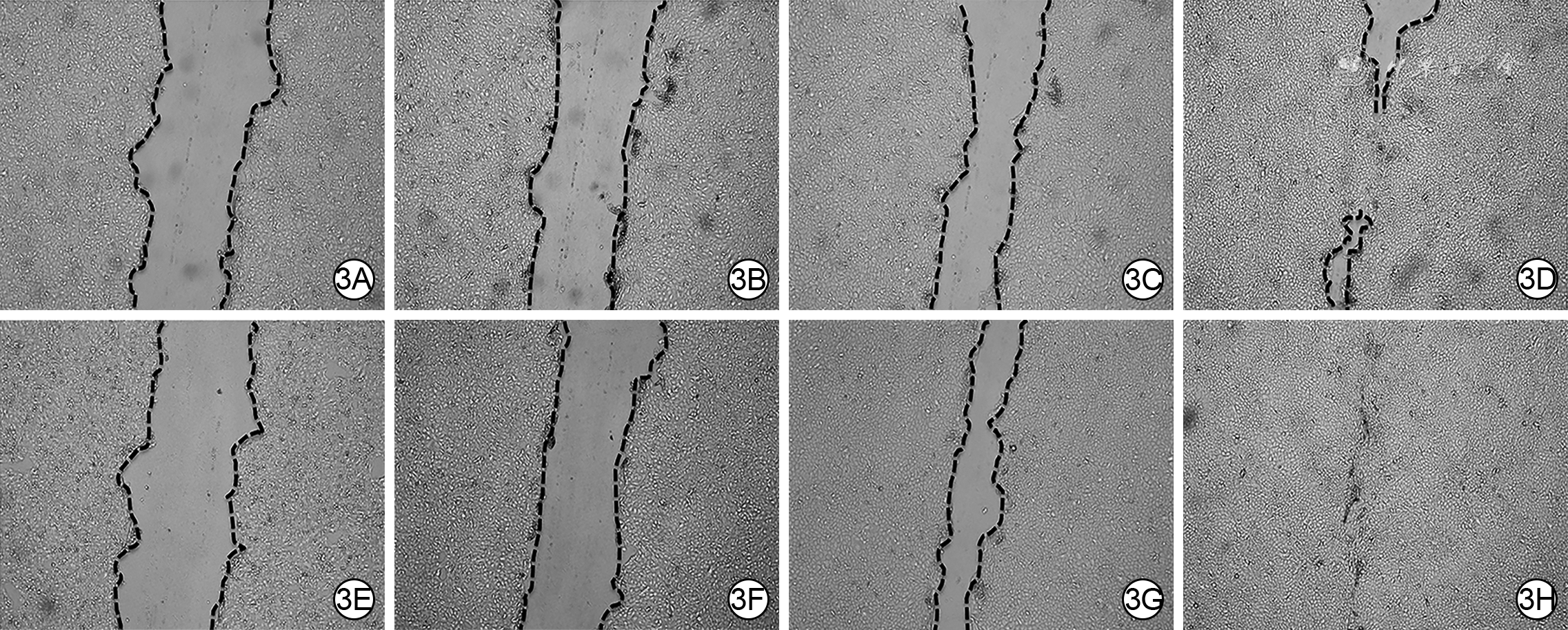
3 2组HaCaT划痕后各时间点划痕面积 倒置荧光显微镜×100。3A、3B、3C、3D.分别为PBS组划痕后0(即刻)、24、48、72 h的划痕面积;3E、3F、3G、3H.分别为hAEC-Exo组划痕后0、24、48、72 h的划痕面积,图3F、3G、3H划痕面积分别明显小于图3B、3C、3D
注:PBS为磷酸盐缓冲液,hAEC-Exo为人羊膜上皮细胞外泌体
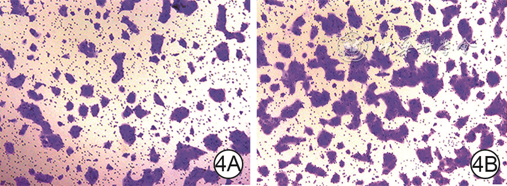
4 Transwell实验观察2组HaCaT培养48 h穿膜迁移情况 苏木精×50。4A、4B.分别为PBS组和hAEC-Exo组,图4B穿膜细胞数明显多于图4A
注:蓝色指示穿膜细胞;PBS为磷酸盐缓冲液,hAEC-Exo为人羊膜上皮细胞外泌体
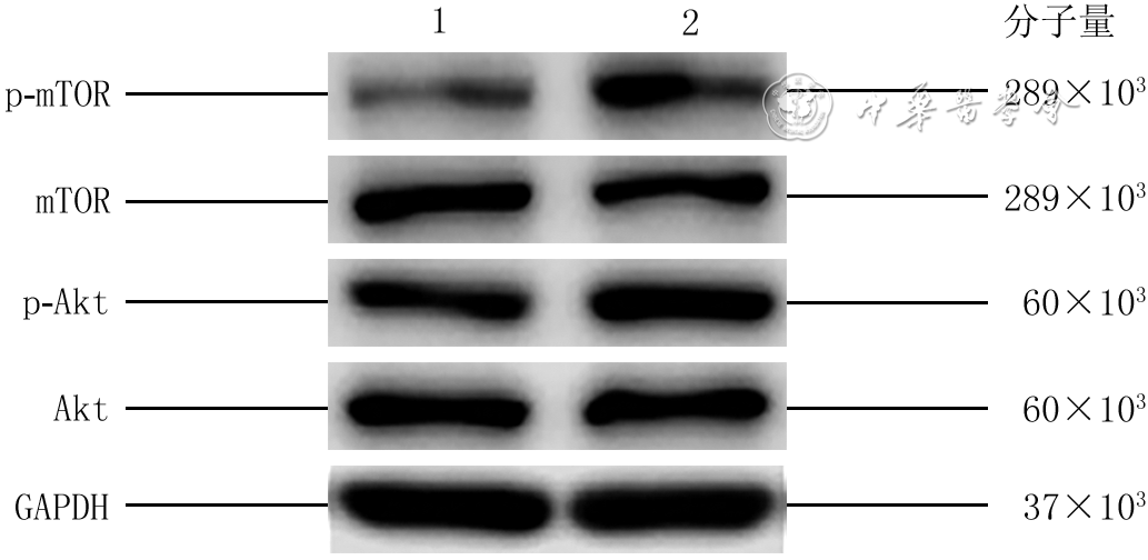
5 蛋白质印迹法检测2组HaCaT培养24 h PI3K-Akt-mTOR通路相关蛋白的表达水平
注:PI3K为磷脂酰肌醇3-激酶,mTOR为哺乳动物雷帕霉素靶蛋白,p-mTOR为磷酸化mTOR,Akt为蛋白激酶B,p-Akt为磷酸化Akt,GAPDH为3-磷酸甘油醛脱氢酶;1、2分别为磷酸盐缓冲液组和人羊膜上皮细胞外泌体组

6 蛋白质印迹法检测3组HaCaT培养24 h PI3K-Akt-mTOR通路相关蛋白的表达量
注:PI3K为磷脂酰肌醇3-激酶,mTOR为哺乳动物雷帕霉素靶蛋白,p-mTOR为磷酸化mTOR,Akt为蛋白激酶B,p-Akt为磷酸化Akt,GAPDH为3-磷酸甘油醛脱氢酶;1、2、3分别为二甲基亚砜(DMSO)+磷酸盐缓冲液组,DMSO+人羊膜上皮细胞外泌体(hAEC-Exo)组,LY294002+hAEC-Exo组
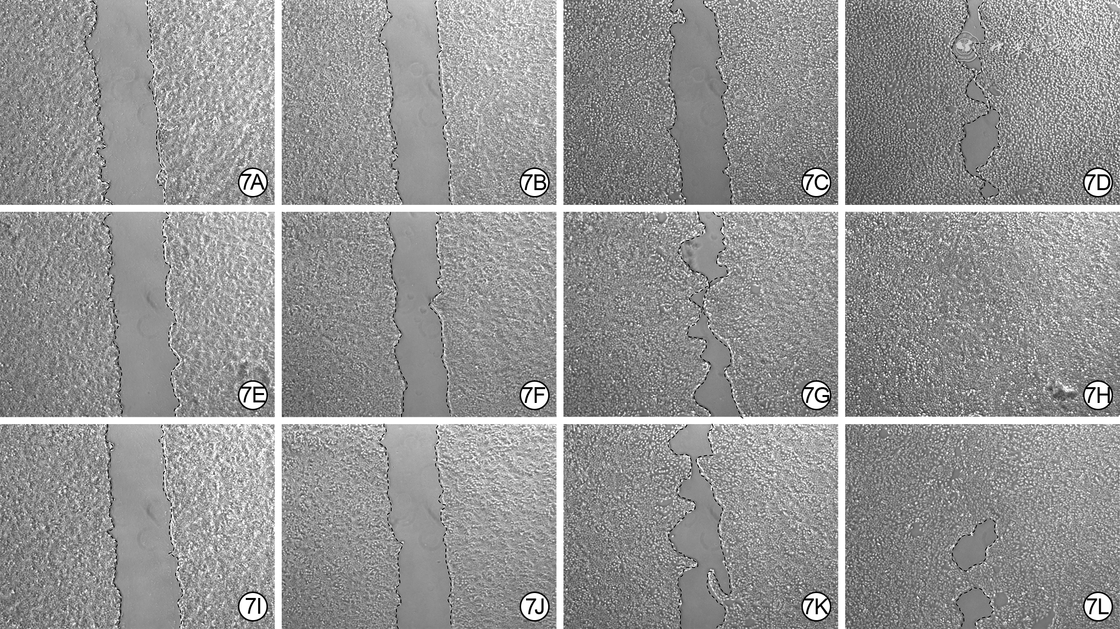
7 3组HaCaT划痕后各时间点划痕面积。7A、7B、7C、7D.分别为DMSO+PBS组划痕后0(即刻)、24、48、72 h的划痕面积;7E、7F、7G、7H.分别为DMSO+hAEC-Exo组划痕后0、24、48、72 h的划痕面积,图7F、7G、7H划痕面积明显小于图7B、7C、7D;7I、7J、7K、7L.分别为LY294002+hAEC-Exo组划痕后0、24、48、72 h的划痕面积,图7J、7K、7L划痕面积明显大于图7F、7G、7H
注:DMSO为二甲基亚砜,PBS为磷酸盐缓冲液,hAEC-Exo为人羊膜上皮细胞外泌体
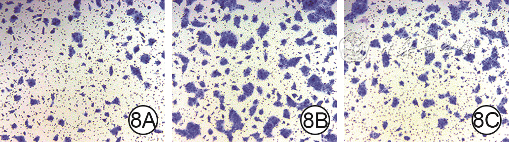
8 Transwell实验观察3组HaCaT培养48 h穿膜迁移情况 苏木精×50。8A、8B、8C.分别为DMSO+PBS组、DMSO+hAEC-Exo组和LY294002+hAEC-Exo组,图8B穿膜迁移细胞数明显多于图8A、8C
注:图中蓝色指示穿膜细胞;DMSO为二甲基亚砜, PBS为磷酸盐缓冲液,hAEC-Exo为人羊膜上皮细胞外泌体
表 1 2组HaCaT培养各时间点增殖活力比较(
组别 样本数 12 h 24 h 36 h 48 h 60 h PBS组 3 0.273±0.025 0.386±0.028 0.494±0.030 0.789±0.060 0.943±0.040 hAEC-Exo组 3 0.404±0.030 0.770±0.041 0.923±0.026 1.161±0.042 1.471±0.038 t值 3.691 10.861 12.121 10.531 14.931 P值 <0.01 <0.01 <0.01 <0.01 <0.01 注:PBS为磷酸盐缓冲液,hAEC-Exo为人羊膜上皮细胞外泌体;处理因素主效应,F=783.2,P<0.01;时间因素主效应,F=494.7,P<0.01;两者交互作用,F=27.0,P<0.01  下载: 导出CSV
表2 2组HaCaT划痕后各时间点划痕愈合率比较
表2. (%,x¯±s)
组别 样本数 24 h 48 h 72 h PBS组 3 22.6±2.4 42.8±4.0 74.5±4.5 hAEC-Exo组 3 34.7±4.9 66.1±4.8 94.4±4.1 t值 3.342 6.427 5.485 P值 <0.05 <0.01 <0.01 注:PBS为磷酸盐缓冲液,hAEC-Exo为人羊膜上皮细胞外泌体;处理因素主效应,F=692.7,P<0.01;时间因素主效应,F=25.4,P<0.01;两者交互作用,F=14.4,P<0.01
下载: 导出CSV
表3 2组HaCaT培养24 h PI3K-Akt-mTOR通路相关蛋白的表达量比较(
组别 样本数 mTOR p-mTOR Akt p-Akt PBS组 3 0.517±0.045 0.267±0.066 0.823±0.068 0.333±0.050 hAEC-Exo组 3 0.537±0.049 0.437±0.046 0.787±0.025 0.610±0.029 t值 0.404 4.240 0.740 5.586 P值 >0.05 <0.01 >0.05 <0.01 注:PBS为磷酸盐缓冲液,hAEC-Exo为人羊膜上皮细胞外泌体;PI3K为磷脂酰肌醇3-激酶,Akt为蛋白激酶B,p-Akt为磷酸化Akt,mTOR为哺乳动物雷帕霉素靶蛋白,p-mTOR为磷酸化mTOR
下载: 导出CSV
表4 3组HaCaT中培养24 h PI3K-Akt-mTOR通路相关蛋白的表达量比较(
组别 样本数 mTOR p-mTOR Akt p-Akt DMSO+PBS组 3 0.716±0.017 0.395±0.038 0.859±0.033 0.228±0.028 DMSO+hAEC-Exo组 3 0.716±0.026 0.600±0.054 0.847±0.029 0.505±0.034 LY294002+hAEC-Exo组 3 0.699±0.027 0.433±0.029 0.865±0.039 0.374±0.033 F值 0.4 13.8 0.1 38.3 P值 >0.05 <0.01 >0.05 <0.01 t1值 0.024 6.155 0.353 8.338 P1值 >0.05 <0.01 >0.05 <0.01 t2值 0.501 5.030 0.517 3.960 P2值 >0.05 <0.01 >0.05 <0.01 注:DMSO为二甲基亚砜,PBS为磷酸盐缓冲液,hAEC-Exo为人羊膜上皮细胞外泌体,PI3K为磷脂酰肌醇3-激酶,Akt为蛋白激酶B,p-Akt为磷酸化Akt,mTOR为哺乳动物雷帕霉素靶蛋白,p-mTOR为磷酸化mTOR;t1值、P1值,t2值、P2值分别DMSO+hAEC-Exo组与DMSO+PBS组、LY294002+hAEC-Exo组比较所得
下载: 导出CSV
表5 3组HaCaT培养各时间点增殖活力比较(
组别 样本数 12 h 24 h 36 h 48 h 60 h DMSO+PBS组 3 0.46±0.04 0.69±0.07 0.98±0.08 1.16±0.08 1.26±0.11 DMSO+hAEC-Exo组 3 0.78±0.05 1.23±0.07 1.60±0.09 1.86±0.09 2.03±0.08 LY294002+hAEC-Exo组 3 0.59±0.03 0.96±0.09 1.20±0.08 1.39±0.08 1.55±0.10 t1值 4.376 7.398 8.488 9.766 10.730 P1值 <0.01 <0.01 <0.01 <0.01 <0.01 t2值 2.691 3.639 5.447 6.605 6.693 P2值 >0.05 <0.05 <0.01 <0.01 <0.01 注:DMSO为二甲基亚砜,PBS为磷酸盐缓冲液,hAEC-Exo为人羊膜上皮细胞外泌体;处理因素主效应,F=141.3,P<0.01;时间因素主效应,F=279.1,P<0.01;两者交互作用,F=8.2,P<0.01;t1值、P1值,t2值、P2值分别DMSO+hAEC-Exo组与DMSO+PBS组、LY294002+hAEC-Exo组比较所得
下载: 导出CSV
表6 3组HaCaT培养各时间点划痕愈合率比较(%,
组别 样本数 24 h 48 h 72 h DMSO+PBS组 3 11.2±1.5 35.2±4.4 71.2±3.4 DMSO+hAEC-Exo组 3 23.0±2.0 54.0±3.9 94.0±2.9 LY294002+hAEC-Exo组 3 11.8±1.8 40.1±2.5 82.0±2.9 t1值 4.003 6.349 7.714 P1值 <0.01 <0.01 <0.01 t2值 3.805 4.676 4.067 P2值 <0.05 <0.01 <0.01 注:DMSO为二甲基亚砜,PBS为磷酸盐缓冲液,hAEC-Exo为人羊膜上皮细胞外泌体;处理因素主效应,F=779.4,P<0.01;时间因素主效应,F=57.1,P<0.01;两者交互作用,F=2.2,P=0.11;t1值、P1值,t2值、P2值分别DMSO+hAEC-Exo组与DMSO+PBS组、LY294002+hAEC-Exo组比较所得
下载: 导出CSV
-


 下载:
下载:
计量
- 文章访问数: 470
- HTML全文浏览量: 112
- PDF下载量: 47
- 被引次数: 0
