


Differences of water barrier function between keloid and its surrounding normal skin in patients with keloids and its related mechanism
-
摘要:
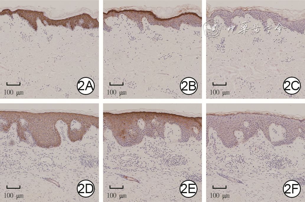
目的 比较瘢痕疙瘩患者的瘢痕疙瘩与其周围正常皮肤的皮肤水屏障功能的差异并初步探索相关机制。 方法 采用横断面观察性研究方法。2020年10月—2021年3月,上海交通大学医学院附属第九人民医院接诊30例符合入选标准的瘢痕疙瘩患者,其中男12例、女18例,年龄20~48岁。接诊当日采用多功能皮肤测试仪测定30例患者瘢痕疙瘩皮肤及其周围正常皮肤的经皮水分丢失(TEWL)。对5例患者瘢痕疙瘩修复术后的瘢痕疙瘩皮肤及其周围正常皮肤行苏木精-伊红染色观测表皮厚度,取其中3例患者标本采用免疫组织化学法检测瘢痕疙瘩皮肤及正常皮肤细胞角蛋白10、内披蛋白和聚丝蛋白的表达。对数据行配对样本 t检验或独立样本 t检验。 结果 接诊当日,30例患者瘢痕疙瘩皮肤TEWL为9.0(6.9,13.4)g·m -2·h -1,正常皮肤的TEWL为8.1(6.4,18.1)g·m -2·h -1,二者比较差异无统计学意义( t=0.44, P>0.05)。瘢痕疙瘩修复术后,5例患者瘢痕疙瘩皮肤表皮厚度为(194±44)μm,明显厚于正常皮肤的(44±11)μm( t=6.88, P<0.01);且瘢痕疙瘩区域均存在角质形成细胞(KC)数量增多、正常皮肤的表皮脊结构缺乏、表皮明显增厚等现象。瘢痕疙瘩修复术后,3例患者瘢痕疙瘩皮肤细胞角蛋白10表达明显低于正常皮肤( t=8.50, P<0.01),而瘢痕疙瘩皮肤表皮的内披蛋白和聚丝蛋白表达均与正常皮肤相近( t值分别为0.07、0.96, P>0.05)。 结论 瘢痕疙瘩患者瘢痕疙瘩皮肤组织中存在KC增多、表皮增厚的现象,但瘢痕疙瘩区域的水屏障功能与周围正常皮肤组织相近,TEWL可能并不是影响瘢痕疙瘩持续发展的主要机制。 Abstract:Objective To compare the differences of water barrier function between keloids and its surrounding normal skin in patients with keloids, and to explore the primary mechanism. Methods A cross-sectional observational study was conducted. From October 2020 to March 2021, 30 patients with keloids who met the inclusion criteria visited Shanghai Ninth People's Hospital Affiliated to Shanghai Jiao Tong University School of Medicine, including 18 females and 12 males, aged 20-48 years. The transepidermal water loss (TEWL) of their keloids and the surrounding normal skin of the 30 patients were measured by multi probe adapter on the reception day. The keloid tissues and normal skin of 5 patients after keloid repair surgery were processed for hematoxylin-eosin staining to measure the thickness of epidermis. Immunohistochemistry was performed on samples from 3 of those 5 patients to detect the expressions of cytokeratin-10, involucrin, and filaggrin in keloids and normal skin. Data were statistically analyzed with paired sample ttest and independent sample t test. Results On the reception day, the TEWL of keloids of 30 patients was 9.0 (6.9, 13.4) g·m -2·h -1 and the TEWL of the normal skin was 8.1 (6.4, 18.1) g·m -2·h -1, between which the difference was not statistically significant ( t=0.44, P>0.05). After keloid repair surgery, the thickness of epidermis in the keloids of 5 patients was (194±44) μm, which was significantly thicker than that of the normal skin (44±11) μm, ( t=6.88, P<0.01). Furthermore, increased keratinocytes, lack of normal epidermal ridge structures, and thickened stratum corneum were observed in the keloid area. After keloid repair surgery, the expression level of cytokeratin-10 in keloids was significantly lower than that in normal skin of 3 patients ( t=8.50, P<0.01), but there were no statistically significant differences in the expression levels of involucrin or filaggrin between keloids and normal skin (with t values of 0.07 and 0.96, respectively, P>0.05). Conclusions Keloid tissue from patients with keloids displays increased number of keratinocytes and thickened epidermis. But the water barrier function in keloid area is similar to the surrounding normal skin, suggesting that TEWL may not be the main mechanism lead to the persistent development of keloids. -
Key words:
- Keloid /
- Epidermis /
- Skin water barrier function /
- Keratinocyte /
- Transepidermal water loss
-
于磊:实施研究、采集数据、分析/解释数据;杨雅婷:起草文章,对文章的知识性内容作批判性审阅、统计分析;刘伟:酝酿和设计实验、分析/解释数据、对文章的知识性内容作批判性审阅、统计分析、获取研究经费、论文修改和指导所有作者均声明不存在利益冲突该研究旨在探讨血红素加氧酶1在烧伤早期急性肾损伤中的作用及其相关机制。采用100 ℃水浴建立经典的大鼠烧伤模型,伤后即刻腹腔注射血红素加氧酶1。采用基于结构变化和功能的组织病理学和血生物化学评估早期急性肾损伤进展,ELISA、免疫染色、实时荧光定量PCR和蛋白质印迹法检测血红素加氧酶1、氧化应激、促炎症介质和Toll样受体4(TLR4)相关信号通路。使用TLR4抑制剂环己烯衍生物TAK242和诱导剂LPS检测血红素加氧酶1在烧伤大鼠肾脏炎症及TLR4信号通路中的作用。研究表明,氯化高铁血红素诱导的血红素加氧酶1通过减少肾氧化应激和释放促炎症介质,下调TLR4的表达及下游核因子κB抑制剂(IκB)激酶α/β、IκBα和核因子κB p65的磷酸化,改善烧伤大鼠的肾损伤和功能障碍;TAK242的作用与血红素加氧酶1相似但较弱;LPS抑制了血红素加氧酶1的抗炎及TLR4信号的调节作用。结果证实血红素加氧酶1通过TLR4/核因子κB信号途径保护肾脏。吴学洁,编译自《Burns》, 2021: S0305-4179(21)00094-2;申传安,审校
-
参考文献
(36) [1] TanS, KhumaloN, BayatA. Understanding keloid pathobiology from a quasi-neoplastic perspective: less of a scar and more of a chronic inflammatory disease with cancer-like tendencies[J]. Front Immunol,2019, 10:1810.DOI: 10.3389/fimmu.2019.01810. [2] AndrewsJP, MarttalaJ, MacarakE, et al. Keloids: the paradigm of skin fibrosis-pathomechanisms and treatment[J]. Matrix Biol,2016,51:37-46.DOI: 10.1016/j.matbio.2016.01.013. [3] OgawaR. Keloid and hypertrophic scars: are the result of chronic inflammation in the reticular dermis[J]. Int J Mol Sci,2017,18(3):606.DOI: 10.3390/ijms18030606. [4] LimandjajaGC, NiessenFB, ScheperRJ, et al. The keloid disorder: heterogeneity, histopathology, mechanisms and models[J]. Front Cell Dev Biol,2020,8:360.DOI: 10.3389/fcell.2020.00360. [5] TsaiCH, OgawaR. Keloid research: current status and future directions[J]. Scars Burn Heal, 2019,5:2059513119868659.DOI: 10.1177/2059513119868659. [6] UehaS, ShandFHW, MatsushimaK. Cellular and molecular mechanisms of chronic inflammation-associated organ fibrosis[J]. Front Immunol,2012,3:71.DOI: 10.3389/fimmu.2012.00071. [7] WangZC, ZhaoWY, CaoYY, et al. The roles of inflammation in keloid and hypertrophic scars[J]. Front Immunol,2020,11:603187.DOI: 10.3389/fimmu.2020.603187. [8] TandaraAA, MustoeTA. The role of the epidermis in the control of scarring:evidence for mechanism of action for silicone gel[J]. J Plast Reconstr Aesthet Surg,2008,61(10):1219-1225.DOI: 10.1016/j.bjps.2008.03.022. [9] MustoeTA. Evolution of silicone therapy and mechanism of action in scar management[J]. Aesthetic Plast Surg,2008,32(1):82-92.DOI: 10.1007/s00266-007-9030-9. [10] OgawaR, DohiT, TosaM, et al. The latest strategy for keloid and hypertrophic scar prevention and treatment: the Nippon Medical School (NMS) protocol[J]. J Nippon Med Sch,2021,88(1):2-9.DOI: 10.1272/jnms.JNMS.2021_88-106. [11] O'BrienL, JonesDJ. Silicone gel sheeting for preventing and treating hypertrophic and keloid scars[J]. Cochrane Database Syst Rev,2013,2013(9):CD003826.DOI: 10.1002/14651858.CD003826.pub3. [12] AlexanderH, BrownS, DanbyS, et al. Research techniques made simple: transepidermal water loss measurement as a research tool[J]. J Invest Dermatol,2018,138(11):2295-2300.e1.DOI: 10.1016/j.jid.2018.09.001. [13] DiniV, BarbaneraS, RomanelliM. Quantitative evaluation of maceration in venous leg ulcers by transepidermal water loss(TEWL) measurement[J]. Int J Low Extrem Wounds,2014,13(2):116-119.DOI: 10.1177/1534734614536035. [14] GardienKLM, BaasDC, de VetHCW, et al. Transepidermal water loss measured with the Tewameter TM300 in burn scars[J]. Burns,2016,42(7):1455-1462.DOI: 10.1016/j.burns.2016.04.018. [15] KuniiT, HiraoT, KikuchiK, et al. Stratum corneum lipid profile and maturation pattern of corneocytes in the outermost layer of fresh scars: the presence of immature corneocytes plays a much more important role in the barrier dysfunction than do changes in intercellular lipids[J]. Br J Dermatol,2003, 149(4):749-756.DOI: 10.1046/j.1365-2133.2003.05545.x. [16] EhrlichHP, DesmoulièreA, DiegelmannRF, et al. Morphological and immunochemical differences between keloid and hypertrophic scar[J]. Am J Pathol,1994,145(1):105-113. [17] BetarbetU, BlalockTW. Keloids: a review of etiology, prevention, and treatment[J]. J Clin Aesthet Dermatol,2020,13(2):33-43. [18] BaroniA, BuomminoE, De GregorioV, et al. Structure and function of the epidermis related to barrier properties[J]. Clin Dermatol,2012,30(3):257-262.DOI: 10.1016/j.clindermatol.211.08.007. [19] MenonGK, ClearyGW, LaneME. The structure and function of the stratum corneum[J]. Int J Pharm, 2012,435(1):3-9.DOI: 10.1016/j.ijpharm.2012.06.005. [20] RawlingsAV, HardingCR. Moisturization and skin barrier function[J]. Dermatol Ther,2004,17 (Suppl 1):S43-48.DOI: 10.1111/j.1396-0296.2004.04s1005.x. [21] SuetakeT, SasaiS, ZhenYX, et al. Functional analyses of the stratum corneum in scars. Sequential studies after injury and comparison among keloids, hypertrophic scars, and atrophic scars[J]. Arch Dermatol,1996,132(12):1453-1458. [22] LimandjajaGC, van den BroekLJ, WaaijmanT, et al. Increased epidermal thickness and abnormal epidermal differentiation in keloid scars[J]. Br J Dermatol,2017,176(1):116-126. DOI: 10.1111/bjd.14844. [23] OuseyK, CuttingKF, RogersAA, et al. The importance of hydration in wound healing: reinvigorating the clinical perspective[J]. J Wound Care,2016,25(3):122,124-130.DOI: 10.12968/jowc.2016.25.3.122. [24] SawadaY, SoneK. Hydration and occlusion treatment for hypertrophic scars and keloids[J]. Br J Plast Surg,1992,45(8):599-603.DOI: 10.1016/0007-1226(92)90027-u. [25] SuetakT, SasaiS, ZhenYX, et al. Effects of silicone gel sheet on the stratum corneum hydration[J]. Br J Plast Surg,2000,53(6):503-507.DOI: 10.1054/bjps.2000.3388. [26] KumarAS, KamalasananK. Drug delivery to optimize angiogenesis imbalance in keloid: a review[J]. J Control Release,2021,329:1066-1076.DOI: 10.1016/j.jconrel.2020.10.035. [27] MariW, AlsabriSG, TabalN, et al. Novel insights on understanding of keloid scar: article review[J]. J Am Coll Clin Wound Spec,2016,7(1/2/3):1-7.DOI: 10.1016/j.jccw.2016.10.001. [28] LeeHJ, JangYJ. Recent understandings of biology, prophylaxis and treatment strategies for hypertrophic scars and keloids[J]. Int J Mol Sci,2018,19(3):711.DOI: 10.3390/ijms19030711. [29] SogabeY, AkimotoS, AbeM, et al. Functions of the stratum corneum in systemic sclerosis as distinct from hypertrophic scar and keloid functions[J]. J Dermatol Sci,2002,29(1):49-53.DOI: 10.1016/s0923-1811(02)00006-3. [30] SandlandsA, SutherlandC, IrvineAD, et al. Filaggrin in the frontline: role in skin barrier function and disease[J]. J Cell Sci,2009,122(Pt 9):1285-1294.DOI: 10.1242/jcs.033969. [31] BrettmannEA, de Guzman StrongC. Recent evolution of the human skin barrier[J]. Exp Dermatol, 2018,27(8):859-866.DOI: 10.1111/exd.13689. [32] AnS, ChaHJ, KoJM, et al. Kinetin improves barrier function of the skin by modulating keratinocyte differentiation markers[J]. Ann Dermatol,2017,29(1):6-12.DOI: 10.5021/ad.2017.29.1.6. [33] VisscherMO, SaidD, WickettR. Stratum corneum cytokines, structural proteins, and transepidermal water loss: effect of hand hygiene[J]. Skin Res Technol,2010,16(2):229-236.DOI: 10.1111/j.1600-0846.2009.00411.x. [34] MollR, DivoM, LangbeinL. The human keratins: biology and pathology[J]. Histochem Cell Biol, 2008,129(6):705-733.DOI: 10.1007/s00418-008-0435-6. [35] JumperN, PausR, BayatA. Functional histopathology of keloid disease[J]. Histol Histopathol,2015, 30(9):1033-1057. DOI: 10.14670/HH-11-624. [36] LeeJYY, YangCC, ChaoSC, et al. Histopathological differential diagnosis of keloid and hypertrophic scar[J]. Am J Dermatopathol,2004,26(5): 379-384.DOI: 10.1097/00000372-200410000-00006. -

1 1例瘢痕疙瘩患者瘢痕疙瘩皮肤和正常皮肤的组织形态学观察 苏木精-伊红×100,图中标尺为200 µm。1A.正常皮肤;1B.瘢痕疙瘩皮肤,较图1A表皮增厚,细胞层数增多
-

 下载:
下载:
图(2)
计量
- 文章访问数: 453
- HTML全文浏览量: 249
- PDF下载量: 48
- 被引次数: 0
