


Effect of human decidua mesenchymal stem cells-derived exosomes on the function of high glucose-induced senescent human dermal fibroblasts and its possible mechanism
-
摘要:
目的 建立人真皮成纤维细胞(HDF)高糖老化模型,探讨人蜕膜间充质干细胞(dMSC)来源外泌体对高糖老化HDF增殖、迁移、凋亡的影响及其可能机制。 方法 采用实验研究方法。收集2021年1—3月解放军总医院第四医学中心收治的4例男性包茎患者(18~22岁)环切术后废弃包皮组织,分离培养获取原代HDF。取第6代HDF,按照随机数字表法分为低糖组和高糖组,分别采用低糖完全培养基和高糖完全培养基进行每72小时换液、不传代培养,10 d后取细胞,于接种后24 h,采用β-半乳糖苷酶试剂盒检测细胞衰老情况;于接种后48 h,采用蛋白质印迹法检测细胞衰老相关蛋白p16、p53表达情况;于接种后24、48、72 h,采用细胞计数试剂盒8(CCK-8)法检测细胞增殖情况;于接种后48 h,采用脱氧尿嘧啶核苷(EdU)染色法检测细胞增殖情况,采用流式细胞术检测细胞周期及凋亡情况;于接种后24 h,采用Transwell实验测定细胞迁移能力。取人dMSC培养48~72 h,采用差速高速离心法获取其外泌体,采用透射电子显微镜观察dMSC外泌体形态,采用纳米颗粒追踪分析法检测dMSC外泌体的粒径分布,采用蛋白质印迹法检测dMSC外泌体标志蛋白CD9、肿瘤易感基因101(TSG101)的表达。取dMSC外泌体及前述高糖完全培养基诱导老化的HDF共孵育24 h,采用PKH67试剂盒检测细胞摄取外泌体的情况。取前述高糖完全培养基诱导老化的HDF,同前分为单纯高糖组、高糖+低浓度外泌体组、高糖+高浓度外泌体组,分别于高糖完全培养基中加入等体积的磷酸盐缓冲液、终质量浓度为50 μg/mL dMSC外泌体、终质量浓度为100 μg/mL dMSC外泌体进行常规细胞培养。分组后同前于对应时间点采用CCK-8法和EdU染色法、流式细胞术、Transwell实验分别检测细胞增殖、细胞周期和凋亡及细胞迁移情况。根据前述结果,另取经高糖完全培养基诱导老化的HDF,分为单纯高糖组、高糖+高浓度外泌体组并同前处理。分组培养48 h,采用实时荧光定量反转录PCR法检测单纯高糖组和高糖+高浓度外泌体组细胞衰老相关的微小RNA-145-5p(miR-145-5p)、miR-498、miR-503-5p及其靶基因钙/钙调素依赖性蛋白激酶1D(CAMK1D)、人第10号染色体缺失的磷酸酶及张力蛋白同源的基因(PTEN基因)和细胞周期蛋白D1的mRNA表达情况。对数据行析因设计方差分析、单因素方差分析、LSD-t检验和独立样本t检验。 结果 接种后24 h,高糖组HDF β-半乳糖苷酶阳性染色率为(38.4±4.2)%,明显高于低糖组的(16.5±2.2)%(t=4.65,P<0.01)。接种后48 h,高糖组HDF的衰老相关蛋白p16和p53的表达量均明显高于低糖组(t值分别为11.85、3.02,P<0.05或P<0.01)。接种后24、48、72 h,高糖组HDF的增殖活性均明显低于低糖组(t值分别为4.13、9.90、15.12,P<0.01)。接种后48 h,高糖组HDF的EdU阳性染色率明显低于低糖组(t=3.83, P<0.05)。接种后48 h,高糖组HDF周期的G2/M+S亚群在3个亚群(G0/G1、S和G2/M)的占比明显低于低糖组(t=8.74,P<0.01)。接种后24 h,高糖组HDF穿过Transwell滤膜到达下室的细胞数量为(37±6)个,明显少于低糖组的(74±7)个(t=8.42,P<0.01)。接种后48 h,高糖组HDF凋亡率明显高于低糖组(t=8.48,P<0.01)。dMSC外泌体为边缘清晰、大小分布均匀的杯状或者圆形囊泡,粒径基本处于80~200 nm。dMSC外泌体标志性蛋白CD9、TSG101表达均呈阳性。共孵育24 h,外泌体被HDF摄入胞内,主要分布于细胞核周围。分组培养24、48、72 h,高糖+低浓度外泌体组和高糖+高浓度外泌体组HDF增殖活性均明显高于单纯高糖组(t值分别为6.36、6.10、7.76,8.92、12.17、10.74,P<0.01),高糖+高浓度外泌体组HDF增殖活性均明显高于高糖+低浓度外泌体组(t值分别为7.92、4.82、4.72,P<0.01)。分组培养48 h,与单纯高糖组比较,高糖+低浓度外泌体组和高糖+高浓度外泌体组HDF EdU阳性染色率均明显升高(t值分别为5.32、9.88,P<0.01);与高糖+低浓度外泌体组比较,高糖+高浓度外泌体组HDF EdU阳性染色率显著升高(t=5.27,P<0.01)。分组培养48 h,与单纯高糖组比较,高糖+低浓度外泌体组和高糖+高浓度外泌体组HDF中的G0/G1期亚群占比均显著降低(t值分别为3.81、4.31,P<0.05),G2/M+S亚群占比均明显升高(t值分别为3.81、4.31,P<0.05)。分组培养24 h,与单纯高糖组相比,高糖+低浓度外泌体组和高糖+高浓度外泌体组HDF穿过滤膜的数量均明显增多(t值分别为10.14、13.39,P<0.01);与高糖+低浓度外泌体组相比,高糖+高浓度外泌体组HDF穿过滤膜的数量明显增多(t=6.27,P<0.01)。分组培养48 h,与单纯高糖组比较,高糖+低浓度外泌体组和高糖+高浓度外泌体组HDF凋亡率均明显降低(t值分别为3.72、5.53,P<0.05或P<0.01)。分组培养48 h,与单纯高糖组相比,高糖+高浓度外泌体组HDF的miR-145-5p、miR-498 mRNA表达量均明显上升(t值分别为13.03、8.90,P<0.01),miR-503-5p mRNA表达量明显下降(t=3.85,P<0.05);高糖+高浓度外泌体组中HDF的CAMK1D、PTEN基因mRNA表达量均明显低于单纯高糖组(t值分别为8.83、5.97,P<0.01),细胞周期蛋白D1 mRNA表达量明显高于单纯高糖组(t=4.03,P<0.05)。 结论 人dMSC来源外泌体可显著提高高糖老化HDF的增殖和迁移能力,抑制其凋亡。这可能与HDF内miR-145-5p和miR-498表达增高抑制了CAMK1D和PTEN基因的表达及miR-503-5p表达下降促进了细胞周期蛋白D1的表达有关。 Abstract:Objective To establish a high glucose senescent model of human dermal fibroblasts (HDFs), and to investigate the effects of exosomes derived from human decidua mesenchymal stem cells (dMSCs) on the proliferation, migration, and apoptosis of senescent HDFs and possible mechanism. Methods The experimental research method was used. From January to March 2021, discarded foreskin tissue was collected for isolation and culture of primary HDFs from 4 male phimosis patients (aged 18-22 years) admitted for circumcision in the Fourth Medical Center of the PLA General Hospital. The 6th passage of HDFs were taken and divided into low glucose group and high glucose group according to the random number table, and subsequently cultured in low-glucose complete medium and high-glucose complete medium, respectively, with medium changed every 72 h without subculturing. After 10 days of culture, the cells were taken and measured for cellular senescence using the β-galactosidase kit at 24 h after seeding; the expression of senescence-related proteins p16 and p53 was assessed by Western blotting at 48 h after seeding; cell proliferation was detected at 24, 48, and 72 h after seeding using the cell counting kit 8 (CCK-8) method; the cell proliferation was evaluated by 5-ethynyl-2'-deoxyuridine (EdU) staining method, cell cycle and apoptosis were measured by flow cytometry after 48 h of seeding; Transwell experiment was used for the calculation of cell migration rate at 24 h after seeding. The human dMSCs were taken and cultured for 48-72 h from which the exosomes were extracted by differential high speed centrifugal method. The morphology of dMSC exosomes was observed by transmission electron microscopy, the particle size distribution of dMSC exosomes was measured by nanoparticle tracking analysis, and the expression of dMSC-exosomes marker proteins CD9 and tumor susceptibility gene101 (TSG101) were detected by Western blotting. The dMSC exosomes and high-glucose complete medium-induced senescent HDFs were co-cultured for 24 hours, then PKH67 kit was used to detect the uptake of exosomes by HDFs. High-glucose complete medium-induced senescent HDFs were taken and divided into high glucose alone group, high glucose+low concentration of exosomes group, and high glucose+high concentration of exosomes group according to the same method above. The high-glucose complete medium with equal volume of phosphate buffered saline, dMSC exosomes with final concentration of 50 μg/mL, and dMSC exosomes with final concentration of 100 μg/mL were added to the corresponding groups for conventional cell culture, respectively. After grouped, the cell proliferation, cell cycle and apoptosis as well as cell migration were detected by CCK-8 method and EdU staining method, flow cytometry, and Transwell experiment at the corresponding time points as before, respectively. Based on the previous results, high-glucose complete medium-induced senescent HDFs were taken and divided into high glucose alone group and high glucose+high concentration of exosomes group for the same treatment. After being grouped and cultured for 48 h, real-time fluorescent quantitative polymerase chain reaction was used to evaluate the mRNA expression of senescent-related microRNA (miR)-145-5p, miR-498, miR-503-5p, calcium/calmodulin dependent protein kinase 1D (CAMK1D), phosphates and tensin homologue deleted on chromosome ten (PTEN) gene, and Cyclin D1 in high glucose alone group and high glucose+high concentration of exosomes group. Data were statistically analyzed with analysis of variance for factorial design, one-way analysis of variance, least significant difference t test, and independent sample t test. Results At 24 h after seeding, the rate of β-galactosidase-positive staining of HDF in high glucose group was (38.4±4.2)%, which was significantly higher than (16.5±2.2)% of low glucose group (t=4.65, P<0.01). At 48 h after seeding, the expression levels of senescence-related proteins p16 and p53 both were significantly higher in HDFs of high glucose group than those in low glucose group (with t values of 11.85 and 3.02, respectively, P<0.05 or P<0.01). At 0, 24, 48, and 72 h after seeding, the cell proliferation viability of HDFs in high glucose group was all significantly lower than in low glucose group (with t values of 4.13, 9.90, and 15.12, respectively, P<0.01). At 48 h after seeding, the rate of EdU-positive staining of HDFs in high glucose group was obviously lower than that of low glucose group (t=3.83, P<0.05). At 48 h after seeding, the percentage of G2/M+S subpopulations in three subpopulations (G0/G1, S, and G2/M) of HDF cycle was significantly lower in high glucose group than that in low glucose group (t=8.74, P<0.01). At 24 h after seeding, the number of HDFs migrated through the filter membrane to the lower chamber was 37±6 in high glucose group, which was significantly less than 74±7 in low glucose group (t=8.42, P<0.01). At 48 h after seeding, the HDF apoptosis rate was significantly higher in high glucose group than in low glucose group (t=8.48, P<0.01). The dMSC exosomes were cup-shaped or round vesicles with well-defined edges and uniform size distribution. The size of dMSC exosomes was basically in the range of 80-200 nm. Exosomal markers including CD9 and TSG101 were positively presented on the dMSC exosomes. After being co-cultured for 24 hours, the dMSC exosomes were taken up intracellularly by HDFs and mainly distributed around the nucleus of HDFs. After being grouped and cultured for 24, 48, and 72 h, the HDF proliferation viabilities in high glucose+low concentration of exosomes group and high glucose+high concentration of exosomes group were both significantly higher than in high glucose alone group (with t values of 6.36, 6.10, 7.76, 8.92, 12.17, and 10.74, respectively, P<0.01), the HDF proliferation viability in high glucose+high concentration of exosomes group was significantly higher than in high glucose+low concentration of exosomes group (with t values of 7.92, 4.82, and 4.72, respectively, P<0.01). After being grouped and cultured for 48 h, the percentages of EdU-positive HDFs in high glucose+low concentration of exosomes group and high glucose+high concentration of exosomes group were both significantly higher than in high glucose alone group (with t values of 5.32 and 9.88, respectively, P<0.01), the percentage of EdU-positive HDFs in high glucose+high concentration of exosomes group was notably higher than in high glucose+low concentration of exosomes group (t=5.27, P<0.01). After being grouped and cultured for 48 h, the proportion of G0/G1 subpopulation in both high glucose+low concentration of exosomes group and high glucose+high concentration of exosomes group was distinctly lower (with t values of 3.81 and 4.31, respectively, P<0.05), while the proportion of G2/M+S subpopulation was markedly higher (with t values of 3.81, 4.31, respectively, P<0.05) than in high glucose alone group. After being grouped and cultured for 24 h, the number of HDFs migrated through the filter membrane in both high glucose+low concentration of exosomes group and high glucose+high concentration of exosomes group was significantly higher than in high glucose alone group (with t values of 10.14 and 13.39, respectively, P<0.01), the number of HDFs migrated through the filter membrane in high glucose+high concentration of exosomes group was significantly increased than in high glucose+low concentration of exosomes group (t=6.27, P<0.01). After being grouped and cultured for 48 h, the HDF apoptosis rates in high glucose+low concentration of exosomes group and high glucose+high concentration of exosomes group were both significantly lower than in high glucose alone group (with t values of 3.72 and 5.53, respectively, P<0.05 or P<0.01). After being grouped and cultured for 48 h, compared with those in high glucose alone group, the mRNA expression levels of miR-145-5p and miR-498 were both obviously higher (with t values of 13.03 and 8.90, respectively, P<0.01), while the mRNA expression level of miR-503-5p was significantly lower (t=3.85, P<0.05) in high glucose+high concentration of exosomes group. After being grouped and cultured for 48 h, compared with those in high glucose alone group, the mRNA expression levels of CAMK1D and PTEN gene were both significantly lower (with t values of 8.83 and 5.97, respectively, P<0.01), while the mRNA expression level of Cyclin D1 was significantly higher in high glucose+high concentration of exosomes group (t=4.03, P<0.05). Conclusions The dMSC exosomes are capable of improving cell proliferation and migration, and inhibiting cell apoptosis of high-glucose senescent HDFs. This may be related to the mechanism by which the increased expressions of intracellular miR-145-5p and miR-498 inhibit the expression of CAMK1D and PTEN gene, and the decreased expression of miR-503-5p promote the expression of Cyclin D1. -
Key words:
- Mesenchymal stem cells /
- Exosomes /
- Cell aging /
- Glucose solution, hypertonic /
- Fibroblasts
-
(1)重点阐述了瘢痕整复设计的基本原则,提出应当积极稳妥规范开展瘢痕整复相关新技术,并重视整复后随访和评估工作。
(2)针对人工智能、三维生物打印、基础研究成果转化等瘢痕整复领域未来研究方向提出了建议。
Highlights:
(1)Basic principles of scar reconstruction design were emphasized and it was proposed that the new technologies related to scar reconstruction should be developed actively and steadily in a standardized manner, with emphasis on the follow-up and evaluation work after reconstruction.
(2)Suggestions were put forward for future research directions in the field of scar reconstruction, such as artificial intelligence, three-dimensional bioprinting, and translation of basic research achievements.
瘢痕是组织遭受损伤后不能完全再生,进而在组织不完全性修复中形成的,能引起机体功能及外观改变的结缔组织。皮肤是体表的最外层,发挥着重要的屏障功能,同时也容易因外界因素而受损。根据最近的一项大型跨国网络调查,48.5%的受访者表示自己至少有1处皮肤瘢痕,且这些瘢痕中约22%为最近1年新发 [ 1] 。国内最近一项基于住院瘢痕患者的全国性数据调查显示,瘢痕并发挛缩、畸形、感染等概率高,烧伤是造成瘢痕的主要病因 [ 2] 。皮肤瘢痕的整复是烧伤与整形外科最主要的工作之一,但也面临着患者情况多样、治疗手段复杂、效果不尽如人意等诸多挑战。随着生活水平的不断提高,广大患者对瘢痕整复的期望越来越高,因此如何不断提高瘢痕整复效果是值得思考和研究的。
1. 精心设计瘢痕整复方案
整复方案设计是实施瘢痕整复前的关键一步,决定着整复成败和效果好坏。每例瘢痕患者的病情都有其自身特点,必须高度重视整复方案设计问题,应当在充分评估和沟通的基础上,结合医师能力实际,制订出对患者而言最有利的方案。
1.1 总体原则
1.1.1 全面评估患者情况
主要从瘢痕情况和全身情况2个方面进行。除详细了解瘢痕的病因、部位、面积、形状、数量、类型、合并症等情况外,还应使用瘢痕评估量表对瘢痕整体情况进行全面评估。目前,临床常用的瘢痕评估量表/方法是温哥华瘢痕量表、视觉模拟评分法和患者与观察者瘢痕评估量表 [ 3] 。为了在治疗前后对瘢痕有较为客观的比较,须对患处拍摄标准化照片。此外,随着技术的进步,三维摄影作为评估瘢痕的手段也显示出良好的可靠性和客观性 [ 4] 。患者全身情况也是评估内容的重要方面,重点在于评估患者营养状况、精神心理状态、慢性病情况、皮肤疾病情况等;需要手术治疗的,还应特别关注有无手术禁忌证。
1.1.2 主要依据瘢痕类型确定整复方案
不同类型的瘢痕,整复方案差异较大。本课题组认为,瘢痕类型是决定瘢痕整复方法的主要因素,主要应从瘢痕形状、面积、成熟情况等方面考虑。对于表浅性瘢痕,可用皮肤磨削术和激光治疗;线状或小面积片状的增生性瘢痕的治疗通常首先使用非手术方法,例如外用硅凝胶产品、压力治疗和激光治疗;如果线状或小面积片状的增生性瘢痕持续增生,还可向瘢痕内注射糖皮质激素。对于大面积片状增生性瘢痕,特别是严重挛缩和造成局部畸形的瘢痕,待其稳定成熟后,应果断采用各类皮片、皮瓣移植手术进行整复。对于小范围凹陷性瘢痕,可行切除缝合、注射填充、激光治疗;广泛的凹陷性瘢痕需手术治疗,具体方式要根据凹陷和畸形程度决定。瘢痕疙瘩是一类特殊的瘢痕,特征是无自愈趋势且面积越来越大,应以手术切除病变、瘢痕内药物注射和放射治疗为主要方法,施行动态综合疗法,并加强随访以预防复发 [ 5] 。如瘢痕出现溃疡,一般无须等待创面完全愈合而应尽早进行瘢痕切除手术。
1.1.3 注意不同部位差异
不同部位瘢痕整复的主要关注点不同,设计时应“因地制宜”。头面部是外露部位,对美学效果要求高,应注意按亚单位进行整复,应用皮肤软组织扩张术整复后美学效果好 [ 6] 。颈部瘢痕挛缩严重影响颈部外观及功能,宜采用颈横动脉颈段皮支扩张皮瓣等胸前区扩张皮瓣予以整复;对于无扩张条件者,则采用皮片移植整复 [ 7] 。躯干部皮肤面积较广、移动度大,且日常生活中多被衣物遮盖,因此采用局部皮瓣整复躯干小面积瘢痕,多可获得较为满意的效果 [ 8] 。四肢瘢痕整复的重点是解除挛缩畸形以恢复运动功能,常用的手术方法包括“Z”成形术、皮片移植术、皮瓣移植术等。会阴区瘢痕常常伴有严重挛缩,如果会阴区周围有可供利用的正常皮肤,进行局部皮瓣+游离皮片移植的效果优于单纯游离皮片移植;在轴型皮瓣整复会阴瘢痕的应用中,可选用髂腰部皮瓣,此皮瓣旋转弧度大、修复面积广,术后效果好 [ 9] 。
1.1.4 充分尊重患者意愿
不同瘢痕患者情况和诉求各异,医师应当耐心倾听、仔细询问、全面评估,充分尊重并考虑患者意愿,切不可为了追求提高个人技术或受经济利益驱使而劝说患者采取某种特定整复方式。瘢痕形成时间长者,经过长期生活,心理上逐渐接受,通常预期较为合理,往往要求稍加改善即可;对于瘢痕形成时间短者,如其心理压力较大且瘢痕尚未成熟,应适当推迟手术整复;对于瘢痕牵拉周围组织器官导致功能障碍者,特别是儿童患者,应当尽早通过手术松解;对于期望过高或期望不合理者,整复治疗前应当反复沟通、慎之又慎。
1.1.5 综合考虑费效比
在保证疗效的前提下,瘢痕整复方案设计应当先简再繁、先单一再联合。应当避免“大处方、大联合”,综合考虑患者经济承受能力和期望效果,尽量降低整复方案的费效比。目前,国外学者对瘢痕整复领域的卫生经济学问题已进行了探索。McPhail等 [ 10] 认为,在患者获益程度相似的情况下,单用硅凝胶制剂的花费低于压力疗法或二者联合;另一项涉及脉冲染料激光干预早期瘢痕形成的成本效益分析研究正在计划开展中 [ 11] 。如何在不显著增加患者费用负担的情况下,进一步提高瘢痕治疗效果,是未来努力方向。
1.2 手术治疗原则
1.2.1 把握合适时机
通常认为,对于经过12个月或更长时间的保守治疗无效的瘢痕,可采用手术切除 [ 3, 12] 。这主要是考虑到瘢痕成熟后再手术时,层次清楚,术中出血少,术后发生瘢痕再次增生的概率也相对较小 [ 13] 。但有的瘢痕形成1年余还明显充血发红,此时也不宜手术;而对于影响生长发育、导致严重功能障碍或长期糜烂的瘢痕,应尽早手术。因此,应当综合判断分析后决定手术时机,不可教条呆板。
1.2.2 方法尽量简便
在保证整复效果前提下,尽量选择简便的方法。能直接切除缝合者,就不用移植皮瓣或者皮片;能用局部皮瓣、邻位皮瓣修复者,就不应该移植皮片、远位皮瓣或者游离皮瓣;单纯皮片移植就能够达到功能恢复目的者,就不要移植远位皮瓣或游离皮瓣等 [ 13] 。
1.2.3 减少额外损伤
开展任何手术都需要关注手术损伤问题,瘢痕整复手术也不例外。首先,需要减少供区的损伤和继发畸形。在皮片移植术中,可采用厚中厚皮替代全厚皮进行瘢痕切除后创面修复,为供区保留一定真皮组织。皮肤扩张术的供区通常能直接缝合,一般不继发畸形和功能障碍,因此皮肤扩张术是减少供区损伤的有效方法 [ 14, 15] 。此外,应用真皮替代物能减少创面愈合过程中异常瘢痕的形成,同时有效降低瘢痕切除修复术后病理性瘢痕的发生率 [ 16] 。其次是减少受区损伤,主要通过术前选择合适的体位、切口位置以及手术器材,术中操作轻柔、注意层次、严格无菌。
1.2.4 提前预防并发症
在整复方案设计之初就应该考虑并发症预防问题,重视并发症危险因素的评估和识别,提前做好预防措施 [ 17] 。应注意观察供区和受区皮肤有无破损、炎症或者感染情况;注意明确术区重要神经和血管分布情况,防止整复后神经、血管损伤或肢端血运障碍;完善各项常规和特殊的检查检验,详细询问并了解患者既往史和家族史,特别是前期治疗经过。
1.3 非手术治疗原则
非手术治疗是瘢痕早期预防的重要手段和瘢痕手术治疗后的必要补充,应当落实“早期”“联合”“全程”的基本原则。“早期”是指应树立创面愈合期就开始预防瘢痕的理念,在创面愈合的不同阶段予以合适的处理,维护创面愈合正常程序,从而从源头控制瘢痕 [ 18] ;创面愈合后即可干预瘢痕形成,越早干预,效果可能越理想。“联合”是指由于目前尚没有消除瘢痕的特效方法,非手术治疗需配合手术治疗共同或序贯施行,才能达到较佳的瘢痕整复效果。“全程”则是指非手术治疗通常不是一蹴而就的,需要在一定时间段内多次或持续使用。以目前较为推荐的脉冲染料激光+剥脱性点阵二氧化碳激光序贯治疗为例,通常是在创面愈合后1周即可开始使用脉冲染料激光,每月1次;当瘢痕充血程度明显减轻后,开始使用剥脱性点阵二氧化碳激光,每3个月1次 [ 19] 。
2. 积极稳妥规范开展瘢痕整复新技术
2.1 积极开展,持续研究
瘢痕整复领域新技术层出不穷,近年来研究较多的是脂肪注射、肉毒毒素注射和光电治疗 [ 20] 。Kwon等 [ 21] 进行双盲对照试验后认为,从脂肪组织中提取的血管基质组分能有效抑制瘢痕形成。Disphanurat等 [ 22] 的研究表明,向隆胸术切口皮下注射A型肉毒毒素,可明显降低瘢痕宽度和高度。Kim等 [ 23] 观察到,使用基于830 nm发光二极管设备的光生物调节疗法可有效促进甲状腺手术切口愈合,并能防止瘢痕增生,且没有明显不良反应。此外,硅胶棒、曲安奈德溶解微针贴片、点阵微针射频等方法也在瘢痕治疗的临床试验中显示了较好疗效,值得推广应用 [ 24, 25, 26] 。这些治疗和预防瘢痕的新技术应用已愈加广泛和成熟,需对其加强经验总结,不断优化细节,并持续拓展相关研究。
2.2 稳妥开展,尊重实际
应用上述新技术时,也应当基于医患双方实际,不可盲目急躁跟风。首先需把握好各项技术的适应证,选择合适的施治对象;其次要熟知技术原理,熟练掌握基本操作,熟悉并发症处置,真正做到理论指导实践;还应立足本人及本单位实际,优先掌握常规方法,以常规方法为基础,注意新技术与常规技术的相互配合。
2.3 遵章开展,建立规范
瘢痕整复新技术的开展离不开相关制度规范的保驾护航。医师需加强对新技术标准规范的学习,严格遵守操作规程,谨记“无菌、无创、无无效腔、无创面外露”的整形外科基本原则。所在单位要加强新技术开展的把关,严格按照临床新技术开展的有关规定进行审批和监管。此外,本课题组呼吁制订更多权威性高、科学性好、操作性强的瘢痕整复技术教程,供广大基层医师学习参考。
3. 重视瘢痕整复后随访和评估工作
3.1 做好长期随访工作
坚持长期随访对所有研究者而言都是艰巨的挑战,主要原因是重视程度不够、患者依从性参差不齐、随访制度和手段缺乏等。本课题组认为,要想持续做好随访工作,必须要增强随访意识,认识到坚持长期随访对远期治疗效果评判的重要意义;应当加强患者教育,积极取得患者信任和配合;还需推动随访相关规范化制度和操作流程的制订工作。本课题组曾对355例经皮肤扩张术修复面颈部病损的患者(绝大部分为瘢痕患者)进行了随访观察,随访时间最长达10年,这项工作虽然工作量巨大,但对于总结经验、提高技术、改进细节大有裨益 [ 27] 。
3.2 注重客观评估方法的应用
目前,常常通过主观性的评分或量表评估瘢痕治疗效果,在一定程度上能够反映医患双方对治疗效果的评价,但不足之处在于评价标准不统一、且不便于不同研究之间相互比较。因此,本课题组更加推荐客观评估方法的应用,可从皮肤的感觉功能,皮瓣/皮片回缩率、弹性、颜色,瘢痕宽度、体积等方面进行评估 [ 28] 。还可从触觉、温度觉、痛觉、两点辨别觉等维度测量感觉功能;使用皮肤诊疗系统和肤质诊断专家系统,还可实现回缩率、弹性、颜色、瘢痕体积等的精确测量。
3.3 应用病例管理数据库
在随访评估和临床科研中,病例的丰富性与病例数据收集整理不完善之间的矛盾十分突出,建议利用数据库技术对瘢痕整复患者临床资料进行管理。可以建立小型专题数据库,也可以依托大型专业病例数据库,还可以利用医院现有病历系统进行资料管理。本课题组的经验是,通过Access数据库软件构建专病数据库,即可达到预期的数据管理效果 [ 29] 。好的专病数据库的建立不是一朝一夕之功,需要临床专家和信息工程师密切合作,需要不断提高模式化水平、维护数据完整性,还可能需要多家单位、多个中心的相互协作。
4. 对瘢痕整复未来方向的展望
虽然到目前为止,尚无完全预防和根治瘢痕的方法,但随着整形外科手术技术的日臻成熟,特别是皮肤软组织扩张术的开展应用,瘢痕整复的效果愈加令人满意。本课题组认为,未来瘢痕整复领域的发展方向仍是以外科手术为基础的综合治疗,重点应关注以下几个方面。
4.1 关注人工智能在瘢痕整复领域的潜在应用
随着人工智能的蓬勃发展,该技术被逐渐应用于医学领域,例如放射学、病理学、眼科学和皮肤科学领域等 [ 30] 。在瘢痕整复领域,人工智能已被用于提高瘢痕疙瘩诊疗决策的精准性 [ 31] 。本课题组认为,人工智能很有可能在瘢痕整复手术设计、术中规划、治疗后评估等多个方面具有应有价值。值得强调的是,瘢痕整复的对象毕竟不是冰冷的机器,因此人工智能的职能定位始终应当是辅助者,而不是替代者。
4.2 重视用三维生物打印技术开发皮肤替代品
三维生物打印是通过逐层打印生物功能材料及其支持成分,以创建具有所需三维细胞结构和功能的复杂活组织和器官的一种计算机辅助技术,是目前皮肤再生领域的前沿研究热点 [ 32] 。目前三维生物打印的皮肤替代品已经初步制作成功,具有少量正常皮肤功能 [ 33] 。需要进行瘢痕整复的患者本质特征是体表正常皮肤的缺失,如能用三维生物打印技术制造出功能和美学修复效果兼备,且成本可接受的皮肤替代品,必将为瘢痕患者带来福音。
4.3 加强瘢痕领域基础研究成果向临床应用转化
随着单细胞测序、类器官、免疫学技术等的进步和应用,研究者对于瘢痕形成机制的认知也愈加深入。基于瘢痕发生发展的分子机制,一些特异性治疗靶点正在被逐步开发,例如抗瘢痕内异常炎症、抗瘢痕内血管生成、向瘢痕内移植自体干细胞等 [ 34] 。但也应当注意到,还有许多瘢痕研究仅仅停留在基础层面,并未很好向临床应用转化。应当以一张蓝图绘到底的决心,一体推进、一体筹划基础与临床研究工作,使二者相互促进、相互转化,最终实现造福瘢痕患者的美好愿望。
苏建隆:实施研究、采集数据、分析/解释数据、起草文章、统计分析;马奎:对文章的知识性内容作批评性审阅;张翠萍、付小兵:酝酿和设计研究、研究指导、行政支持、技术支持、材料支持所有作者均声明不存在利益冲突巨噬细胞是代谢血红素的主要效应细胞,当受损或衰老红细胞的处理需求增加时,肝脏中巨噬细胞数量会一过性地增加。巨噬细胞在抵御微生物威胁方面也很重要,但血红素过量的病理状态很可能导致免疫抑制的发生。在此,作者揭示了在感染肺炎克雷伯菌后,巨噬细胞需清除的衰老红细胞急剧增多,并最终转变为免疫抑制表型的机制,而免疫抑制表型巨噬细胞的存在,使得肺炎克雷伯菌感染特征呈现为肺外细菌增殖增加,小鼠患脓毒症后的存活率降低。该研究观察到感染缺乏铁载体功能的肺炎克雷伯菌突变体的小鼠与感染野生型菌株的小鼠对过量衰老红细胞的清理结果相似,主要表现为肺细菌负荷没有差异,因此认为肺炎克雷伯菌相关的免疫受损和细菌铁载体获取铁无关。然而,在肺炎克雷伯菌相关脓毒症中,对衰老红细胞的处理导致肝脏转录组组谱呈现出显著的信号转导及转录激活因子1(STAT1)抑制和干扰素相关反应。在感染时,巨噬细胞过度处理血红素致STAT1抑制,这一过程需要核因子E2相关因子1和2的协同激活,但无须血红素加氧酶1诱导。尽管该免疫受损与铁的获取无关,但血红素的卟啉部分足以介导抑制人和小鼠巨噬细胞中STAT1依赖的反应,并促进肺炎克雷伯菌在体内肝脏传播。因此,细胞血红素代谢障碍负向调节STAT1通路与严重感染有关。杨云稀、高晞,编译自《J Clin Invest》, 2021,131(1):e137468;孙炳伟,审校 -
参考文献
(40) [1] LiB, BianX, HuW, et al. Regenerative and protective effects of calcium silicate on senescent fibroblasts induced by high glucose[J]. Wound Repair Regen,2020,28(3):315-325. DOI: 10.1111/wrr.12794. [2] LiM,ZhaoY,HaoH,et al.Umbilical cord-derived mesenchymal stromal cell-conditioned medium exerts in vitro antiaging effects in human fibroblasts[J].Cytotherapy,2017,19(3):371-383.DOI: 10.1016/j.jcyt.2016.12.001. [3] RaniS,RyanAE,GriffinMD,et al.Mesenchymal stem cell-derived extracellular vesicles: toward cell-free therapeutic applications[J].Mol Ther,2015,23(5):812-823.DOI: 10.1038/mt.2015.44. [4] NikfarjamS,RezaieJ,ZolbaninNM,et al.Mesenchymal stem cell derived-exo somes: a modern approach in translational medicine[J].J Transl Med,2020,18(1):449.DOI: 10.1186/s12967-020-02622-3. [5] KomakiM,NumataY,MoriokaC,et al.Exosomes of human placenta-derived mesenchymal stem cells stimulate angiogenesis[J].Stem Cell Res Ther,2017,8(1):219.DOI: 10.1186/s13287-017-0660-9. [6] ZhaoB,ZhangY,HanS,et al.Exosomes derived from human amniotic epithelial cells accelerate wound healing and inhibit scar formation[J].J Mol Histol,2017,48(2):121-132.DOI: 10.1007/s10735-017-9711-x. [7] DashBC,XuZ,LinL,et al.Stem cells and engineered scaffolds for regenerative wound healing[J].Bioengineering (Basel),2018, 5(1):23. DOI: 10.3390/bioengineering5010023. [8] LermanOZ,GalianoRD,ArmourM,et al.Cellular dysfunction in the diabetic fibroblast: impairment in migration, vascular endothelial growth factor production, and response to hypoxia[J].Am J Pathol,2003,162(1):303-312.DOI: 10.1016/S0002-9440(10)63821-7. [9] ThomasK,KiwitM,KernerW.Glucose concentration in human subcutaneous adipose tissue: comparison between forearm and abdomen[J].Exp Clin Endocrinol Diabetes,1998,106(6):465-469.DOI: 10.1055/s-0029-1212017. [10] WeiQ,WangY,MaK,et al.Extracellular vesicles from human umbilical cord mesenchymal stem cells facilitate diabetic wound healing through miR-17-5p-mediated enhancement of angiogenesis[J/OL].Stem Cell Rev Rep,2021(2021-05-04)[2021-10-13]. https://pubmed.ncbi.nlm.nih.gov/33942217/. DOI:10.1007/s12015-021-10176-0. [published online ahead of print]. [11] ChengL,ZhangK,WuS,et al.Focus on mesenchymal stem cell-derived exosomes: opportunities and challenges in cell-free therapy[J].Stem Cells Int,2017,2017:6305295.DOI: 10.1155/2017/6305295. [12] LuK,LiHY,YangK,et al.Exosomes as potential alternatives to stem cell therapy for intervertebral disc degeneration: in-vitro study on exosomes in interaction of nucleus pulposus cells and bone marrow mesenchymal stem cells[J].Stem Cell Res Ther,2017,8(1):108.DOI: 10.1186/s13287-017-0563-9. [13] Casado-DíazA,Quesada-GómezJM,DoradoG.Extracellular vesicles derived from mesenchymal stem cells (MSC) in regenerative medicine: applications in skin wound healing[J].Front Bioeng Biotechnol,2020,8:146.DOI: 10.3389/fbioe.2020.00146. [14] HuY,RaoSS,WangZX,et al.Exosomes from human umbilical cord blood accelerate cutaneous wound healing through miR-21-3p-mediated promotion of angiogenesis and fibroblast function[J].Theranostics,2018,8(1):169-184.DOI: 10.7150/thno.21234. [15] PomattoM,GaiC,NegroF,et al.Differential therapeutic effect of extracellular vesicles derived by bone marrow and adipose mesenchymal stem cells on wound healing of diabetic ulcers and correlation to their cargoes[J].Int J Mol Sci,2021, 22(8):3851. DOI: 10.3390/ijms22083851. [16] WangC,WangM,XuT,et al.Engineering bioactive self-healing antibacterial exosomes hydrogel for promoting chronic diabetic wound healing and complete skin regeneration[J].Theranostics,2019,9(1):65-76.DOI: 10.7150/thno.29766. [17] McBrideJD,Rodriguez-MenocalL,GuzmanW,et al.Bone marrow mesenchymal stem cell-derived CD63+ exosomes transport Wnt3a exteriorly and enhance dermal fibroblast proliferation, migration, and angiogenesis in vitro[J].Stem Cells Dev,2017,26(19):1384-1398.DOI: 10.1089/scd.2017.0087. [18] YangJ,ChenZ,PanD,et al.Umbilical cord-derived mesenchymal stem cell-derived exosomes combined pluronic F127 hydrogel promote chronic diabetic wound healing and complete skin regeneration[J].Int J Nanomedicine,2020,15:5911-5926.DOI: 10.2147/IJN.S249129. [19] ZhangW,BaiX,ZhaoB,et al.Cell-free therapy based on adipose tissue stem cell-derived exosomes promotes wound healing via the PI3K/Akt signaling pathway[J].Exp Cell Res,2018,370(2):333-342.DOI: 10.1016/j.yexcr.2018.06.035. [20] FangS,XuC,ZhangY,et al.Umbilical cord-derived mesenchymal stem cell-derived exosomal microRNAs suppress myofibroblast differentiation by inhibiting the transforming growth factor-β/SMAD2 pathway during wound healing[J].Stem Cells Transl Med,2016,5(10):1425-1439.DOI: 10.5966/sctm.2015-0367. [21] HuL,WangJ,ZhouX,et al.Exosomes derived from human adipose mensenchymal stem cells accelerates cutaneous wound healing via optimizing the characteristics of fibroblasts[J].Sci Rep,2016,6:32993.DOI: 10.1038/srep32993. [22] WiklanderOP, NordinJZ, O'LoughlinA,et al.Extracellular vesicle in vivo biodistribution is determined by cell source, route of administration and targeting[J].J Extracell Vesicles,2015,4:26316.DOI: 10.3402/jev.v4.26316. [23] YangD,ZhangW,ZhangH,et al.Progress, opportunity, and perspective on exosome isolation - efforts for efficient exosome-based theranostics[J].Theranostics,2020,10(8):3684-3707.DOI: 10.7150/thno.41580. [24] DingM,WangC,LuX,et al.Comparison of commercial exosome isolation kits for circulating exosomal microRNA profiling[J].Anal Bioanal Chem,2018,410(16):3805-3814.DOI: 10.1007/s00216-018-1052-4. [25] KusumaGD,CarthewJ,LimR,et al.Effect of the microenvironment on mesenchymal stem cell paracrine signaling: opportunities to engineer the therapeutic effect[J].Stem Cells Dev,2017,26(9):617-631.DOI: 10.1089/scd.2016.0349. [26] WuD,KangL,TianJ,et al.Exosomes derived from bone mesenchymal stem cells with the stimulation of Fe3O4 nanoparticles and static magnetic field enhance wound healing through upregulated miR-21-5p[J].Int J Nanomedicine,2020,15:7979-7993.DOI: 10.2147/IJN.S275650. [27] YangH,LinJ,JiangJ,et al.miR-20b-5p functions as tumor suppressor microRNA by targeting cyclinD1 in colon cancer[J].Cell Cycle,2020,19(21):2939-2954.DOI: 10.1080/15384101.2020.1829824. [28] DimitrovaN,GochevaV,BhutkarA,et al.Stromal expression of miR-143/145 promotes neoangiogenesis in lung cancer development[J].Cancer Discov,2016,6(2):188-201.DOI: 10.1158/2159-8290.CD-15-0854. [29] DimitrovaN,GochevaV,BhutkarA,et al.Stromal expression of miR-143/145 promotes neoangiogenesis in lung cancer development[J].Cancer Discov,2016,6(2):188-201.DOI: 10.1158/2159-8290.CD-15-0854. [30] LawsonJ,DickmanC,MacLellanS,et al.Selective secretion of microRNAs from lung cancer cells via extracellular vesicles promotes CAMK1D-mediated tube formation in endothelial cells[J].Oncotarget,2017,8(48):83913-83924.DOI: 10.18632/oncotarget.19996. [31] GrasC,RatusznyD,HadamitzkyC,et al.miR-145 contributes to hypertrophic scarring of the skin by inducing myofibroblast activity[J].Mol Med,2015,21(1):296-304.DOI: 10.2119/molmed.2014.00172. [32] ChaiC,WuH,WangB,et al.MicroRNA-498 promotes proliferation and migration by targeting the tumor suppressor PTEN in breast cancer cells[J].Carcinogenesis,2018,39(9):1185-1196.DOI: 10.1093/carcin/bgy092. [33] DuanXM,LiuXN,LiYX,et al.MicroRNA-498 promotes proliferation, migration, and invasion of prostate cancer cells and decreases radiation sensitivity by targeting PTEN[J].Kaohsiung J Med Sci,2019,35(11):659-671.DOI: 10.1002/kjm2.12108. [34] LiJ,ZhangF,LiH,et al.Circ_0010220-mediated miR-503-5p/CDCA4 axis contributes to osteosarcoma progression tumorigenesis[J].Gene,2020,763:145068.DOI: 10.1016/j.gene.2020.145068. [35] MarkopoulosGS,RoupakiaE,TokamaniM,et al.Senescence-associated microRNAs target cell cycle regulatory genes in normal human lung fibroblasts[J].Exp Gerontol,2017,96:110-122.DOI: 10.1016/j.exger.2017.06.017. [36] JiangL,ZhaoZ,ZhengL,et al.Downregulation of miR-503 promotes ESCC cell proliferation, migration, and invasion by targeting cyclin D1[J].Genomics Proteomics Bioinformatics,2017,15(3):208-217.DOI: 10.1016/j.gpb.2017.04.003. [37] BovyN,BlommeB,FrèresP,et al.Endothelial exosomes contribute to the antitumor response during breast cancer neoadjuvant chemotherapy via microRNA transfer[J].Oncotarget,2015,6(12):10253-10266.DOI: 10.18632/oncotarget.3520. [38] HouSQ,OuyangM,BrandmaierA,et al.PTEN in the maintenance of genome integrity: From DNA replication to chromosome segregation[J].Bioessays,2017,39(10):10. DOI: 10.1002/bies.201700082. [39] LiB,LuanS,ChenJ,et al.The MSC-derived exosomal incRNA H19 promotes wound healing in diabetic foot ulcers by upregulating PTEN via microRNA-152-3p[J].Mol Ther Nucleic Acids,2020,19:814-826.DOI: 10.1016/j.omtn.2019.11.034. [40] LongJ,OuC,XiaH,et al.MiR-503 inhibited cell proliferation of human breast cancer cells by suppressing CCND1 expression[J].Tumour Biol,2015,36(11):8697-8702.DOI: 10.1007/s13277-015-3623-8. -
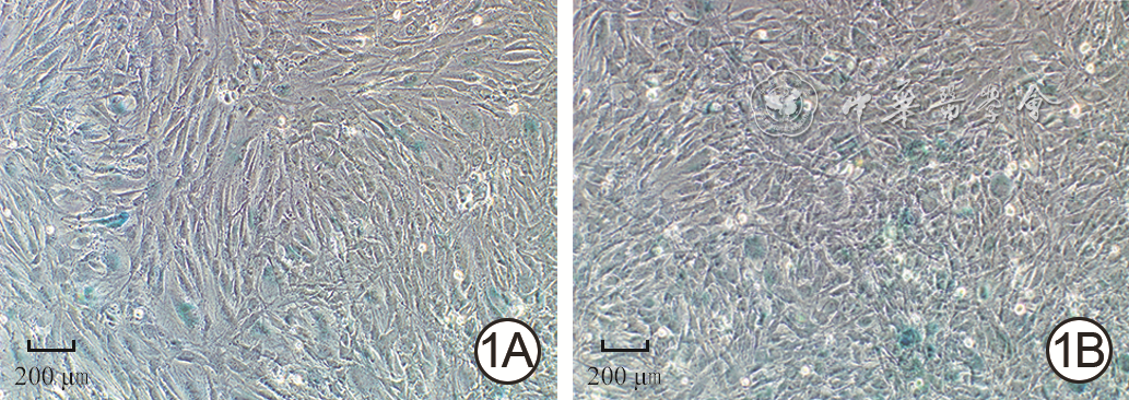
1 2组人真皮成纤维细胞中接种后24 h β-半乳糖苷酶表达情况 光学显微镜×100,图中标尺为200 μm。1A.低糖组β-半乳糖苷酶染色;1B.高糖组β-半乳糖苷酶阳性染色率较图1A高
注:低糖组、高糖组细胞培养基中的葡萄糖物质的量浓度分别为5.5、35.0 mmol/L;图中蓝绿色指示细胞β-半乳糖苷酶阳性染色
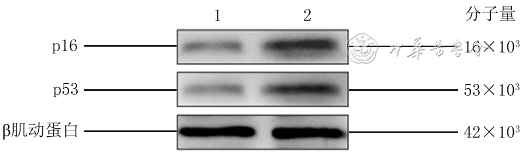
2 蛋白质印迹法检测2组人真皮成纤维细胞接种后48 h衰老相关蛋白表达
注:1为低糖组,2为高糖组;低糖组、高糖组细胞培养基中的葡萄糖物质的量浓度分别为5.5、35.0 mmol/L
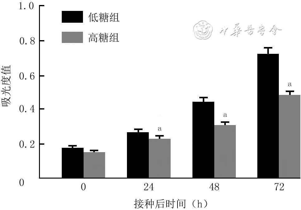
3 细胞计数试剂盒8法检测2组人真皮成纤维细胞接种各时间点的增殖活性(样本数为3,
注:低糖组、高糖组细胞培养基中的葡萄糖物质的量浓度分别为5.5、35.0 mmol/L;处理因素主效应,F=244.90,P<0.001;时间因素主效应,F=892.30,P<0.001;两者交互作用,F=56.23,P<0.001;与低糖组比较,aP<0.01
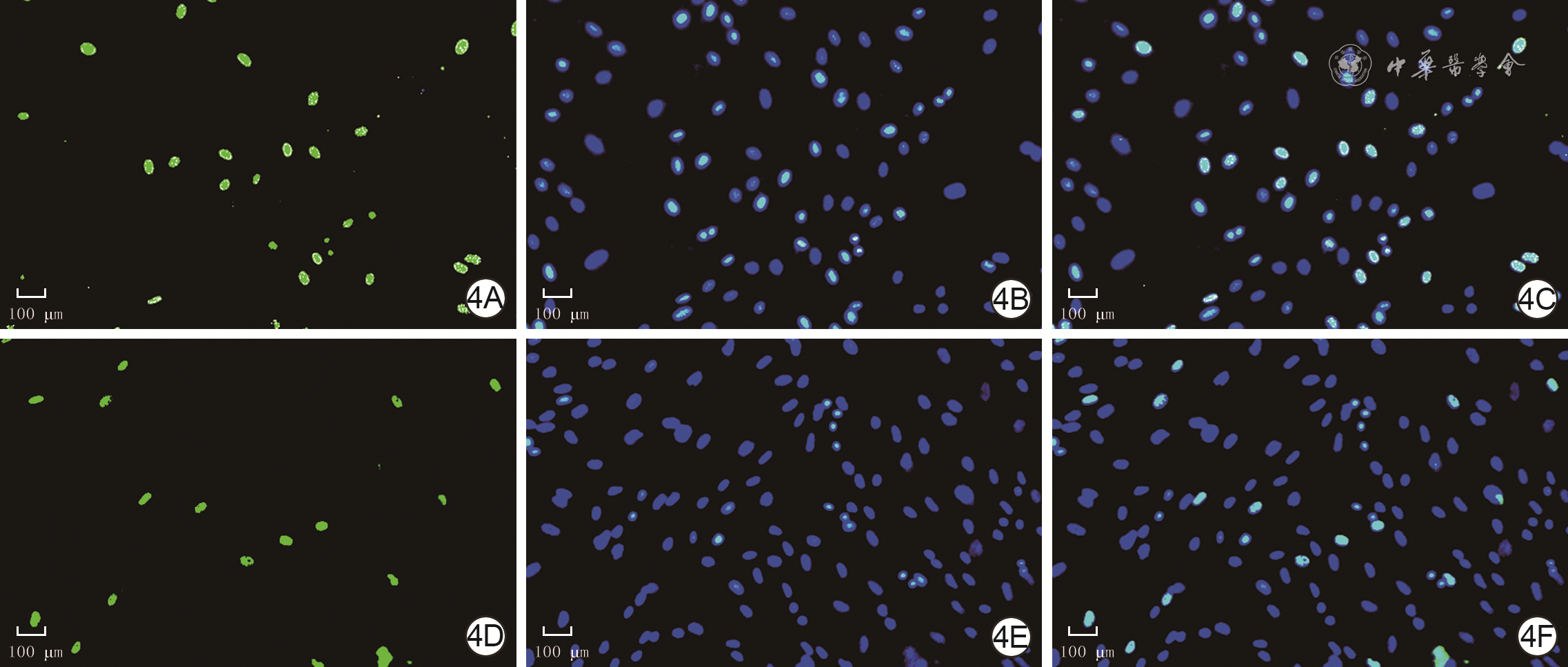
4 EdU染色法检测2组人真皮成纤维细胞培养48 h增殖活力 EdU-hochst×200,图中标尺为100 μm。4A、4B、4C.分别为低糖组细胞EdU染色、细胞核染色、细胞EdU与细胞核共染色情况;4D、4E、4F.分别为高糖组细胞EdU染色、细胞核染色、细胞EdU与细胞核共染色情况,细胞核完整,图4D EdU阳性细胞较图4A明显减少
注:低糖组、高糖组细胞培养基中的葡萄糖浓度分别为5.5、35.0 mmol/L;以细胞脱氧尿嘧啶核苷(EdU)阳性染色为绿色,细胞核阳性染色为蓝色,绿色+蓝色双荧光染色为增殖细胞
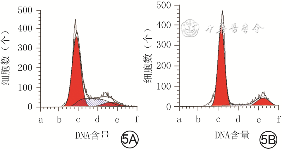
5 流式细胞术检测2组人真皮成纤维细胞培养48 h的细胞周期情况。5A、5B.分别为低糖组、高糖组
注:低糖组、高糖组细胞培养基中的葡萄糖物质的量浓度分别为5.5、35.0 mmol/L;图5A、5B中横坐标a、b、c、d、e、f分别为0、500、1 000、1 600、2 000、2 500;图中第1个红色波峰为G1期,第2个红色波峰为G2期,蓝色斜线区为S期
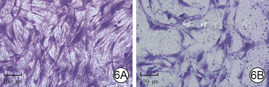
6 Transwell实验观察2组人真皮成纤维细胞培养24 h迁移情况 结晶紫×200,图中标尺为100 μm。6A、6B.分别为低糖组、高糖组,图6B穿膜细胞数较图6A少
注:低糖组、高糖组细胞培养基中的葡萄糖物质的量浓度分别为5.5、35.0 mmol/L;图中紫色指示穿膜细胞
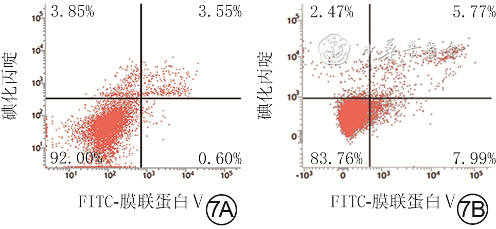
7 流式细胞术检测2组人真皮成纤维细胞培养48 h凋亡情况。7A.低糖组绝大多数细胞处于左下象限,凋亡细胞少;7B.高糖组细胞右上、右下象限凋亡细胞数较图7A明显增多
注:低糖组、高糖组细胞培养基中的葡萄糖物质的量浓度分别为5.5、35.0 mmol/L;FITC为异硫氰酸荧光素;图中左下象限显示活细胞,右下象限显示早期凋亡细胞,右上象限显示晚期凋亡细胞和坏死细胞,左上象限显示细胞收集过程中出现的损伤细胞
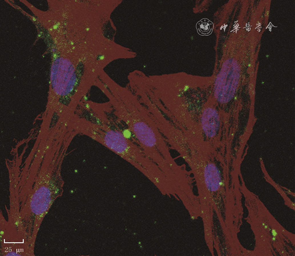
11 人真皮成纤维细胞(HDF)与人dMSC外泌体共培养24 h后,外泌体被HDF内吞入细胞质,并聚集于细胞核周围培养 4',6-二脒基-2-苯基吲哚-罗丹明标记的鬼笔环肽-PKH67×400,图中标尺为25 μm
注:紫色指示细胞核,酱红色指示细胞骨架,绿色为人蜕膜间充质干细胞(dMSC)来源外泌体

12 细胞计数试剂盒8法检测3组人真皮成纤维细胞培养各时间点活力(样本数为3,
注:单纯高糖组、高糖+低浓度外泌体组、高糖+高浓度外泌体组分别于高糖完全培养基中加入等体积的磷酸盐缓冲液、终质量浓度50 μg/mL人蜕膜间充质干细胞外泌体、终质量浓度100 μg/mL 人蜕膜间充质干细胞外泌体进行常规细胞培养;处理因素主效应,F=107.80,P<0.001;时间因素主效应,F=2 992.00,P<0.001;两者交互作用,F=24.99,P<0.001;与单纯高糖组比较,aP<0.01;与高糖+低浓度外泌体组比较,bP<0.01

13 EdU染色法检测3组HDF培养48 h的增殖活力 EdU-hoechst×200,图中标尺为100 μm。13A、13B、13C.分别为单纯高糖组、高糖+低浓度外泌体组、高糖+高浓度外泌体组细胞EdU与细胞核共染色情况,细胞核完整,图13B EdU阳性细胞较图13A明显增多,且图13C EdU阳性染色细胞数最多
注:单纯高糖组、高糖+低浓度外泌体组、高糖+高浓度外泌体组分别于高糖完全培养基中加入等体积的磷酸盐缓冲液、终质量浓度50 μg/mL人蜕膜间充质干细胞外泌体、终质量浓度100 μg/mL人蜕膜间充质干细胞外泌体进行常规细胞培养;细胞脱氧尿嘧啶核苷(EdU)阳性染色为绿色,细胞核阳性染色为蓝色,绿色+蓝色双荧光染色为增殖的人真皮成纤维细胞(HDF)
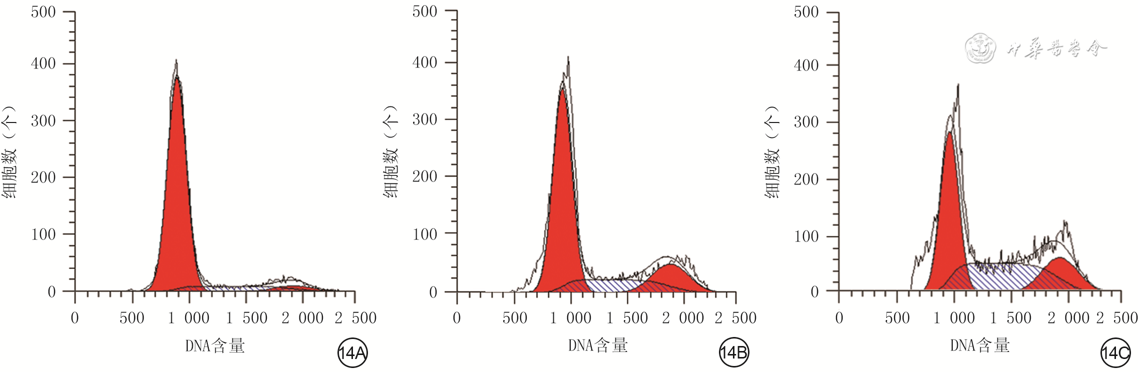
14 流式细胞术检测3组人真皮成纤维细胞培养48 h的细胞周期情况。14A、14B、14C.分别为单纯高糖组、高糖+低浓度外泌体组、高糖+高浓度外泌体组
注:单纯高糖组、高糖+低浓度外泌体组、高糖+高浓度外泌体组分别于高糖完全培养基中加入等体积的磷酸盐缓冲液、终质量浓度50 μg/mL人蜕膜间充质干细胞外泌体、终质量浓度100 μg/mL人蜕膜间充质干细胞外泌体进行常规细胞培养;图中第1个红色波峰为G1期,第2个红色波峰为G2期,蓝色斜线区为S期

15 Transwell实验观察3组人真皮成纤维细胞培养24 h迁移能力 结晶紫×200,图中标尺为100 μm。15A、15B、15C.分别为单纯高糖组、高糖+低浓度外泌体组、高糖+高浓度外泌体组穿膜细胞生长情况,图15B、15C穿膜细胞数均较图15A多,且图15C穿膜细胞数最多
注:单纯高糖组、高糖+低浓度外泌体组、高糖+高浓度外泌体组分别于高糖完全培养基中加入等体积的磷酸盐缓冲液、终质量浓度50 μg/mL人蜕膜间充质干细胞外泌体、终质量浓度100 μg/mL人蜕膜间充质干细胞外泌体进行常规细胞培养;图中紫色指示穿膜细胞
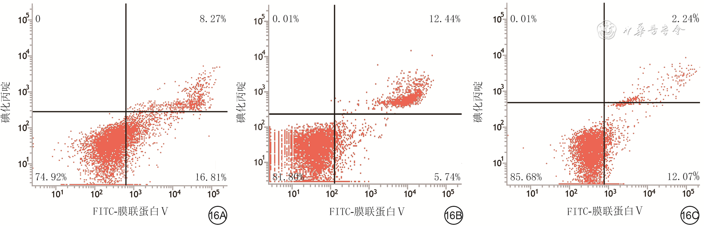
16 流式细胞术检测3组人真皮成纤维细胞(HDF)培养48 h凋亡情况。16A.单纯高糖组HDF绝大多数细胞处于左下象限,凋亡细胞较多;16B.高糖+低浓度外泌体组凋亡HDF较单纯高糖组减少;16C.高糖+高浓度外泌体组右上/下象限凋亡HDF较图16A明显减少,且凋亡细胞数低于图16B
注:单纯高糖组、高糖+低浓度外泌体组、高糖+高浓度外泌体组分别于高糖完全培养基中加入等体积的磷酸盐缓冲液、终质量浓度50 μg/mL人蜕膜间充质干细胞外泌体、终质量浓度100 μg/mL人蜕膜间充质干细胞外泌体进行常规细胞培养;FITC为异硫氰酸荧光素;图中左下象限显示活细胞,右下象限显示早期凋亡细胞,右上象限显示晚期凋亡细胞和坏死细胞,左上象限显示细胞收集过程中出现的损伤细胞
表1 2组高糖诱导老化人真皮成纤维细胞培养48 h衰老相关微小RNA表达情况比较(
组别 样本数 miR-145-5p miR-498 miR-503-5p 单纯高糖组 3 1.01±0.14 1.06±0.21 1.05±0.37 高糖+高浓度外泌体组 3 12.15±1.47 2.89±0.29 0.22±0.03 t值 13.03 8.90 3.85 P值 <0.001 <0.001 0.018 注:单纯高糖组、高糖+高浓度外泌体组分别于高糖完全培养基中加入等体积的磷酸盐缓冲液、终质量浓度100 μg/mL人蜕膜间充质干细胞外泌体进行常规细胞培养;miR为微小RNA  下载: 导出CSV
表2 2组高糖诱导老化人真皮成纤维细胞中衰老相关基因的mRNA表达情况比较(
组别 样本数 CAMK1D PTEN基因 细胞周期蛋白D1 单纯高糖组 3 1.00±0.11 1.00±0.04 1.14±0.18 高糖+高浓度外泌体组 3 0.37±0.06 0.52±0.13 2.85±0.70 t值 8.83 5.97 4.03 P值 <0.001 0.003 <0.015 注:单纯高糖组、高糖+高浓度外泌体组分别于高糖完全培养基中加入等体积的磷酸盐缓冲液、终质量浓度100 μg/mL人蜕膜间充质干细胞外泌体进行常规细胞培养;CAMK1D为钙/钙调素依赖性蛋白激酶1D,PTEN基因为人第10号染色体缺失的磷酸酶及张力蛋白同源的基因
下载: 导出CSV
-


 下载:
下载:


计量
- 文章访问数: 520
- HTML全文浏览量: 153
- PDF下载量: 34
- 被引次数: 0
