


Effects of low-dose photodynamic therapy on the function of human adipose mesenchymal stem cells and its mechanism
-
摘要:
目的 探讨低剂量光动力对人脂肪间充质干细胞(ADSC)的增殖、调节及分泌功能的作用及其相关机制,以期为慢性创面的修复探索新方法。 方法 采用实验研究方法。取2021年2—4月于陆军军医大学(第三军医大学)第一附属医院皮肤科行皮肤外科手术的10例患者(5例男性、5例女性,23~47岁)捐献的术后废弃脂肪组织,提取细胞并鉴定表型。取3批ADSC,各批细胞均分为仅行常规培养的正常对照组,行海姆泊芬处理后常规培养的单纯光敏剂组,行红光照射处理后常规培养的单纯光照组,行海姆泊芬和红光照射处理后再常规培养的光敏剂+光照组,样本数均为3。第1和2批细胞于处理完成后培养24 h,分别采用脱氧尿嘧啶核苷染色法检测ADSC增殖水平,Transwell实验检测与ADSC共培养的HaCaT细胞迁移比例;第3批细胞于处理完成后培养7 d,采用免疫荧光法检测ADSC的细胞外基质蛋白表达。取ADSC分为光动力后0 min组、光动力后15 min组、光动力后30 min组、光动力后60 min组,每组3孔,分别于行光动力处理后相应时间点采用蛋白质印迹法检测并计算磷酸化哺乳动物雷帕霉素靶蛋白(p-mTOR)/哺乳动物雷帕霉素靶蛋白(mTOR)、磷酸化p70核糖体蛋白S6激酶(p-p70 S6K)/p70核糖体蛋白S6激酶(p70 S6K)比值。取2批ADSC,各批细胞均分为正常对照组、单纯光动力组及光动力+雷帕霉素组,每组3孔,并行相应处理。第1批细胞于处理完成后培养15 min,同前检测并计算p-mTOR/mTOR、p-p70 S6K/p70 S6K比值;第2批细胞于处理完成后培养7 d,同前检测细胞外基质蛋白表达。对数据行单因素方差分析、LSD检验。 结果 培养12 d后,细胞鉴定为ADSC。处理完成后培养24 h,单纯光敏剂组和单纯光照组ADSC增殖水平[分别为(4.0±1.0)%、(4.1±0.4)%]和HaCaT细胞迁移比例(分别为1.17±0.14、1.13±0.12)均与正常对照组[分别为(3.7±0.6)%、1.00±0.16]相近(P>0.05),且均明显低于光敏剂+光照组[分别为(34.2±7.0)%、2.55±0.13,P<0.01]。处理完成后培养7 d,与正常对照组相比,单纯光敏剂组ADSC的Ⅲ型胶原表达增加(P<0.05),单纯光照组ADSC的Ⅰ型胶原与Ⅲ型胶原表达均明显增加(P<0.01);与单纯光敏剂组和单纯光照组相比,光敏剂+光照组ADSC的Ⅰ型胶原、Ⅲ型胶原与纤维连接蛋白表达均明显增加(P<0.01)。与光动力后0 min组相比,光动力后15 min组ADSC的p-mTOR/mTOR与p-p70 S6K/p70 S6K比值均明显增加(P<0.01),光动力后30 min组、光动力后60 min组ADSC的p-p70 S6K/p70 S6K比值均明显增加(P<0.01)。处理完成后培养15 min,与正常对照组相比,单纯光动力组ADSC的p-mTOR/mTOR与p-p70 S6K/p70 S6K比值均明显增加(P<0.05或P<0.01);与单纯光动力组相比,光动力+雷帕霉素组ADSC的p-mTOR/mTOR与p-p70 S6K/p70 S6K比值均明显减少(P<0.05)。处理完成后培养7 d,与正常对照组相比,单纯光动力组ADSC的Ⅰ型胶原、Ⅲ型胶原与纤维连接蛋白表达均明显增加(P<0.01);与单纯光动力组相比,光动力+雷帕霉素组ADSC的Ⅰ型胶原、Ⅲ型胶原与纤维连接蛋白表达均明显减少(P<0.01)。 结论 低剂量光动力可以促进ADSC的增殖、提高ADSC调节HaCaT细胞迁移的能力,并通过快速激活mTOR信号通路增强细胞外基质蛋白的分泌。 Abstract:Objective To investigate the effects of low-dose photodynamic therapy on the proliferation, regulation, and secretion functions of human adipose mesenchymal stem cells (ADSCs) and the related mechanism, so as to explore a new method for the repair of chronic wounds. Methods The experimental research methods were adopted. From February to April 2021, 10 patients (5 males and 5 females, aged 23 to 47 years) who underwent cutaneous surgery in the Department of Dermatology of the First Affiliated Hospital of Army Medical University (the Third Military Medical University) donated postoperative waste adipose tissue. The cells were extracted from the adipose tissue and the phenotype was identified. Three batches of ADSCs were taken, with each batch of cells being divided into normal control group with conventional culture only, photosensitizer alone group with conventional culture after being treated with Hemoporfin, irradiation alone group with conventional culture after being treated with red light irradiation, and photosensitizer+irradiation group with conventional culture after being treated with Hemoporfin and red light irradiation, with sample number of 3 in each group. At culture hour of 24 after the treatment of the first and second batches of cells, the ADSC proliferation level was evaluated by 5-ethynyl-2'-deoxyuridine staining method and the migration percentage of HaCaT cells cocultured with ADSCs was detected by Transwell experiment, respectively. On culture day of 7 after the treatment of the third batch of cells, the extracellular matrix protein expression of ADSCs was detected by immunofluorescence method. The ADSCs were divided into 0 min post-photodynamic therapy group, 15 min post-photodynamic therapy group, 30 min post-photodynamic therapy group, and 60 min post-photodynamic therapy group, with 3 wells in each group. Western blotting was used to detect the protein expressions and calculate the phosphorylated mammalian target of rapamycin complex (p-mTOR)/mammalian target of rapamycin (mTOR), phosphorylated p70 ribosomal protein S6 kinase (p-p70 S6K)/p70 ribosomal protein S6 kinase (p70 S6K) ratio at the corresponding time points after photodynamic therapy. Two batches of ADSCs were taken, and each batch was divided into normal control group, photodynamic therapy alone group, and photodynamic therapy+rapamycin group, with 3 wells in each group. At culture minute of 15 after the treatment, p-mTOR/mTOR and p-p70 S6K/p70 S6K ratios of cells from the first batch were calculated and detected as before. On culture day of 7 after the treatment, extracellular matrix protein expression of cells from the second batch was detected as before. Data were statistically analyzed with one-way analysis of variance and least significant difference test. Results After 12 d of culture, the cells were verified as ADSCs. At culture hour of 24 after the treatment, the ADSC proliferation level ((4.0±1.0)% and (4.1±0.4)%, respectively) and HaCaT cell migration percentages (1.17±0.14 and 1.13±0.12, respectively) in photosensitizer alone group and irradiation alone group were similar to those of normal control group ((3.7±0.6)% and 1.00±0.16, respectively, P>0.05), and were significantly lower than those of photosensitizer+irradiation group ((34.2±7.0)% and 2.55±0.13, respectively, P<0.01). On culture day of 7 after the treatment, compared with those in normal control group, the expression of collagen Ⅲ in ADSCs of photosensitizer alone group was significantly increased (P<0.05), and the expressions of collagen Ⅰ and collagen Ⅲ in ADSCs of irradiation alone group were significantly increased (P<0.01). Compared with those in photosensitizer alone group and irradiation alone group, the expressions of collagen Ⅰ, collagen Ⅲ, and fibronectin of ADSCs in photosensitizer+irradiation group were significantly increased (P<0.01). Compared with those in 0 min post-photodynamic therapy group, the ratios of p-mTOR/mTOR and p-p70 S6K/p70 S6K of ADSCs in 15 min post-photodynamic therapy group were significantly increased (P<0.01), the ratios of p-p70 S6K/p70 S6K of ADSCs in 30 min post-photodynamic therapy group and 60 min post-photodynamic therapy group were both significantly increased (P<0.01). At culture minute of 15 after the treatment, compared with those in normal control group, the ratios of p-mTOR/mTOR and p-p70 S6K/p70 S6K of ADSCs in photodynamic therapy alone group were significantly increased (P<0.05 or P<0.01). Compared with those in photodynamic therapy alone group, the ratios of p-mTOR/mTOR and p-p70 S6K/p70 S6K of ADSCs in photodynamic therapy+rapamycin group were significantly decreased (P<0.05). On culture day of 7 after the treatment, compared with those in normal control group, the expressions of collagen Ⅰ, collagen Ⅲ, and fibronectin of ADSCs in photodynamic therapy alone group were significantly increased (P<0.01). Compared with those in photodynamic therapy alone group, the expressions of collagen Ⅰ, collagen Ⅲ, and fibronectin of ADSCs in photodynamic therapy+rapamycin group were significantly decreased (P<0.01). Conclusions Low-dose photodynamic therapy can promote the proliferation of ADSCs, improve the ability of ADSCs to regulate the migration of HaCaT cells, and enhance the secretion of extracellular matrix protein by rapidly activating mTOR signaling pathway. -
参考文献
(35) [1] CastanoAP, DemidovaTN, HamblinMR. Mechanisms in photodynamic therapy: part one-photosensitizers, photochemistry and cellular localization[J]. Photodiagnosis Photodyn Ther,2004,1(4):279-293.DOI: 10.1016/S1572-1000(05)00007-4. [2] GunaydinG, GedikME, AyanS. Photodynamic therapy for the treatment and diagnosis of cancer-a review of the current clinical status[J]. Front Chem,2021,9:686303.DOI: 10.3389/fchem.2021.686303. [3] KimM, JungHY, ParkHJ. Topical PDT in the treatment of benign skin diseases: principles and new applications[J]. Int J Mol Sci,2015,16(10):23259-23278.DOI: 10.3390/ijms161023259. [4] PlaetzerK, KrammerB, BerlandaJ, et al. Photophysics and photochemistry of photodynamic therapy: fundamental aspects[J]. Lasers Med Sci,2009,24(2):259-268.DOI: 10.1007/s10103-008-0539-1. [5] HuCX, ZhaoLF, PengCG, et al. Regulation of the mitochondrial reactive oxygen species: strategies to control mesenchymal stem cell fates ex vivo and in vivo[J]. J Cell Mol Med,2018,22(11):5196-5207.DOI: 10.1111/jcmm.13835. [6] YangZJ, HuXH, ZhouLN, et al. Photodynamic therapy accelerates skin wound healing through promoting re-epithelialization[J/OL]. Burns Trauma,2021,9:tkab008[2022-03-25]. https://pubmed.ncbi.nlm.nih.gov/34514005/.DOI: 10.1093/burnst/tkab008. [7] KaushikK, DasA. Endothelial progenitor cell therapy for chronic wound tissue regeneration[J]. Cytotherapy,2019,21(11):1137-1150.DOI: 10.1016/j.jcyt.2019.09.002. [8] De LucaM, AiutiA, CossuG, et al. Advances in stem cell research and therapeutic development[J]. Nat Cell Biol,2019,21(7):801-811.DOI: 10.1038/s41556-019-0344-z. [9] JoH, BritoS, KwakBM, et al. Applications of mesenchymal stem cells in skin regeneration and rejuvenation[J]. Int J Mol Sci,2021,22(5):2410.DOI: 10.3390/ijms22052410. [10] DasM, MayilsamyK, MohapatraSS, et al. Mesenchymal stem cell therapy for the treatment of traumatic brain injury: progress and prospects[J]. Rev Neurosci,2019,30(8):839-855.DOI: 10.1515/revneuro-2019-0002. [11] DolatiS, YousefiM, MahdipourM, et al. Mesenchymal stem cell and bone marrow mononuclear cell therapy for cardiomyopathy: from bench to bedside[J]. J Cell Biochem,2019,120(1):45-55.DOI: 10.1002/jcb.27531. [12] HaDH, KimHK, LeeJ, et al. Mesenchymal stem/stromal cell-derived exosomes for immunomodulatory therapeutics and skin regeneration[J]. Cells,2020,9(5):1157.DOI: 10.3390/cells9051157. [13] ShuklaL, YuanYN, ShayanR, et al. Fat therapeutics: the clinical capacity of adipose-derived stem cells and exosomes for human disease and tissue regeneration[J]. Front Pharmacol,2020,11:158.DOI: 10.3389/fphar.2020.00158. [14] NaderiN, CombellackEJ, GriffinM, et al. The regenerative role of adipose-derived stem cells (ADSC) in plastic and reconstructive surgery[J]. Int Wound J,2017,14(1):112-124.DOI: 10.1111/iwj.12569. [15] WernerS, GroseR. Regulation of wound healing by growth factors and cytokines[J]. Physiol Rev,2003,83(3):835-870.DOI: 10.1152/physrev.2003.83.3.835. [16] FalangaV. Growth factors and chronic wounds: the need to understand the microenvironment[J]. J Dermatol,1992,19(11):667-672.DOI: 10.1111/j.1346-8138.1992.tb03756.x. [17] ShuFT, GaoHJ, WuWF, et al. Amniotic epithelial cells accelerate diabetic wound healing by protecting keratinocytes and fibroblasts from high-glucose-induced senescence[J]. Cell Biol Int,2022,46(5):755-770.DOI: 10.1002/cbin.11771. [18] BerberichB, ThrieneK, GretzmeierC, et al. Proteomic profiling of fibroblasts isolated from chronic wounds identifies disease-relevant signaling pathways[J]. J Invest Dermatol,2020,140(11):2280-2290.e4.DOI: 10.1016/j.jid.2020.02.040. [19] HardingKG, MooreK, PhillipsTJ. Wound chronicity and fibroblast senescence--implications for treatment[J]. Int Wound J,2005,2(4):364-368.DOI: 10.1111/j.1742-4801.2005.00149.x. [20] DongWP, SongZC, LiuSH, et al. Adipose-derived stem cells based on electrospun biomimetic scaffold mediated endothelial differentiation facilitating regeneration and repair of abdominal wall defects via HIF-1α/VEGF pathway[J]. Front Bioeng Biotechnol,2021,9:676409.DOI: 10.3389/fbioe.2021.676409. [21] LeeCH, ShahB, MoioliEK, et al. CTGF directs fibroblast differentiation from human mesenchymal stem/stromal cells and defines connective tissue healing in a rodent injury model[J]. J Clin Invest,2010,120(9):3340-3349.DOI: 10.1172/JCI43230. [22] Chavez-MunozC, NguyenKT, XuW, et al. Transdifferentiation of adipose-derived stem cells into keratinocyte-like cells: engineering a stratified epidermis[J]. PLoS One,2013,8(12):e80587.DOI: 10.1371/journal.pone.0080587. [23] JiangDS, Scharffetter-KochanekK. Mesenchymal stem cells adaptively respond to environmental cues thereby improving granulation tissue formation and wound healing[J]. Front Cell Dev Biol,2020,8:697.DOI: 10.3389/fcell.2020.00697. [24] HuangYZ, GouM, DaLC, et al. Mesenchymal stem cells for chronic wound healing: current status of preclinical and clinical studies[J]. Tissue Eng Part B Rev,2020,26(6):555-570.DOI: 10.1089/ten.TEB.2019.0351. [25] MaxsonS, LopezEA, YooD, et al. Concise review: role of mesenchymal stem cells in wound repair[J]. Stem Cells Transl Med,2012,1(2):142-149.DOI: 10.5966/sctm.2011-0018. [26] FuXR, LiuG, HalimA, et al. Mesenchymal stem cell migration and tissue repair[J]. Cells,2019,8(8):784.DOI: 10.3390/cells8080784. [27] RaposioE, BertozziN. Isolation of ready-to-use adipose-derived stem cell (ASC) pellet for clinical applications and a comparative overview of alternate methods for ASC isolation[J]. Curr Protoc Stem Cell Biol,2017,41:1F.17.1-1F.17.12.DOI: 10.1002/cpsc.29. [28] BacakovaL, ZarubovaJ, TravnickovaM, et al. Stem cells: their source, potency and use in regenerative therapies with focus on adipose-derived stem cells - a review[J]. Biotechnol Adv,2018,36(4):1111-1126.DOI: 10.1016/j.biotechadv.2018.03.011. [29] SiZZ, WangX, SunCH, et al. Adipose-derived stem cells: sources, potency, and implications for regenerative therapies[J]. Biomed Pharmacother,2019,114:108765.DOI: 10.1016/j.biopha.2019.108765. [30] Díaz-GarcíaD, FilipováA, Garza-VelozI, et al. A beginner's introduction to skin stem cells and wound healing[J]. Int J Mol Sci,2021,22(20):11030.DOI: 10.3390/ijms222011030. [31] RodriguesM, KosaricN, BonhamCA, et al. Wound healing: a cellular perspective[J]. Physiol Rev,2019,99(1):665-706.DOI: 10.1152/physrev.00067.2017. [32] MoritaM, GravelSP, HuleaL, et al. mTOR coordinates protein synthesis, mitochondrial activity and proliferation[J]. Cell Cycle,2015,14(4):473-480.DOI: 10.4161/15384101.2014.991572. [33] CarrascoE, CalvoMI, Blzquez-CastroA, et al. Photoactivation of ROS production in situ transiently activates cell proliferation in mouse skin and in the hair follicle stem cell niche promoting hair growth and wound healing[J]. J Invest Dermatol,2015,135(11):2611-2622.DOI: 10.1038/jid.2015.248. [34] Al-AzabM, WangB, ElkhiderA, et al. Indian hedgehog regulates senescence in bone marrow-derived mesenchymal stem cell through modulation of ROS/mTOR/4EBP1, p70S6K1/2 pathway[J]. Aging (Albany NY),2020,12(7):5693-5715.DOI: 10.18632/aging.102958. [35] GuoW, QiuW, AoX, et al. Low-concentration DMSO accelerates skin wound healing by Akt/mTOR-mediated cell proliferation and migration in diabetic mice[J]. Br J Pharmacol,2020,177(14):3327-3341.DOI: 10.1111/bph.15052. -

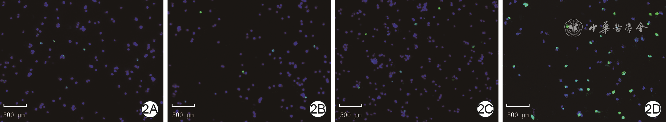
2 4组人脂肪间充质干细胞处理完成后培养24 h的增殖水平 EdU-Hoechst×40。2A、2B、2C、2D.分别为正常对照组、单纯光敏剂组、单纯光照组、光敏剂+光照组染色结果,图2D中的EdU阳性细胞比例明显高于图2A、2B、2C
注:脱氧尿嘧啶核苷(EdU)阳性染色为绿色,细胞核阳性染色为蓝色,绿色+蓝色双荧光染色为增殖细胞

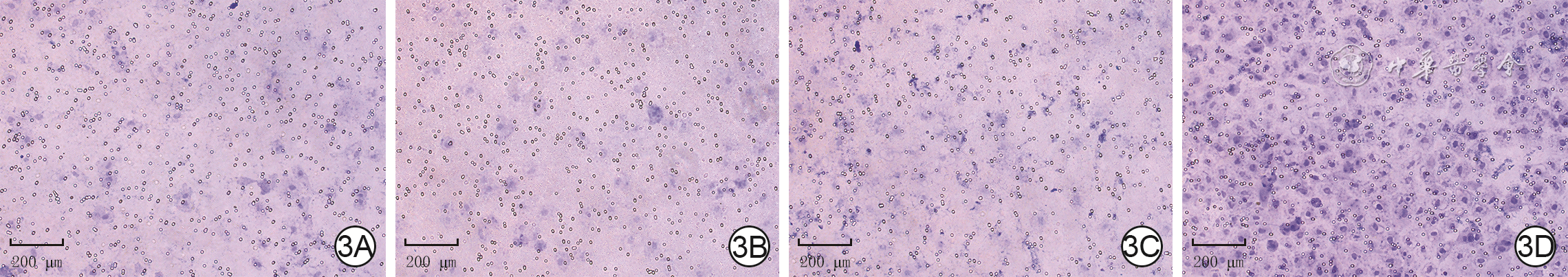
3 4组人脂肪间充质干细胞处理完成后与HaCaT细胞共培养24 h后的HaCaT细胞迁移情况 结晶紫×100。3A、3B、3C、3D.分别为正常对照组、单纯光敏剂组、单纯光照组、光敏剂+光照组HaCaT细胞迁移情况,图3D中的HaCaT细胞迁移数量明显多于图3A、3B、3C

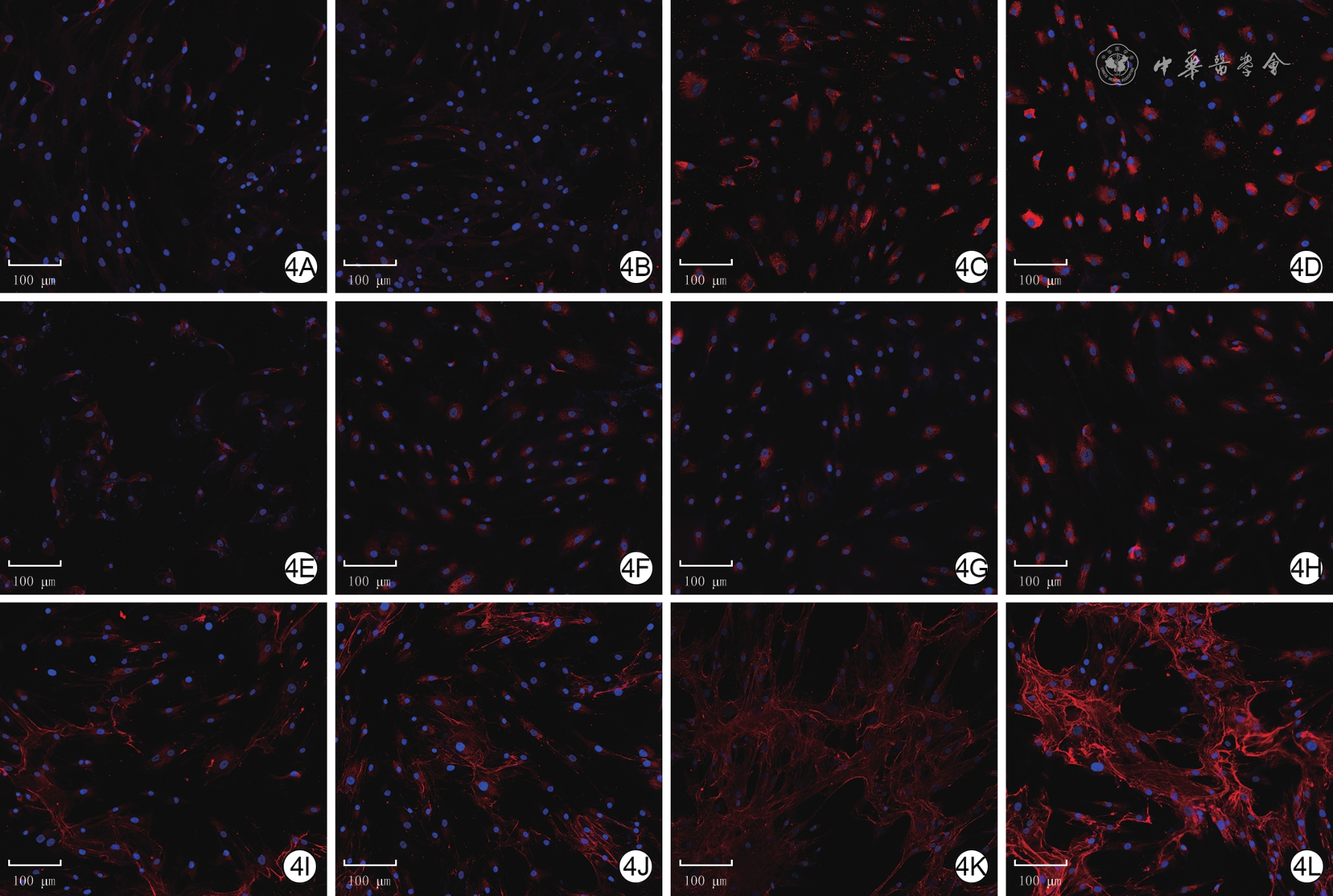
4 4组人脂肪间充质干细胞处理完成后培养7 d表达细胞外基质蛋白的情况 Alexa Fluor 594-4′,6-二脒基-2-苯基吲哚×40。4A、4B、4C、4D与4E、4F、4G、4H及4I、4J、4K、4L.分别为正常对照组、单纯光敏剂组、单纯光照组、光敏剂+光照组Ⅰ型胶原,Ⅲ型胶原,纤维连接蛋白染色情况;其中图4D中Ⅰ型胶原表达最多、图4H中Ⅲ型胶原表达最多、图4L中纤维连接蛋白表达最多
注:Ⅰ型胶原、Ⅲ型胶原、纤维连接蛋白阳性染色为红色,细胞核阳性染色为蓝色

5 4组人脂肪间充质干细胞行光动力处理后培养相应时间p-mTOR/mTOR与p-p70 S6K/p70 S6K比值情况。5A.条带图;5B.条图(样本数为3,
注:p-mTOR为磷酸化哺乳动物雷帕霉素靶蛋白,mTOR为哺乳动物雷帕霉素靶蛋白,p-p70 S6K为磷酸化p70核糖体蛋白S6激酶,p70 S6K为p70核糖体蛋白S6激酶,GAPDH为3-磷酸甘油醛脱氢酶;条带图上方和条图横坐标下1、2、3、4均分别为光动力后0 min组、光动力后15 min组、光动力后30 min组、光动力后60 min组;图5B中4组间p-mTOR/mTOR与p-p70 S6K/p70 S6K比值总体比较,F值分别为65.51、103.09,P值均<0.001;与光动力后0 min组相比,a P<0.01

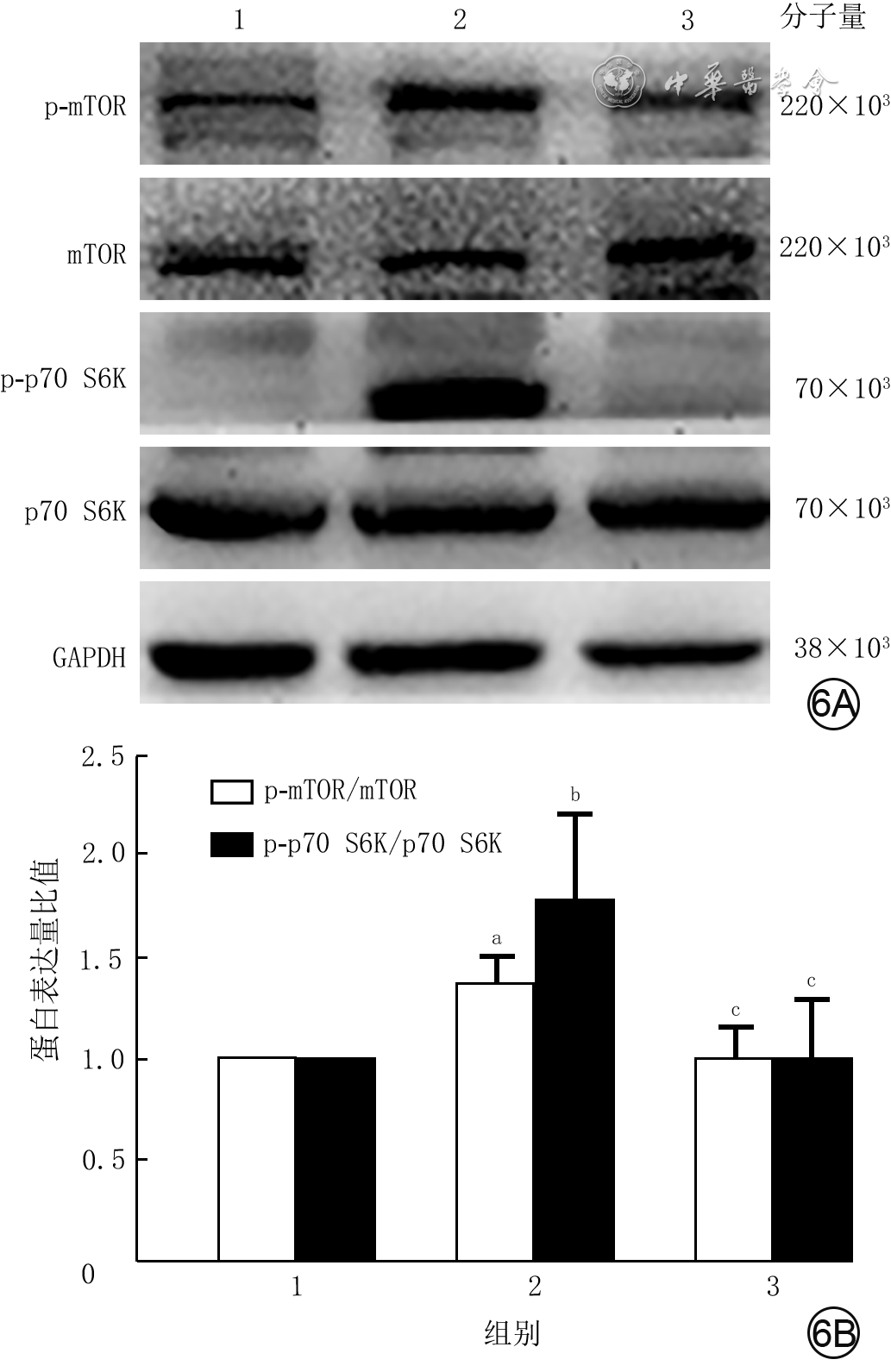
6 4组人脂肪间充质干细胞行光动力处理后培养15 min的p-mTOR/mTOR与p-p70 S6K/p70 S6K比值情况。6A.条带图;6B.条图(样本数为3,
注:p-mTOR为磷酸化哺乳动物雷帕霉素靶蛋白,mTOR为哺乳动物雷帕霉素靶蛋白,p-p70 S6K为磷酸化p70核糖体蛋白S6激酶,p70 S6K为p70核糖体蛋白S6激酶,GAPDH为3-磷酸甘油醛脱氢酶;条带图上方和条图横坐标下1、2、3均分别为正常对照组、单纯光动力组、光动力+雷帕霉素组;图6B中3组间p-mTOR/mTOR与p-p70 S6K/p70 S6K比值总体比较,F值分别为10.41、4.42,P值分别为0.011、0.066;与正常对照组相比,a P<0.01,b P<0.05;与单纯光动力组相比,c P<0.05

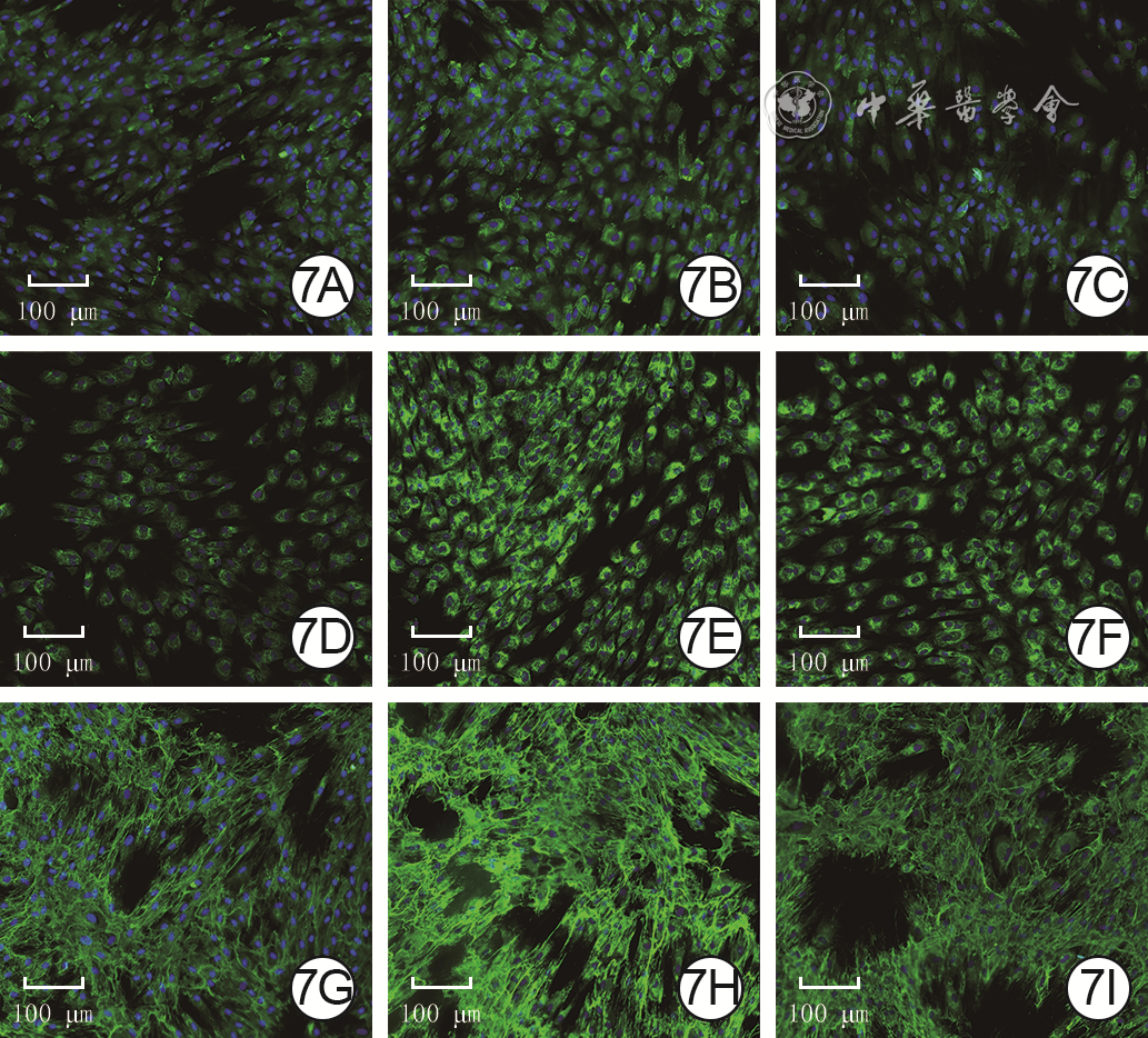
7 3组人脂肪间充质干细胞处理完成后培养7 d表达细胞外基质蛋白的情况 Alexa Fluor 488-4′,6-二脒基-2-苯基吲哚×40。7A、7B、7C.分别为正常对照组、单纯光动力组、光动力+雷帕霉素组Ⅰ型胶原染色情况,图7B中Ⅰ型胶原表达最多;7D、7E、7F.分别为正常对照组、单纯光动力组、光动力+雷帕霉素组Ⅲ型胶原染色情况,图7E中Ⅲ型胶原表达最多;7G、7H、7I.分别为正常对照组、单纯光动力组、光动力+雷帕霉素组纤维连接蛋白染色情况,图7H中纤维连接蛋白表达最多
注:Ⅰ型胶原、Ⅲ型胶原、纤维连接蛋白阳性染色为绿色,细胞核阳性染色为蓝色
表1 4组人脂肪间充质干细胞处理完成后培养7 d细胞外基质蛋白的荧光强度比较(
组别 样本数 Ⅰ型胶原 Ⅲ型胶原 纤维连接蛋白 正常对照组 3 21.2±3.2 35.1±3.7 49.3±7.7 单纯光敏剂组 3 25.2±1.4 42.1±3.8 58.1±1.7 单纯光照组 3 59.0±6.1 45.7±0.4 56.6±4.1 光敏剂+光照组 3 83.3±7.3 67.8±2.5 100.3±10.3 F值 101.33 69.24 34.90 P值 <0.001 <0.001 <0.001 P 1值 0.359 0.020 0.147 P 2值 <0.001 0.002 0.221 P 3值 <0.001 <0.001 <0.001 P 4值 <0.001 <0.001 <0.001 注:F值、P值为4组间各指标总体比较所得;P 1值为单纯光敏剂组与正常对照组各指标比较所得,P 2值为单纯光照组与正常对照组各指标比较所得,P 3值为光敏剂+光照组与单纯光敏剂组各指标比较所得,P 4值为光敏剂+光照组与单纯光照组各指标比较所得  下载: 导出CSV
表2 3组人脂肪间充质干细胞处理完成后培养7 d细胞外基质蛋白的荧光强度比较(
组别 样本数 Ⅰ型胶原 Ⅲ型胶原 纤维连接蛋白 正常对照组 3 69±6 117.2±3.1 109±4 单纯光动力组 3 96±7 138.7±4.8 152±6 光动力+雷帕霉素组 3 64±3 118.4±1.9 111±16 F值 16.02 81.50 18.87 P值 <0.001 <0.001 <0.001 P 1值 0.002 <0.001 0.001 P 2值 0.347 0.623 0.822 P 3值 <0.001 <0.001 0.002 注:F值、P值为3组间各指标总体比较所得;P 1值为正常对照组与单纯光动力组各指标比较所得,P 2值为正常对照组与光动力+雷帕霉素组各指标比较所得,P 3值为单纯光动力组与光动力+雷帕霉素组各指标比较所得
下载: 导出CSV
-

 下载:
下载:


计量
- 文章访问数: 369
- HTML全文浏览量: 172
- PDF下载量: 18
- 被引次数: 0
