Lyu GZ,Zhao P.New bioactive materials for promoting wound repair and skin regeneration[J].Chin J Burns,2021,37(12):1105-1109.DOI: 10.3760/cma.j.cn501120-20211029-00373.
Citation: Liang LT,Song W,Zhang C,et al.Effects of in situ cross-linked graphene oxide-containing gelatin methacrylate anhydride hydrogel on wound vascularization of full-thickness skin defect in mice[J].Chin J Burns Wounds,2022,38(7):616-628.DOI: 10.3760/cma.j.cn501225-20220314-00063.

Effects of in situ cross-linked graphene oxide-containing gelatin methacrylate anhydride hydrogel on wound vascularization of full-thickness skin defect in mice

doi: 10.3760/cma.j.cn501225-20220314-00063
Funds:

Youth Science Foundation of National Natural Science Foundation of China 32000969, 82002056

Chinese Academy of Medical Sciences Innovation Fund for Medical Sciences 2019-I2M-5-059

Military Medical Innovation Research Project of PLA General Hospital CX19026

Wang Zhengguo Foundation for Traumatic Medicine Growth Factor Rejuvenation Plan SZYZ-TR-03

Key Program of Guangzhou Science Research Plan 201904020047

Special Project of Dengfeng Program of Guangdong Provincial People's Hospital DFJH201812, KJ012019119, KJ012019423

More Information
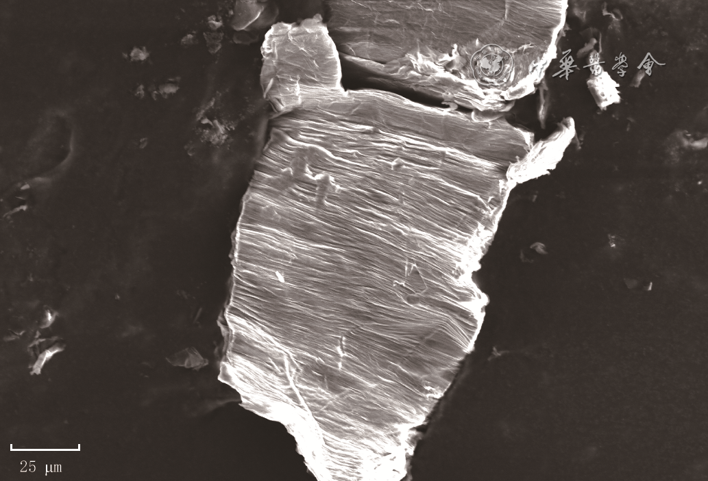
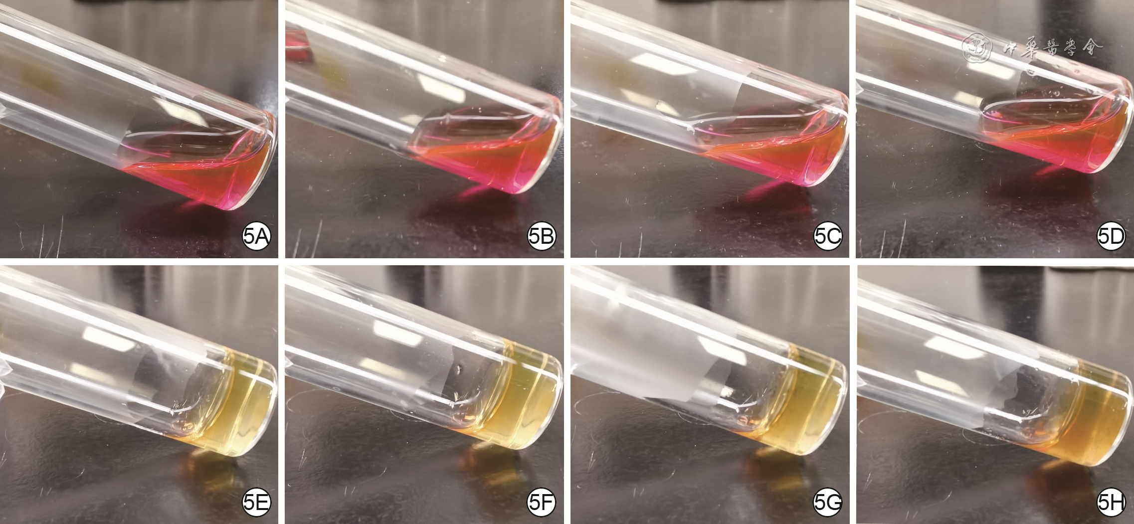
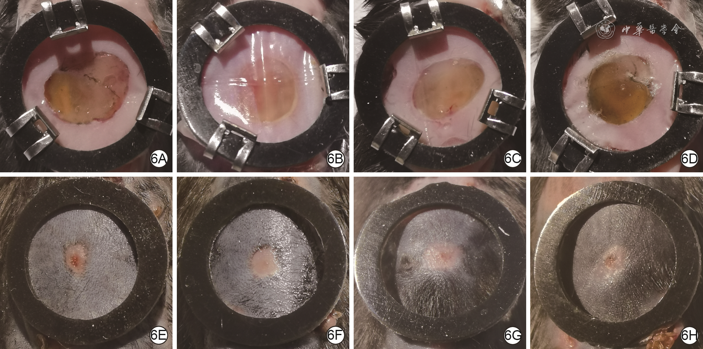
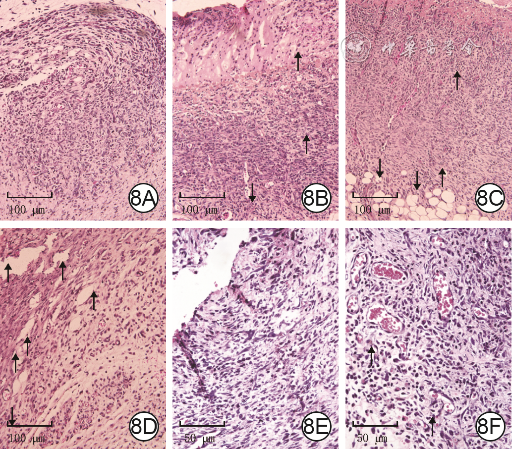
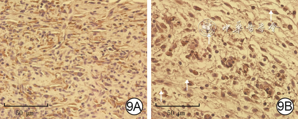
  •   Objective  To prepare graphene oxide (GO)-containing gelatin methacrylate anhydride (GelMA) hydrogel and to investigate the effects of in situ photopolymerized GO-GelMA composite hydrogel in wound vascularization of full-thickness skin defect in mice.  Methods  The experimental study method was used. The 50 μL of 0.2 mg/mL GO solution was evenly applied onto the conductive gel, and the structure and size of GO were observed under field emission scanning electron microscope after drying. Human skin fibroblasts (HSFs) were divided into 0 μg/mL GO (without GO solution, the same as below) group, 0.1 μg/mL GO group, 1.0 μg/mL GO group, 5.0 μg/mL GO group, and 10.0 μg/mL GO group treated with GO of the corresponding final mass concentration, and the absorbance value was detected using a microplate analyzer after 48 h of culture to reflect the proliferation activity of cells (n=6). HSFs and human umbilical vein vascular endothelial cells (HUVECs) were divided into 0 μg/mL GO group, 0.1 μg/mL GO group, 1.0 μg/mL GO group, and 5.0 μg/mL GO group treated with GO of the corresponding final mass concentration, and the migration rates of HSFs at 24 and 36 h after scratching (n=5) and HUVECs at 12 h after scratching (n=3) were detected by scratch test, and the level of vascular endothelial growth factor (VEGF) secreted by HSFs after 4, 6, and 8 h of culture was detected by enzyme-linked immunosorbent assay method (n=3). The prepared GO-GelMA composite hydrogels containing GO of the corresponding final mass concentration were set as 0 μg/mL GO composite hydrogel group, 0.1 μg/mL GO composite hydrogel group, 1.0 μg/mL GO composite hydrogel group, and 5.0 μg/mL GO composite hydrogel group to observe their properties before and after cross-linking, and to detect the release of GO after soaking with phosphate buffer solution for 3 and 7 d (n=3). The full-thickness skin defect wounds were made on the back of 16 6-week-old female C57BL/6 mice. The mice treated with in situ cross-linked GO-GelMA composite hydrogel containing GO of the corresponding final mass concentration were divided into 0 μg/mL GO composite hydrogel group, 0.1 μg/mL GO composite hydrogel group, 1.0 μg/mL GO composite hydrogel group, and 5.0 μg/mL GO composite hydrogel group according to the random number table, with 4 mice in each group. The general condition of wound was observed and the wound healing rate was calculated on 3, 7, and 14 d of treatment, the wound blood perfusion was detected by laser Doppler flowmetry on 3, 7, and 14 d of treatment and the mean perfusion unit (MPU) ratio was calculated, and the wound vascularization on 7 d of treatment was observed after hematoxylin-eosin staining and the vascular density was calculated (n=3). The wound tissue of mice in 0 μg/mL GO composite hydrogel group and 0.1 μg/mL GO composite hydrogel group on 7 d of treatment was collected to observe the relationship between the distribution of GO and neovascularization by hematoxylin-eosin staining (n=3) and the expression of VEGF by immunohistochemical staining. Data were statistically analyzed with analysis of variance for repeated measurement, one-way analysis of variance, and Tukey's method.  Results  GO had a multilayered lamellar structure with the width of about 20 μm and the length of about 50 μm. The absorbance value of HSFs in 10.0 μg/mL GO group was significantly lower than that in 0 μg/mL GO group after 48 h of culture (q=7.64, P<0.01). At 24 h after scratching, the migration rates of HSFs were similar in the four groups (P>0.05); at 36 h after scratching, the migration rate of HSFs in 0.1 μg/mL GO group was significantly higher than that in 0 μg/mL GO group, 1.0 μg/mL GO group, and 5.0 μg/mL GO group (with q values of 7.48, 10.81, and 10.20, respectively, P<0.01). At 12 h after scratching, the migration rate of HUVECs in 0.1 μg/mL GO group was significantly higher than that in 0 μg/mL GO group, 1.0 μg/mL GO group, and 5.0 μg/mL GO group (with q values of 7.11, 8.99, and 14.92, respectively, P<0.01), and the migration rate of HUVECs in 5.0 μg/mL GO group was significantly lower than that in 0 μg/mL GO group and 1.0 μg/mL GO group (with q values of 7.81 and 5.33, respectively, P<0.05 or P<0.01 ). At 4 and 6 h of culture, the VEGF expressions of HSFs in the four groups were similar (P>0.05); at 8 h of culture, the VEGF expression of HSFs in 0.1 μg/mL GO group was significantly higher than that in 0 μg/mL GO group and 5.0 μg/mL GO group (with q values of 4.75 and 4.48, respectively, P<0.05). The GO-GelMA composite hydrogels in the four groups were all red liquid before cross-linking, which turned to light yellow gel after cross-linking, with no significant difference in fluidity. The GO in the GO-GelMA composite hydrogel of 0 μg/mL GO composite hydrogel group had no release of GO at all time points; the GO in the GO-GelMA composite hydrogels of the other 3 groups was partially released on 3 d of soaking, and all the GO was released on 7 d of soaking. From 3 to 14 d of treatment, the wounds of mice in the 4 groups were covered with hydrogel dressings, kept moist, and gradually healed. On 3, 7, and 14 d of treatment, the wound healing rates of mice in the four groups were similar (P>0.05). On 3 d of treatment, the MPU ratio of wound of mice in 0.1 μg/mL GO composite hydrogel group was significantly higher than that in 0 μg/mL GO composite hydrogel group, 1.0 μg/mL GO composite hydrogel group, and 5.0 μg/mL GO composite hydrogel group (with q values of 10.70, 11.83, and 10.65, respectively, P<0.05 or P<0.01). On 7 and 14 d of treatment, the MPU ratios of wound of mice in the four groups were similar (P>0.05). The MPU ratio of wound of mice in 0.1 μg/mL GO composite hydrogel group on 7 d of treatment was significantly lower than that on 3 d of treatment (q=14.38, P<0.05), and that on 14 d of treatment was significantly lower than that on 7 d of treatment (q=27.78, P<0.01). On 7 d of treatment, the neovascular density of wound of mice on 7 d of treatment was 120.7±4.1 per 200 times of visual field, which was significantly higher than 61.7±1.3, 77.7±10.2, and 99.0±7.9 per 200 times of visual field in 0 μg/mL GO composite hydrogel group, 1.0 μg/mL GO composite hydrogel group, and 5.0 μg/mL GO composite hydrogel group (with q values of 12.88, 7.79, and 6.70, respectively, P<0.01), and the neovascular density of wound of mice in 1.0 μg/mL GO composite hydrogel group and 5.0 μg/mL GO composite hydrogel group was significantly higher than that in 0 μg/mL GO composite hydrogel group (with q values of 5.10 and 6.19, respectively, P<0.05). On 7 d of treatment, cluster of new blood vessels in wound of mice in 0.1 μg/mL GO composite hydrogel group was significantly more than that in 0 μg/mL GO composite hydrogel group, and the new blood vessels were clustered near the GO; a large amount of VEGF was expressed in wound of mice in 0.1 μg/mL GO composite hydrogel group in the distribution area of GO and new blood vessels.  Conclusions  GO with mass concentration lower than 10.0 μg/mL had no adverse effect on proliferation activity of HSFs, and GO of 0.1 μg/mL can promote the migration of HSFs and HUVECs, and can promote the secretion of VEGF in HSFs. In situ photopolymerized of GO-GelMA composite hydrogel dressing can promote the wound neovascularization of full-thickness skin defect in mice and increase wound blood perfusion in the early stage, with GO showing an enrichment effect on angiogenesis, and the mechanism may be related to the role of GO in promoting the secretion of VEGF by wound cells.


  • 烧创伤导致的严重体表组织缺损对患者打击巨大,除容易引发感染和脓毒症外,还会导致骨、肌肉等深部组织外露甚至进行性坏死,形成更复杂的难治性创面。快速发展的急救技术显著提高了烧创伤患者的抢救成功率,但烧创伤创面的治疗仍存在难题。首先是创面瘢痕愈合,虽然生命得以挽救,患者仍长期遭受瘢痕带来的困扰,包括毁容、挛缩、瘙痒、无法排汗等。烧伤创面患者增生性瘢痕发生率高达70%1。据估计,全球范围每年用于瘢痕治疗的医疗费用在2027年将达到320亿美元,是沉重的社会负担2。此外,生存质量的下降还给患者造成沉重的心理负担3。其次是复杂创面的修复难题。由于创基条件差,胸骨、肌肉等深部组织外露创面的治疗十分棘手。在感染等因素影响下这些创面会转变为慢性创面,进一步加大治疗难度,最终会导致截肢等。解决这些难题具有重要的临床价值,也一直是创面修复领域的研究热点。结合本课题组的研究经验,本文主要总结了近期国内外有关生物活性材料促进创面修复和皮肤附件再生的研究成果和进展,为后续研究提供参考。

    创面愈合是人体在生物进化过程中获得的一种自我保护功能,使人体免遭外界污染物的危害而得以存活。但是,创面自愈合能力有限,自然状态下,烧创伤创面愈合缓慢,增加进行性加深和感染的风险。而且,自愈创面与临床干预创面相比更容易发生异常愈合。创面深度过深、张力过高、异常炎症反应等均是引发异常愈合,尤其是瘢痕愈合的原因。与成人相比,人类妊娠早期胎儿创面发生无瘢痕愈合。成人和早期胎儿创面微环境差异被认为是决定创面不同愈合模式的关键因素4

    创面微环境是指创面局部及其邻近区域中影响创面愈合的物理、生物化学、细胞等因素的总和,可分为外部微环境和内部微环境5。外部微环境涵盖温度、湿度、张力、酸碱度等因素,内部微环境涵盖ECM和细胞因素。不同原因影响创面愈合的作用最终均体现于对内部微环境的改变,例如,创面张力通过提高ECM刚度促进肌Fb(MFb)分化6。不利于创面再生性修复的内部微环境因素包括异常的炎症反应、生物化学和生物物理信号、细胞表型和功能。调控创面微环境的本质是通过材料、药物、物理疗法等不同方法直接或间接地改善内部微环境,在生物化学、机械力、空间结构等多方面调控免疫和非免疫细胞行为,进而影响创面愈合过程。微环境调控是促进创面再生修复的必由之路7

    生物活性材料在调控创面微环境方面具有显著优势。与局部用药、负压吸引等方法相比,利用生物活性材料可同时实现生物化学、机械力、空间结构等微环境要素的调节和营造,为创面细胞提供三维生长空间的同时,向细胞传递机械力、生物化学等信号。随着第3代生物材料理论体系的建立,创面修复生物活性材料的研发思路发生改变,通过再生医学、材料学、工程学等学科的交叉融合,构建主动、高效调控创面细胞行为的生物活性材料成为研究焦点8。近年来,以创面再生性修复和皮肤附件再生为目标,国内外研究者研发了系列新型生物活性材料,为创面修复提供新材料的同时,进一步加深了人们对创面修复的理解。

    利用生物活性材料可实现对创面免疫细胞行为和炎症反应的调控。损伤修复和组织重建的过程与炎症反应密切相关,因为免疫细胞不仅发挥吞噬病原体和坏死组织的作用9,还通过分泌细胞因子、生长因子以及基质金属蛋白酶(MMP)等参与组织重建过程。一些生物活性材料通过诱导巨噬细胞发生M2型极化,营造促再生炎症反应微环境。葡聚糖-甲基丙烯酸异氰基乙酯-乙胺水凝胶诱导巨噬细胞发生M2型极化,其促进真皮功能性重建和毛囊再生的作用分别在小鼠和猪全层皮肤缺损创面模型中得到验证10。膀胱脱细胞基质(urinary bladder-decellularized matrix,UBM)和小肠黏膜下层基质等猪组织脱细胞基质诱导巨噬细胞向M2型极化,在烧创伤急性创面、下肢复杂性创面的修复中显示出与胶原蛋白基合成真皮替代物相比更强的促愈合能力11, 12。笔者课题组研究显示,糖尿病小鼠全层皮肤缺损创面巨噬细胞较快迁入并广泛分布于UBM中,UBM诱导迁入的巨噬细胞发生M2型极化,表达FGF-2、VEGF等促愈和促血管新生生长因子13。脱细胞基质材料含有基质结合纳米囊,后者含有的微小RNA被证实是调控巨噬细胞向抗炎表型极化的关键分子14。临床证据与上述结论相符,UBM单独应用即可促进肌腱和骨外露复杂创面愈合和真皮重建15,而胶原蛋白基合成真皮替代物需联合VSD系统以提高复杂创面愈合速度16。虽然缺少临床组织学证据,但大量的体外和动物模型研究证据提示,调控巨噬细胞行为并产生促再生微环境是ECM材料增强创面愈合的关键机制之一。

    除调控炎症反应之外,近期研究证实了生物活性材料诱导创面发生适应性免疫反应的功能。Jennifer Elisseeff课题组较早注意到UBM激活适应性免疫反应的作用17,并证实UBM诱导小鼠角膜上皮缺损创面发生适应性免疫反应,促进角膜再生和视力恢复18。此外,一些合成生物活性材料也被证实具有激活创面适应性免疫反应的功能。Griffin等19对比了分别含有D型和L型氨基酸多肽的微孔退火颗粒水凝胶对小鼠全层皮肤缺损创面炎症反应的影响。结果显示,与L型相比,含有D型氨基酸多肽的微孔退火颗粒水凝胶可显著增强IL-33+骨髓细胞的募集,抑制瘢痕形成,并促进毛囊、皮脂腺再生。目前,与炎症反应相比,生物活性材料调控适应性免疫反应的机制有待明确,主要包括抗原产生、呈递过程及其与材料组成、结构等性质的关系20

    利用生物活性材料可实现对创面非免疫细胞行为的调控。在高张力、真皮缺失等因素的影响下,深度创面中的MFb过度活跃,大量合成胶原蛋白等基质分子,最终形成瘢痕组织。因此,调控创基细胞行为,抑制其向MFb分化是减轻瘢痕形成的重要策略21。然而,单独应用药物或物理的方法难以达到这一目的,因为调控MFb行为的因素多且复杂,不仅涉及炎症反应,还涉及细胞所处微环境的结构、力学性能和生物化学组成等。生物活性材料在调控MFb行为方面具有显著优势。已上市的Integra®、PELNAC®等胶原蛋白基真皮替代物显示出抑制瘢痕形成的功能,其关键作用机制之一即是通过支架中GFOGER和GLOGER多肽调控MFb的黏附行为,抑制MFb聚集及其收缩创面的作用22。但是,Integra®等真皮替代物未显示出对MFb分化的抑制性,而一些脱细胞基质材料则显示出阻碍创面MFb分化的作用18。例如,UBM抑制小鼠角膜上皮缺损创面MFb分化18。人羊膜脱细胞基质抑制兔耳全层皮肤缺损创面MFb分化,可降低胶原蛋白表达量和沉积量并提高MMP-1的表达和分泌,有效促进皮肤再生23。利用载药生物活性材料调控创面细胞行为是促进创面再生性修复的另一途径。含高渗葡萄糖酸钾的胶原蛋白水凝胶可抑制小鼠全层皮肤缺损创面MFb分化,促进皮肤再生24。载氧化锌-姜黄素纳米复合物的胶原蛋白支架可显著上调大鼠烧伤创面细胞表达的TGF-β3的活性,促进创面无瘢痕愈合25。含TGF-β1小干扰RNA的胶原蛋白-壳聚糖支架可长效抑制猪全层皮肤缺损创面细胞TGF-β1信号通路,促进皮肤再生26

    利用生物活性材料的空间结构和力学性能可实现对细胞存活、分化、空间排布等行为的调控。聚己内酯电纺丝支架与聚己内酯多孔支架相比,显示出促进脂肪间充质干细胞表达核因子κB的作用,增强细胞旁分泌功能,使大鼠全层皮肤缺损创面中M2型巨噬细胞增加27。笔者课题组构建了柔性丝蛋白纳米纤维支架,研究显示仅通过纳米结构和机械特性的优化即可诱导大鼠骨髓间充质干细胞向内皮细胞分化28。Xu等29利用聚乳酸和生物玻璃构成的纳米纤维支架向共培养的Fb和内皮细胞同时传递结构信号和生物化学信号,显著增强2种细胞的旁分泌功能,促进小鼠全层皮肤缺损创面血管化和再生性修复。生物活性材料通过黏附受体整合素、机械门控离子通道Piezo1蛋白等受体向细胞施加力学和空间结构的调控作用。在材料应力刺激下,巨噬细胞、Fb的整合素受体被激活,使潜在TGF-β1得到释放30。另外,生物活性材料通过肌动蛋白微丝连接整合素的瞬时受体电位M7和C1在细胞膜张力增加时促进钙离子内流,将力学刺激转化为电化学信号,影响细胞行为31。对接种于聚丙烯酰胺水凝胶的小鼠骨髓来源巨噬细胞,细胞Piezo1受体与整合素相比在应力作用下直接向胞内输送钙离子32。材料空间结构对细胞行为的影响是基于细胞黏附配体的空间分布和密度而实现的。在材料刚度一定的条件下,特定微结构如纳米纤维结构、取向结构、孔结构可改变力学刺激的方向和强度,进而调控细胞迁移、增殖和分化等行为33;细胞间连接及其相互作用也受到材料空间结构的影响33

    此外,生物活性材料在皮肤附件再生医学技术中显示出很高的应用潜能。首先,特定小分子药物联合生物可降解三维支架通过多因素协同作用促进皮肤附件再生。Geoffrey Gurtner团队以普鲁兰多糖-胶原蛋白复合水凝胶作为局部黏着斑激酶(focal adhesion kinase,FAK)抑制剂(化合物VS6062)的载体,显著增强杜洛克猪深度创面愈合作用的同时,实现了毛囊和汗腺的再生34。抑制FAK使Fb蛋白激酶B(Akt)信号放大,进而上调早期生长应答因子1 (EGR1)和乳脂球EGF8(MFGE8)这2种蛋白的表达,Akt-EGR1信号通路被认为与细胞再生性表型有关,而MFGE8具有促进胶原蛋白分子被吞噬的作用35。其次,通过化学手性异构激活适应性免疫反应的D型氨基酸多肽的微孔退火颗粒水凝胶支架可诱导小鼠全层皮肤缺损创面毛囊新生,此外,研究者进一步证实了毛囊新生对支架激活适应性免疫的依赖性19,但相关分子机制有待明确。

    生物活性材料与三维生物打印技术的结合为皮肤附件再生提供了新方案。以明胶和透明质酸衍生化合物作为三维生物打印墨水,可维持人Fb和人脐静脉内皮细胞团块在支架中的空间排布,维持打印组织的活力,促进大鼠和猪全层皮肤缺损创面血管化、再生性修复和毛囊、皮脂腺等附件再生36。以脱细胞基质来源材料作为生物墨水成分,可显著增强细胞对细胞因子、生长因子信号的响应性,为皮肤附件再生提供基础37。2020年,付小兵院士团队将足趾垫匀浆蛋白和生物墨水共同作为骨髓间充质干细胞的载体,应用三维打印技术在小鼠足趾垫烫伤创面实现了汗腺的原位再生,并证实了足趾匀浆蛋白在三维环境下诱导骨髓间充质干细胞表达胶原三螺旋重复蛋白1和血红素加氧酶基因1,促进汗腺再生38

    综上,生物活性材料已发展成为创面修复技术的基础性要素39。近年来,系列主动调控创面微环境与细胞的新型生物活性材料得以研发,其调控创面愈合的新机制进一步加深了人们对创面修复的理解,为其临床应用提供了坚实的理论依据。然而,至今,大部分生物活性材料都是针对创面愈合的单一微环境要素或时间而设计和应用的。为了最终达到创面再生性修复的目的,生物活性材料应朝着整合多种作用机制的多功能材料发展,更有效地发挥其通过不同特性协同调控创面愈合的优势。因此,如何在同一体系中整合调控炎症反应、呈递适宜生物化学和生物物理信号的材料基元是关键的科学问题。此外,生物活性材料的研发和应用应更加重视创面的精准修复,针对创面的不同特点、调控创面微环境的不同目的和时机优化材料性能。相信随着再生医学、生物材料学和组织工程学的快速发展,将产生更多的新型生物活性材料40,推动创面修复技术朝着精准、高效调控创面微环境的方向发展,为实现创面“完美修复”和皮肤再生奠定基础。

    脱细胞真皮基质(ADM)重症监护病房(ICU)动脉血氧分压(PaO2
    丙氨酸转氨酶(ALT)白细胞介素(IL)磷酸盐缓冲液(PBS)
    急性呼吸窘迫综合征(ARDS)角质形成细胞(KC)反转录-聚合酶链反应(RT-PCR)
    天冬氨酸转氨酶(AST)半数致死烧伤面积(LA50)全身炎症反应综合征(SIRS)
    集落形成单位(CFU)内毒素/脂多糖(LPS)超氧化物歧化酶(SOD)
    细胞外基质(ECM)丝裂原活化蛋白激酶(MAPK)动脉血氧饱和度(SaO2
    表皮生长因子(EGF)最低抑菌浓度(MIC)体表总面积(TBSA)
    酶联免疫吸附测定(ELISA)多器官功能障碍综合征(MODS)转化生长因子(TGF)
    成纤维细胞(Fb)多器官功能衰竭(MOF)辅助性T淋巴细胞(Th)
    成纤维细胞生长因子(FGF)一氧化氮合酶(NOS)肿瘤坏死因子(TNF)
    3-磷酸甘油醛脱氢酶(GAPDH)负压伤口疗法(NPWT)血管内皮生长因子(VEGF)
    苏木精-伊红(HE)动脉血二氧化碳分压(PaCO2负压封闭引流(VSD)
    下载: 导出CSV 
    | 显示表格
  • [1]
    GuillemotF,SouquetA,CatrosS,et al.High-throughput laser printing of cells and biomaterials for tissue engineering[J].Acta Biomater,2010,6(7):2494-2500.DOI: 10.1016/j.actbio.2009.09.029.
    [2]
    CampbellPG,WeissLE.Tissue engineering with the aid of inkjet printers[J].Expert Opin Biol Ther,2007,7(8):1123-1127.DOI: 10.1517/14712598.7.8.1123.
    [3]
    OzbolatIT.Bioprinting scale-up tissue and organ constructs for transplantation[J].Trends Biotechnol,2015,33(7):395-400.DOI: 10.1016/j.tibtech.2015.04.005.
    [4]
    LiX, LianQ, LiD, et al. Development of a robotic arm based hydrogel additive manufacturing system for in-situ printing[J]. Applied Sciences, 2017, 7(1):73. DOI: 10.3390/app7010073.
    [5]
    SinghS,ChoudhuryD,YuF,et al.In situ bioprinting - bioprinting from benchside to bedside?[J].Acta Biomater,2020,101:14-25.DOI: 10.1016/j.actbio.2019.08.045.
    [6]
    HuangR,HuJ,QianW,et al.Recent advances in nanotherapeutics for the treatment of burn wounds[J/OL].Burns Trauma,2021,9:tkab026[2022-03-14].https://pubmed.ncbi.nlm.nih.gov/34778468/.DOI: 10.1093/burnst/tkab026.
    [7]
    WangH,LiuY,CaiK,et al.Antibacterial polysaccharide-based hydrogel dressing containing plant essential oil for burn wound healing[J/OL].Burns Trauma,2021,9:tkab041[2022-03-14].https://pubmed.ncbi.nlm.nih.gov/34988231/.DOI: 10.1093/burnst/tkab041.
    [8]
    AfewerkiS,SheikhiA,KannanS,et al.Gelatin-polysaccharide composite scaffolds for 3D cell culture and tissue engineering: towards natural therapeutics[J].Bioeng Transl Med,2018,4(1):96-115.DOI: 10.1002/btm2.10124.
    [9]
    LiMN,YuHP,KeQF,et al.Gelatin methacryloyl hydrogels functionalized with endothelin-1 for angiogenesis and full- thickness wound healing[J].J Mater Chem B,2021,9(23):4700-4709.DOI: 10.1039/d1tb00449b.
    [10]
    VandoorenJ,Van den SteenPE,OpdenakkerG.Biochemistry and molecular biology of gelatinase B or matrix metalloproteinase-9 (MMP-9): the next decade[J].Crit Rev Biochem Mol Biol,2013,48(3):222-272.DOI: 10.3109/10409238.2013.770819.
    [11]
    JangMJ,BaeSK,JungYS,et al.Enhanced wound healing using a 3D printed VEGF-mimicking peptide incorporated hydrogel patch in a pig model[J].Biomed Mater,2021,16(4):605.DOI: 10.1088/1748-605X/abf1a8.
    [12]
    LoessnerD,MeinertC,KaemmererE,et al.Functionalization, preparation and use of cell-laden gelatin methacryloyl-based hydrogels as modular tissue culture platforms[J].Nat Protoc,2016,11(4):727-746.DOI: 10.1038/nprot.2016.037.
    [13]
    KlotzBJ,GawlittaD,AJWPRosenberg,et al.Gelatin-methacryloyl hydrogels: towards biofabrication-based tissue repair[J].Trends Biotechnol,2016,34(5):394-407.DOI: 10.1016/j.tibtech.2016.01.002.
    [14]
    NuutilaK,SamandariM,EndoY,et al.In vivo printing of growth factor-eluting adhesive scaffolds improves wound healing[J].Bioact Mater,2022,8:296-308.DOI: 10.1016/j.bioactmat.2021.06.030.
    [15]
    ZhouX,ChenJ,SunH,et al.Spatiotemporal regulation of angiogenesis/osteogenesis emulating natural bone healing cascade for vascularized bone formation[J].J Nanobiotechnology,2021,19(1):420.DOI: 10.1186/s12951-021-01173-z.
    [16]
    ChenYC,LinRZ,QiH,et al.Functional human vascular network generated in photocrosslinkable gelatin methacrylate hydrogels[J].Adv Funct Mater,2012,22(10):2027-2039.DOI: 10.1002/adfm.201101662.
    [17]
    LinRZ,ChenYC,Moreno-LunaR,et al.Transdermal regulation of vascular network bioengineering using a photopolymerizable methacrylated gelatin hydrogel[J].Biomaterials,2013,34(28):6785-6796.DOI: 10.1016/j.biomaterials.2013.05.060.
    [18]
    FangH,LuoC,LiuS,et al.A biocompatible vascularized graphene oxide (GO)-collagen chamber with osteoinductive and anti- fibrosis effects promotes bone regeneration in vivo[J].Theranostics,2020,10(6):2759-2772.DOI: 10.7150/thno.42006.
    [19]
    NorahanMH,AmroonM,GhahremanzadehR,et al.Electroactive graphene oxide-incorporated collagen assisting vascularization for cardiac tissue engineering[J].J Biomed Mater Res A,2019,107(1):204-219.DOI: 10.1002/jbm.a.36555.
    [20]
    ChakrabortyS,PonrasuT,ChandelS,et al.Reduced graphene oxide-loaded nanocomposite scaffolds for enhancing angiogenesis in tissue engineering applications[J].R Soc Open Sci,2018,5(5):172017.DOI: 10.1098/rsos.172017.
    [21]
    MukherjeeS,SriramP,BaruiAK,et al.Graphene oxides show angiogenic properties[J].Adv Healthc Mater,2015,4(11):1722-1732.DOI: 10.1002/adhm.201500155.
    [22]
    ChangTK,LuYC,YehST,et al.In vitro and in vivo biological responses to graphene and graphene oxide: a murine calvarial animal study[J].Int J Nanomedicine,2020,15:647-659.DOI: 10.2147/IJN.S231885.
    [23]
    LiuW,LuoH,WeiQ,et al.Electrochemically derived nanographene oxide activates endothelial tip cells and promotes angiogenesis by binding endogenous lysophosphatidic acid[J].Bioact Mater,2022,9:92-104.DOI: 10.1016/j.bioactmat.2021.07.007.
    [24]
    焦德龙功能化石墨烯/光凝胶复合载药体系用于骨组织再生修复研究上海上海交通大学2019DOI:10.27307/d.cnki.gsjtu.2019.004439

    焦德龙.功能化石墨烯/光凝胶复合载药体系用于骨组织再生修复研究[D].上海:上海交通大学,2019.DOI:10.27307/d.cnki.gsjtu.2019.004439.

    [25]
    YueK,Trujillo-de SantiagoG,AlvarezMM,et al.Synthesis, properties, and biomedical applications of gelatin methacryloyl (GelMA) hydrogels[J].Biomaterials,2015,73:254-271.DOI: 10.1016/j.biomaterials.2015.08.045.
    [26]
    CoentroJQ,PuglieseE,HanleyG,et al.Current and upcoming therapies to modulate skin scarring and fibrosis[J].Adv Drug Deliv Rev,2019,146:37-59.DOI: 10.1016/j.addr.2018.08.009.
    [27]
    VedakumariSW,Veda JancySJ,PravinYR,et al.Facile synthesis of sericin modified graphene oxide nanocomposites for treating ischemic diseases[J].Environ Res,2022,209:112925.DOI: 10.1016/j.envres.2022.112925.
    [28]
    VerdeV,LongoA,CucciLM,et al.Anti-angiogenic and anti-proliferative graphene oxide nanosheets for tumor cell therapy[J].Int J Mol Sci,2020,21(15):5571.DOI: 10.3390/ijms21155571.
    [29]
    LaiPX,ChenCW,WeiSC,et al.Ultrastrong trapping of VEGF by graphene oxide: anti-angiogenesis application[J].Biomaterials,2016,109:12-22.DOI: 10.1016/j.biomaterials.2016.09.005.
    [30]
    ZhangY,AliSF,DervishiE,et al.Cytotoxicity effects of graphene and single-wall carbon nanotubes in neural phaeochromocytoma-derived PC12 cells[J].ACS Nano,2010,4(6):3181-3186.DOI: 10.1021/nn1007176.
    [31]
    LiangY,ChenB,LiM,et al.Injectable antimicrobial conductive hydrogels for wound disinfection and infectious wound healing[J].Biomacromolecules,2020,21(5):1841-1852.DOI: 10.1021/acs.biomac.9b01732.
    [32]
    GurtnerGC,WernerS,BarrandonY,et al.Wound repair and regeneration[J].Nature,2008,453(7193):314-321.DOI: 10.1038/nature07039.
    [33]
    LiM,ZhaoY,HaoH,et al.Theoretical and practical aspects of using fetal fibroblasts for skin regeneration[J].Ageing Res Rev,2017,36:32-41.DOI: 10.1016/j.arr.2017.02.005.
    [34]
    WangM,WangC,ChenM,et al.Efficient angiogenesis-based diabetic wound healing/skin reconstruction through bioactive antibacterial adhesive ultraviolet shielding nanodressing with exosome release[J].ACS Nano,2019,13(9):10279-10293.DOI: 10.1021/acsnano.9b03656.
    [35]
    RidiandriesA,TanJ,BursillCA.The role of chemokines in wound healing[J].Int JTM Mol Sci,2018,19(10):3217.DOI: 10.3390/ijms19103217.
    [36]
    JamiesonD,ChanceB,CadenasE,et al.The relation of free radical production to hyperoxia[J].Annu Rev Physiol,1986,48:703-719.DOI: 10.1146/annurev.ph.48.030186.003415.
    [37]
    AllenRG,BalinAK.Oxidative influence on development and differentiation: an overview of a free radical theory of development[J].Free Radic Biol Med,1989,6(6):631-661.DOI: 10.1016/0891-5849(89)90071-3.
    [38]
    CalabreseV,MancusoC,CalvaniM,et al.Nitric oxide in the central nervous system: neuroprotection versus neurotoxicity[J].Nat Rev Neurosci,2007,8(10):766-775.DOI: 10.1038/nrn2214.
    [39]
    SemenzaGL.HIF-1 and mechanisms of hypoxia sensing[J].Curr Opin Cell Biol,2001,13(2):167-171.DOI: 10.1016/s0955-0674(00)00194-0.
    [40]
    ChongY,GeC,YangZ,et al.Reduced cytotoxicity of graphene nanosheets mediated by blood-protein coating[J].ACS Nano,2015,9(6):5713-5724.DOI: 10.1021/nn5066606.
    [41]
    WeiXQ,HaoLY,ShaoXR,et al.Insight into the interaction of graphene oxide with serum proteins and the impact of the degree of reduction and concentration[J].ACS Appl Mater Interfaces,2015,7(24):13367-13374.DOI: 10.1021/acsami.5b01874.
    [42]
    FengR,YuY,ShenC,et al.Impact of graphene oxide on the structure and function of important multiple blood components by a dose-dependent pattern[J].J Biomed Mater Res A,2015,103(6):2006-2014.DOI: 10.1002/jbm.a.35341.
    [43]
    LiS,MulloorJJ,WangL,et al.Strong and selective adsorption of lysozyme on graphene oxide[J].ACS Appl Mater Interfaces,2014,6(8):5704-5712.DOI: 10.1021/am500254e.
  • 加载中

Catalog

    通讯作者: 陈斌, bchen63@163.com
    • 1. 

      沈阳化工大学材料科学与工程学院 沈阳 110142

    1. 本站搜索
    2. 百度学术搜索
    3. 万方数据库搜索
    4. CNKI搜索

    Figures(10)  / Tables(4)

    Article Metrics

    Article views (5888) PDF downloads(1355) Cited by()
    Proportional views
    Related

    /

    DownLoad:  Full-Size Img  PowerPoint
    Return
    Return