2022 Vol. 38, No. 2

Guidelines and Consensuses
National expert consensus on the aeromedical trans- portation of burn patients (2022 version)
, Liu Yan
2022, 38(2): 101-108. doi: 10.3760/cma.j.cn501120-20211025-00366
Abstract:
The development of burn units in our country is now undergoing a trend of geographic centralization and regionalization. To solve the problems like severe burn patients are too far away from burn units, overloaded operation in regional burn centers when mass burn accidents happen, and growing requirement for aeromedical transportation, etc., it is now the top priority to improve national aeromedical transportation system for burn patients. Expert teams from Chinese Burn Association, National Aeromedical Rescue Base, and China Association for Disaster & Emergency Rescue Medicine discussed and reached a consensus on the key points of aeromedical transportation of burn patients, including organizational structure, staff and materials, and three links before, during, and after aeromedical transportation. The consensus aims to provide guidance for a safe, efficient, and standardized operation of aeromedical transportation for burn patients in China.
Expert Forum
Re-understanding the physiological and pathophysiological roles of neutrophils
Sun Bingwei, Huang Jiamin
2022, 38(2): 109-113. doi: 10.3760/cma.j.cn501120-20211122-00391
Abstract:
Neutrophils have always been considered as a short-lived and homogeneous cell type in the innate immune system, which have limited pro-inflammatory or anti-inflammatory effects. However, in recent 10 years, the understanding of neutrophils has been undergoing some kind of revival as researches progressed. The researches on the heterogeneity of neutrophils and the mechanism of their interaction with other immune cells have promoted the researchers to re-understand the physiological and pathophysiological roles of neutrophils. In the following decades, with the development of single-cell sequencing technology, spatial transcriptome sequencing technology, and multi-omics combined sequencing technology, researchers will have a better understanding of the biological behaviors of neutrophils. This paper briefly reviews the biological behaviors of neutrophils and their roles in various diseases in recent years.
Regulatory role and related mechanism of skin gamma-delta T cell subsets in wound re-epithelialization
He Weifeng
2022, 38(2): 114-118. doi: 10.3760/cma.j.cn501120-20211210-00411
Abstract:

Re-epithelialization is one of the core links that determines the healing process of skin wounds.  The proliferation and differentiation of epidermal stem cells to form new epidermal tissue is the histological basis of re-epithelialization, and the smooth progress of the cell differentiation process of epidermal stem cells-precursor cells-terminal cells is the cytological basis for the continuous formation of new epidermal tissue. The proliferation of stem cells and their differentiation into precursor cells are the determinants of the proliferative potential of newly formed epidermal tissue, while the expansion and differentiation of precursor cells into terminal cells are key factors determining the rate of new epidermal tissue formation. The tissue microenvironment plays a key regulatory role in the process of wound re-epithelialization, and cell growth factor and inflammatory mediators are the two main components of tissue microenvironment, which play regulatory role in different aspects of proliferation and differentiation of epidermal stem cells, jointly promoting the smooth progress of wound re-epithelialization  As an important part of skin immune system, the subsets of gamma-delta (γδ) T cells play crucial role in dynamically shaping early wound microenvironment via secreting different cell growth factors and inflammatory factors. From the prospective of immune microenvironment of wound, this paper discusses the role of skin γδ T cells in maintaining the balance of stem cell proliferation and differentiation and regulating wound re-epithelialization, providing a new direction for the prevention and treatment of refractory wound.

Original Articles · Inflammation and Immunity after Burn
Effects of P311 on the angiogenesis ability of human microvascular endothelial cell 1 in vitro and its molecular mechanism
Wang Song, Li Haisheng, Qian Wei, Zhang Xiaorong, He Weifeng, Luo Gaoxing
2022, 38(2): 119-129. doi: 10.3760/cma.j.cn501120-20211210-00410
Abstract:
      Objective     To explore the effects of P311 on the angiogenesis ability of human microvascular endothelial cell 1 (HMEC-1) in vitro and the potential molecular mechanism.      Methods     The experimental research method was used. HMEC-1 was collected and divided into P311 adenovirus group and empty adenovirus group according to the random number table (the same grouping method below), which were transfected correspondingly for 48 h. The cell proliferation activity was detected using the cell counting kit 8 on 1, 3, and 5 days of culture. The residual scratch area of cells at post scratch hour 6 and 11 was detected by scratch test, and the percentage of the residual scratch area was calculated. The blood vessel formation of cells at 8 h of culture was observed by angiogenesis experiment in vitro, and the number of nodes and total length of the tubular structure were measured. The protein expressions of vascular endothelial growth factor receptor 2 (VEGFR2), phosphorylated VEGFR2 (p-VEGFR2), extracellular signal-regulated kinase 1/2 (ERK1/2), and phosphorylated ERK1/2 (p-ERK1/2) in cells were detected by Western blotting. HMEC-1 was collected and divided into P311 adenovirus+small interfering RNA (siRNA) negative control group, empty adenovirus+siRNA negative control group, P311 adenovirus+siRNA-VEGFR2 group, and empty adenovirus+siRNA-VEGFG2 group, which were treated correspondingly. The protein expressions of VEGFR2, p-VEGFR2, ERK1/2, and p-ERK1/2 in cells were detected by Western blotting at 24 h of transfection. The blood vessel formation of cells at 24 h of transfection was observed by angiogenesis experiment in vitro, and the number of nodes and total length of the tubular structure were measured. HMEC-1 was collected and divided into P311 adenovirus+dimethylsulfoxide (DMSO) group, empty adenovirus+DMSO group, P311 adenovirus+ERK1/2 inhibitor group, and empty adenovirus+ERK1/2 inhibitor group, which were treated correspondingly. The protein expressions of ERK1/2 and p-ERK1/2 in cells were detected by Western blotting at 2 h of treatment. The blood vessel formation of cells at 2 h of treatment was observed by angiogenesis experiment in vitro, and the number of nodes and total length of the tubular structure were measured. The sample number at each time point in each group was 6. Data were statistically analyzed with independent sample t test, analysis of variance for repeated measurement, one-way analysis of variance, and least significant difference test.      Results     Compared with that of empty adenovirus group, the proliferation activity of cells in P311 adenovirus group did not show significant difference on 1, 3, and 5 days of culture (with t values of -0.23, -1.30, and -1.52, respectively, P>0.05). The residual scratch area percentages of cells in P311 adenovirus group were significantly reduced at post scratch hour 6 and 11 compared with those of empty adenovirus group (with t values of -2.47 and -2.62, respectively, P<0.05). At 8 h of culture, compared with those of empty adenovirus group, the number of nodes and total length of the tubular structure of cells in P311 adenovirus group were significantly increased (with t values of 4.49 and 4.78, respectively, P<0.01). At 48 h of transfection, compared with those of empty adenovirus group, the protein expressions of VEGFR2 and ERK1/2 of cells in P311 adenovirus group showed no obvious changes (P>0.05), and the protein expressions of p-VEGFR2 and p-ERK1/2 of cells in P311 adenovirus group were significantly increased (with t values of 17.27 and 16.08, P<0.01). At 24 h of transfection, the protein expressions of p-VEGFR2 and p-ERK1/2 of cells in P311 adenovirus+siRNA negative control group were significantly higher than those in empty adenovirus+siRNA negative control group (P<0.01). The protein expressions of VEGFR2, p-VEGFR2, and p-ERK1/2 of cells in P311 adenovirus+siRNA negative control group were significantly higher than those in P311 adenovirus+siRNA-VEGFR2 group (P<0.01). The protein expressions of VEGFR2 and p-ERK1/2 of cells in empty adenovirus+siRNA negative control group were significantly higher than those in empty adenovirus+siRNA-VEGFR2 group (P<0.05 or P<0.01). At 24 h of transfection, the number of nodes of the tubular structure in cells of P311 adenovirus+siRNA negative control group was 720±62, which was significantly more than 428±38 in empty adenovirus+siRNA negative control group and 364±57 in P311 adenovirus+siRNA-VEGFR2 group (with P values both<0.01). The total length of the tubular structure of cells in P311 adenovirus+siRNA negative control group was (21 241±1 139) μm, which was significantly longer than (17 005±1 156) μm in empty adenovirus+siRNA negative control group and (13 494±2 465) μm in P311 adenovirus+siRNA-VEGFR2 group (with P values both<0.01). The number of nodes of the tubular structure in cells of empty adenovirus+siRNA negative control group was significantly more than 310±75 in empty adenovirus+siRNA-VEGFR2 group (P<0.01),  and the total length of the tubular structure of cells in empty adenovirus+siRNA negative control group was significantly longer than (11 600±2 776) μm in empty adenovirus+siRNA-VEGFR2 group (P<0.01). At 2 h of treatment, the protein expression of p-ERK1/2 of cells in P311 adenovirus+DMSO group was significantly higher than that in empty adenovirus+DMSO group and P311 adenovirus+ERK1/2 inhibitor group (with P values both<0.01), and the protein expression of p-ERK1/2 of cells in empty adenovirus+DMSO group was significantly higher than that in empty adenovirus+ERK1/2 inhibitor group (P<0.05). At 2 h of treatment, the number of nodes of the tubular structure in cells of P311 adenovirus+DMSO group was 726±72, which was significantly more than 421±39 in empty adenovirus+DMSO group and 365±41 in P311 adenovirus+ERK1/2 inhibitor group (with P values both<0.01). The total length of the tubular structure of cells in P311 adenovirus+DMSO group was (20 318±1 433) μm, which was significantly longer than (16 846±1 464) μm in empty adenovirus+DMSO group and (15 114±1 950) μm in P311 adenovirus+ERK1/2 inhibitor group (with P values both<0.01). The number of nodes of the tubular structure in cells of empty adenovirus+DMSO group was significantly more than 317±67 in empty adenovirus+ERK1/2 inhibitor group (P<0.01), and the total length of the tubular structure of cells in empty adenovirus+DMSO group was significantly longer than (13 188±2 306) μm in empty adenovirus+ERK1/2 inhibitor group (P<0.01).      Conclusions     P311 can enhance the angiogenesis ability of HMEC-1 by activating the VEGFR2/ERK1/2 signaling pathway.
Early predictive value of high density lipoprotein cholesterol for secondary acute kidney injury in sepsis patients
Li Jingyan, Yao Yongming, Tian Yingping
2022, 38(2): 130-136. doi: 10.3760/cma.j.cn501120-20210917-00325
Abstract:
  Objective  To investigate the changes of high density lipoprotein cholesterol (HDL-C) in sepsis patients and its early predictive value for secondary acute kidney injury (AKI) in such patients.  Methods  A retrospective case series study was conducted. From June 2019 to June 2021, 232 sepsis patients who met the inclusion criteria were admitted to the Second Hospital of Hebei Medical University, including 126 males and 106 females, aged 24 to 71 years. According to whether complicating secondary AKI, the patients were divided into non-AKI group (n=158) and AKI group (n=74). Data of patients between the two groups were compared and statistically analyzed with independent sample t test or chi-square test, including the sex, age, body mass index (BMI), body temperature, heart rate, primary infection site, combined underlying diseases, acute physiology and chronic health evaluation Ⅱ (APACHE Ⅱ) score and sepsis-related organ failure assessment (SOFA) score at admission, and the serum levels of C-reactive protein (CRP), procalcitonin, creatinine, cystatin C, and HDL-C measured at diagnosis of sepsis. The multivariate logistic regression analysis was performed on the indicators with statistically significant differences between the two groups to screen the independent risk factors for developing secondary AKI in 232 sepsis patients, and the joint prediction model was established based on the independent risk factors. The receiver operating characteristic (ROC) curve of the independent risk factors and the joint prediction model predicting secondary AKI in 232 sepsis patients were drawn, and the area under the curve (AUC), the optimal threshold, and the sensitivity and specificity under the optimal threshold were calculated. The quality of the above-mentioned AUC was compared by Delong test, and the sensitivity and specificity under the optimal threshold were compared using chi-square test.  Results  The sex, age, BMI, body temperature, heart rate, primary infection site, combined underlying diseases, and CRP level of patients between the two groups were similar (P>0.05). The procalcitonin, creatinine, cystatin C, and scores of APACHE Ⅱ and SOFA of patients in AKI group were all significantly higher than those in non-AKI group (with t values of -3.21, -16.14, -12.75, -11.13, and -12.88 respectively, P<0.01), while the HDL-C level of patients in AKI group was significantly lower than that in non-AKI group (t=6.33, P<0.01). Multivariate logistic regression analysis showed that creatinine, cystatin C, and HDL-C were the independent risk factors for secondary AKI in 232 sepsis patients (with odds ratios of 2.45, 1.68, and 2.12, respectively, 95% confidence intervals of 1.38-15.35, 1.06-3.86, and 0.86-2.56, respectively, P<0.01). The AUCs of ROC curves of creatinine, cystatin C, HDL-C, and the joint prediction model for predicting secondary AKI in 232 sepsis patients were 0.69, 0.79, 0.89, and 0.93, respectively (with 95% confidence intervals of 0.61-0.76, 0.72-0.85, 0.84-0.92, and 0.89-0.96, respectively, P values all below 0.01); the optimal threshold were 389.53 μmol/L, 1.56 mg/L, 0.63 mmol/L, and 0.48, respectively; the sensitivity under the optimal threshold were 76.6%, 81.4%, 89.7%, and 95.5%, respectively; the specificity under the optimal threshold values were 78.6%, 86.7%, 88.6%, and 96.6%, respectively. The AUC quality of cystatin C was significantly better than that of creatinine (z=2.34, P<0.05), the AUC quality and sensitivity and specificity under the optimal threshold of HDL-C were all significantly better than those of cystatin C (z=3.33, with χ2 values of 6.43 and 7.87, respectively, P<0.01) and creatinine (z=5.34, with χ2 values of 6.32 and 6.41, respectively, P<0.01); the AUC quality and sensitivity and specificity under the optimal threshold of the joint prediction model were all significantly better than those of creatinine, cystatin C, and HDL-C (with z values of 6.18, 4.50, and 2.06, respectively, χ2 values of 5.31, 7.23, 3.99, 6.56, 7.34, and 4.00, respectively, P<0.05 or P<0.01).  Conclusions  HDL-C level in sepsis patients with secondary AKI is significantly lower than that in patients without secondary AKI. This is an independent risk factor for secondary AKI in sepsis patients with a diagnostic value being superior to that of creatinine and cystatin C. The combination of the aforementioned three indicators would have higher predicative valuable for secondary AKI in sepsis patients.
Analysis of genomic information and biological characteristics of a bacteriophage against methicillin-resistant Staphylococcus aureus in patients with median sternal incision infection
Zhang Jian, Yan Rongshuai, Yang Zichen, Shi Xi, Li Xiang, Mao Tongchun, Zhang Yiming
2022, 38(2): 137-146. doi: 10.3760/cma.j.cn501120-20211130-00400
Abstract:
  Objective  To isolate and purify a bacteriophage against methicillin-resistant Staphylococcus aureus (MRSA), and to analyze its genomic information and biological characteristics.  Methods  The experimental research methods were adopted. MRSA (hereinafter referred to as host bacteria) solution was collected from the wound of a 63-year-old female patient with the median sternum incision infection admitted to the Second Affiliated Hospital of Army Medical University (the Third Military Medical University). The bacteriophage, named bacteriophage SAP23 was isolated and purified from the sewage of the Hospital by sewage co-culture method and double-layer agar plate method, and the plaque morphology was observed. The morphology of bacteriophage SAP23 was observed by transmission electron microscope after phosphotungstic acid negative staining. The whole genome of bacteriophage SAP23 was sequenced with NovaSeq PE15 platform after its DNA was prepared by sodium dodecyl sulfonate/protease cleavage scheme, and genomic analysis including sequence assembly, annotation, and phylogenetic tree were completed. The bacteriophage SAP23 solution was co-incubated with the host bacterial solution for 4 h at the multiplicity of infection (MOI) of 10.000 0, 1.000 0, 0.100 0, 0.010 0, 0.001 0, and 0.000 1, respectively, and then the bacteriophage titer was measured by the drip plate method to select the optimal MOI, with here and the following sample numbers of 3. The bacteriophage SAP23 solution was co-incubated with the host bacterial solution at the optimal MOI for 5, 10, and 15 min, respectively, and the bacteriophage titer was measured by the same method as mentioned above to select the optimal adsorption time. After the bacteriophage SAP23 solution was co-incubated with the host bacterial solution at the optimal MOI for the optimal adsorption time, the bacteriophage titers were measured by the same method as mentioned above at 0 (immediately), 5, 10, 15, 20, 30, 40, 50, 60, 80, 100, and 120 min after culture, respectively, and a one-step growth curve was drawn. The bacteriophage SAP23 solution was incubated at 4, 37, 50, 60, 70, and 80 ℃ and pH 2, 3, 4, 5, 6, 7, 8, 9, 10, 11, and 12 for 1 h, respectively, to determine its stability. A total of 41 MRSA strains stored in the Department of Microbiology of Army Medical University (the Third Military Medical University) were used to determine the host spectrum of bacteriophage SAP23.  Results  The bacteriophage SAP23 could form a transparent plaque on the host bacteria double-layer agar plate. The bacteriophage SAP23 has a polyhedral head with (88±4) nm in diameter and a tail with (279±21) nm in length and (22.6±2.6) nm in width. The bacteriophage SAP23 has a linear, double-stranded DNA with a full length of 151 618 bp and 11 681 bp long terminal repeats sequence in the sequence ends. There were 220 open reading frames predicted and the bacteriophage could encode 4 transfer RNAs, while no resistance genes or virulence factors were found. The annotation function of bacteriophage SAP23 genes could be divided into 5 groups. The GenBank accession number was MZ427930. According to the genomic collinearity analysis, there were 5 local collinear blocks in the whole genome between the bacteriophage SAP23 and the chosen 6 Staphylococcus bacteriophages, while within or outside the local collinear region, there were still some differences. The bacteriophage SAP23 belonged to the Herelleviridae family, Twortvirinae subfamily, and Kayvirus genus. The optimal MOI of bacteriophage SAP23 was 0.010 0, and the optimal adsorption time was 10 min. The bacteriophage SAP23 had a latent period of 20 min, and a growth phase of 80 min. The bacteriophage SAP23 was able to remain stable at the temperature between 4 and 37 ℃ and at the pH values between 4 and 9. The bacteriophage SAP23 could lyse 3 of the 41 tested MRSA strains.  Conclusions  The bacteriophage SAP23 is a member of the Herelleviridae family, Twortvirinae subfamily, and Kayvirus genus. The bacteriophage SAP23 has a good tolerance for temperature and acid-base and a short latent period, and can lyse MRSA effectively. The bacteriophage SAP23 is a new type of potent narrow-spectrum bacteriophage without virulence factors and resistance genes.
Original Articles
Changes of heparin-binding protein in severe burn patients during shock stage and its effects on human umbilical vein endothelial cells and neutrophils
Qi Xinxin, Liu Lu, Yang Yunxi, Huang Jiamin, Sun Bingwei
2022, 38(2): 147-155. doi: 10.3760/cma.j.cn501120-20210805-00269
Abstract:
  Objective  To investigate the changes of heparin-binding protein (HBP) in severe burn patients during shock stage and its effects on human umbilical vein endothelial cells (HUVECs) and neutrophils in vitro.  Methods  Prospective observational and experimental research methods were used. Twenty severe burn patients who met the inclusion criteria and were admitted to the Department of Burns and Plastic Surgery of Affiliated Suzhou Hospital of Nanjing Medical University from August to November 2020 were included in severe burn group (12 males and 8 females, aged 44.5 (31.0, 58.0) years). During the same period, 20 healthy volunteers with normal physical examination results in the unit's Physical Examination Center were recruited into healthy control group (13 males and 7 females, aged 39.5 (26.0, 53.0) years). Enzyme-linked immunosorbent assay (ELISA) method was used to detect the protein expression levels of HBP and tissue inhibitor of metalloproteinase 1 (TIMP-1) in plasma of patients within 48 hours after injury in severe burn group and in plasma of volunteers in healthy control group. The correlation between protein expression of HBP and that of TIMP-1 in the plasma in the two groups was analyzed by Pearson correlation analysis. The fourth passage of HUVECs in logarithmic growth phase were used for the experiment. The HUVECs were divided into normal control group with routine culture (the same treatment below) and recombinant HBP (rHBP)-treated 12 h group, rHBP-treated 24 h group, and rHBP-treated 48 h group with corresponding treatment according to the random number table (the same grouping method below), and the mRNA expression of TIMP-1 in cells was detected by real-time fluorescence quantitative reverse transcription polymerase chain reaction. The HUVECs were divided into normal control group and rHBP-treated 48 h group with corresponding treatment, and the protein expression of TIMP-1 in the cells was detected by Western blotting. The HUVECs were divided into normal control group, rHBP alone group, aprotinin alone group, and rHBP+aprotinin group treated with the corresponding reagents (with the final molarity of rHBP being 200 nmol/L and the final concentration of aprotinin being 20 μg/mL, respectively), cultured for 48 h, and ELISA was used to detect the protein expression of TIMP-1 in the culture supernatant of cells. The neutrophils were isolated from the peripheral venous blood of the aforementioned 10 healthy volunteers by immunomagnetic bead sorting, and the cells were divided into normal control group, recombinant TIMP-1 (rTIMP-1) alone group, phorbol acetate (PMA) alone group, and rTIMP-1+PMA group treated with corresponding reagents (with the final concentration of rTIMP-1 being 500 ng/mL and the final molarity of PMA being 10 nmol/L, respectively). After being cultured for 1 h, the expression of CD63 protein in cells was detected by immunofluorescence method, the positive expression rate of CD63 protein in cells was detected by flow cytometry, and the protein expression levels of HBP and myeloperoxidase (MPO) in the culture supernatant of cells were detected by ELISA. The normal control group underwent the above-mentioned related tests at appropriate time points. The number of samples was 3 in each group of cell experiment. Data were statistically analyzed with chi-square test, Mann-Whitney U test, Kruskal-Wallis H test, and Tamhane's T2 test.  Results  The protein expression levels of HBP and TIMP-1 in the plasma of patients in severe burn group were 404.9 (283.1, 653.2) and 262.1 (240.6, 317.4) ng/mL, respectively, which were both significantly higher than 61.6 (45.0, 68.9) and 81.0 (66.3, 90.0) ng/mL of volunteers in healthy control group (with Z values of -5.41 and -5.21, respectively, P<0.01). The correlation between the protein expression of HBP and that of TIMP-1 in the plasma of volunteers in healthy control group was not strong (P>0.05). The protein expression of HBP was significantly positively correlated with that of TIMP-1 in the plasma of patients in severe burn group (r=0.64, P<0.01). Compared with that in normal control group, the mRNA expression of TIMP-1 in HUVECs was significantly increased in rHBP-treated 12 h group, rHBP-treated 24 h group, and rHBP-treated 48 h group (with t values of -3.58, -2.25, and -1.26, respectively, P<0.05). Western blotting detection showed that compared with that in normal control group, the protein expression of TIMP-1 in HUVECs in rHBP-treated 48 h group was significantly enhanced. After 48 h of culture, compared with that in normal control group, the protein expression level of TIMP-1 in the culture supernatant of HUVECs in rHBP alone group was significantly increased (t=9.43, P<0.05), while the protein expression level of TIMP-1 in the culture supernatant of HUVECs didn't change significantly in aprotinin alone group or rHBP+aprotinin group (P>0.05); compared with that in rHBP alone group, the protein expression level of TIMP-1 in the culture supernatant of HUVECs in rHBP+aprotinin group was significantly decreased (t=4.76, P<0.01). After 1 h of culture, the trend of CD63 protein expression in neutrophils detected by immunofluorescence method and that by flow cytometry were consistent in each group. After 1 h of culture, compared with that in normal control group, the positive expression rate of CD63 protein in the neutrophils and the protein expression levels of HBP and MPO in the culture supernatant of cells in rTIMP-1 alone group all had no significant changes (P>0.05), while the positive expression rate of CD63 protein in the neutrophils and the protein expression levels of HBP and MPO in the culture supernatant of cells were all significantly increased in PMA alone group and rTIMP-1+PMA group (with t values of 2.41, 3.82, 5.73, 1.05, 4.16, and 1.08, respectively, P<0.05 or P<0.01); compared with that in PMA alone group, the positive expression rate of CD63 protein in the neutrophils and the protein expression levels of HBP and MPO in the culture supernatant of cells in rTIMP-1+PMA group were all significantly decreased (with t values of 5.26, 2.83, and 1.26, respectively, P<0.05 or P<0.01).  Conclusions  The expression level of HBP in the plasma of severe burn patients is increased during shock stage. HBP can induce HUVECs to secrete TIMP-1 in vitro, and TIMP-1 can reduce the expression of CD63 molecule in human neutrophils.
Epidemiological characteristics and treatment outcome analysis of 229 patients with hydrofluoric acid burns in hands
Zhang Manjia, Mao Shulei, Zhang Jianfen, Wang Xingang, Ni Liangfang, Zhang Yuanhai
2022, 38(2): 156-164. doi: 10.3760/cma.j.cn501120-20210517-00188
Abstract:
    Objective   To explore the epidemiological characteristics and treatment outcomes of patients with hydrofluoric acid burns in hands.    Methods   A retrospective observational study was conducted. The medical records of 229 patients with hydrofluoric acid burns in hands who were admitted to Zhejiang Quhua Hospital from January 2008 to December 2020 and met the inclusion criteria were collected. The following statistical data of patients were collected, including gender, age, type of affiliated enterprise, hydrofluoric acid mass fraction, injury site, total burn area, prehospital time, length of hospital stay, length of wound healing, whether hypocalcemia and hypomagnesemia occurred or not on admission, whether surgery intervention was performed or not, and whether scar sequelae occurred or not. Single factor and multivariate logistic regression analysis were used to screen out the risk factors impacting surgery intervention and scar sequelae of all the patients and patients whose hydrofluoric acid mass fraction was known. Single factor and multivariate linear regression analysis were used to screen out the risk factors impacting the length of wound healing of all the patients and patients whose hydrofluoric acid mass fraction was known.    Results   The 229 patients included 206 males and 23 females, with the majority aged 30 to 50 years (139 patients). The type of affiliated enterprise of majority patients was non-fluorine chemical enterprise. The hydrofluoric acid mass fraction was known in only 91 patients, mainly medium. The majority injury site was in the middle and end of finger. The total burn area was below or equal to 1% total body surface area. The prehospital time was 19 (9, 29) h. The length of hospital stay was 2 (1, 7) d. The length of wound healing was 12 (8, 18) d. The proportions of hypocalcemia and hypomagnesemia were 0.9% (2/229) and 1.3% (3/229) on admission, respectively. Thirty-six patients had surgeries and 83 patients had scar sequelae. In 229 patients, single factor logistic regression analysis showed that both type of affiliated enterprise and prehospital time were the factors impacting surgery intervention (with odds ratio values of 7.86 and 51.35, respectively, 95% confidence intervals of 1.83-33.76 and 11.89-221.78, respectively, P<0.01) and scar sequelae of patients (with odds ratio values of 3.62 and 27.40, respectively, 95% confidence intervals of 1.76-7.43 and 13.25-56.68, respectively, P<0.01); multivariate logistic regression analysis showed that prehospital time was the independent risks factor impacting surgery intervention and scar sequelae of patients (with odds ratio values of 43.00 and 24.55, respectively, 95% confidence intervals of 9.89-187.03 and 11.78-51.16, respectively, P<0.01); single factor linear regression analysis showed that both type of affiliated enterprise and prehospital time were the factors impacting the length of wound healing of patients (with β values of 6.16 and 12.83, respectively, 95% confidence intervals of 3.38-8.93 and 10.72-14.93, respectively, P<0.01); multivariate linear regression analysis showed that both type of affiliated enterprise and prehospital time were the independent risk factors impacting the length of wound healing of patients (with β values of 2.81 and 12.16, respectively, 95% confidence intervals of 0.50-5.13 and 10.00-14.31, respectively, P<0.05 or P<0.01). In 91 patients whose hydrofluoric acid mass fraction was known, single factor logistic regression analysis showed that type of affiliated enterprise, hydrofluoric acid mass fraction (low and high), and prehospital time were all the factors impacting surgery intervention of patients (with odds ratio values of 9.10, 11.25, 10.69, and 0.04, respectively, 95% confidence intervals of 1.15-72.25, 1.39-90.93, 1.32-86.59, and 0.01-0.19, respectively, P<0.05 or P<0.01), type of affiliated enterprise, hydrofluoric acid mass fraction, and prehospital time were all the factors impacting scar sequelae of patients (with odds ratio values of 0.32, 0.21, and 36.80, respectively, 95% confidence intervals of 0.11-0.92, 0.06-0.73, and 11.03-122.79, respectively, P<0.05 or P<0.01); multivariate logistic regression analysis showed that both hydrofluoric acid mass fraction and prehospital time were the independent risk factors impacting surgery intervention of patients (with odds ratio values of 11.51 and 0.04, respectively, 95% confidence intervals of 1.22-108.26 and 0.01-0.25, respectively, P<0.05 or P<0.01), prehospital time was the independent risk factor impacting scar sequelae of patients (odds ratio=37.71, with 95% confidence interval of 9.97-142.69, P<0.01); single factor linear regression analysis showed that type of affiliated enterprise, hydrofluoric acid mass fraction (low and high), and prehospital time were all the factors impacting the length of wound healing of patients (with β values of 7.12, -5.63, -9.74, and 13.50, respectively, 95% confidence intervals of 2.43-11.81, -10.59--0.68, -14.78--4.70, and 10.14-16.86, respectively, P<0.05 or P<0.01); multivariate linear regression analysis showed that both hydrofluoric acid mass fraction and prehospital time were the independent risk factors impacting the length of wound healing of patients (with β values of -5.84 and 0.09, respectively, 95% confidence intervals of -10.59--1.08 and 0.05-0.12, respectively, P<0.05 or P<0.01).    Conclusions   The majority of patients with hydrofluoric acid burns in hands are young and middle-aged males. Type of affiliated enterprise, hydrofluoric acid mass fraction and prehospital time are the risk factors that affect the treatment outcomes of patients with hydrofluoric acid burns in hands.
Clinical application effects of two longitudes three transverses method in perforator location of thoracodorsal artery perforator flap and deep wound repair
Huang Guangtao, Wei Zairong, Huang Li, Li Shujun, Chen Wei, Yang Chenglan, Nie Kaiyu, Deng Chengliang, Wang Dali
2022, 38(2): 165-169. doi: 10.3760/cma.j.cn501120-20201207-00519
Abstract:
  Objective  To explore the clinical application value of two longitudes three transverses method in the location of the perforator of thoracodorsal artery perforator and deep wound repair.  Methods  The retrospectively observational study was conducted. From December 2018 to June 2020, 17 patients with deep wounds who were admitted to the Affiliated Hospital of Zunyi Medical University met the inclusion criteria and were included in this study, including 7 males and 10 females, aged 12 to 72 years. The wound areas of patients after debridement were 7 cm×3 cm to 11 cm×7 cm. Two longitudinal lines were located through the midpoint of the armpit, the posterior superior iliac spine, and the protruding point of the sacroiliac joint, and three transverse lines were located 5, 10, and 15 cm below the midpoint of the armpit between the two longitudinal lines, i.e. two longitudes three transverses method, resulting in two trapezoidal areas. And then the thoracodorsal artery perforators in two trapezoidal areas were explored by the portable Doppler blood flow detector. On this account, a single or lobulated free thoracodorsal artery perforator flap or flap that carrying partial latissimus dorsi muscle, with an area of 7 cm×4 cm to 12 cm×8 cm was designed and harvested to repair the wound. The donor sites were all closed by suturing directly. The number and location of thoracodorsal artery perforators, and the distance from the position where the first perforator (the perforator closest to the axillary apex) exits the muscle to the lateral border of the latissimus dorsi in preoperative localization and intraoperative exploration, the diameter of thoracodorsal artery perforator measured during operation, and the flap types were recorded. The survivals of flaps and appearances of donor sites were followed up.  Results  The number and location of thoracodorsal artery perforators located before operation in each patient were consistent with the results of intraoperative exploration. A total of 42 perforators were found in two trapezoidal areas, with 2 or 3 perforators each patient. The perforators were all located in two trapezoid areas, and a stable perforator (the first perforator) was located and detected in the first trapezoidal area. There were averagely 1.47 perforators in the second trapezoidal area. The position where the first perforator exits the muscle was 2.1-3.1 cm away from the lateral border of the latissimus dorsi. The diameters of thoracodorsal artery perforators were 0.4-0.6 mm. In this group, 12 cases were repaired with single thoracodorsal artery perforator flap, 3 cases with lobulated thoracodorsal artery perforator flap, and 2 cases with thoracodorsal artery perforator flap carrying partial latissimus dorsi muscle. The patients were followed up for 6 to 16 months. All the 17 flaps survived with good elasticity, blood circulation, and soft texture. Only linear scar was left in the donor area.  Conclusions  The two longitudes three transverses method is helpful to locate the perforator of thoracodorsal artery perforator flap. The method is simple and reliable. The thoracodorsal artery perforator flap designed and harvested based on this method has good clinical effects in repairing deep wound, with minimal donor site damage.
Effect of human decidua mesenchymal stem cells-derived exosomes on the function of high glucose-induced senescent human dermal fibroblasts and its possible mechanism
Su Jianlong, Ma Kui, Zhang Cuiping, Fu Xiaobing
2022, 38(2): 170-183. doi: 10.3760/cma.j.cn501120-20210925-00330
Abstract:
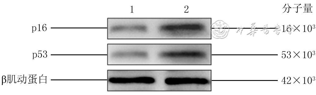
      Objective     To establish a high glucose senescent model of human dermal fibroblasts (HDFs), and to investigate the effects of exosomes derived from human decidua mesenchymal stem cells (dMSCs) on the proliferation, migration, and apoptosis of senescent HDFs and possible mechanism.      Methods     The experimental research method was used. From January to March 2021, discarded foreskin tissue was collected for isolation and culture of primary HDFs from 4 male phimosis patients (aged 18-22 years) admitted for circumcision in the Fourth Medical Center of the PLA General Hospital. The 6th passage of HDFs were taken and divided into low glucose group and high glucose group according to the random number table, and subsequently cultured in low-glucose complete medium and high-glucose complete medium, respectively, with medium changed every 72 h without subculturing. After 10 days of culture, the cells were taken and measured for cellular senescence using the β-galactosidase kit at 24 h after seeding; the expression of senescence-related proteins p16 and p53 was assessed by Western blotting at 48 h after seeding; cell proliferation was detected at 24, 48, and 72 h after seeding using the cell counting kit 8 (CCK-8) method; the cell proliferation was evaluated by 5-ethynyl-2'-deoxyuridine (EdU) staining method, cell cycle and apoptosis were measured by flow cytometry after 48 h of seeding; Transwell experiment was used for the calculation of cell migration rate at 24 h after seeding. The human dMSCs were taken and cultured for 48-72 h from which the exosomes were extracted by differential high speed centrifugal method. The morphology of dMSC exosomes was observed by transmission electron microscopy, the particle size distribution of dMSC exosomes was measured by nanoparticle tracking analysis, and the expression of dMSC-exosomes marker proteins CD9 and tumor susceptibility gene101 (TSG101) were detected by Western blotting. The dMSC exosomes and high-glucose complete medium-induced senescent HDFs were co-cultured for 24 hours, then PKH67 kit was used to detect the uptake of exosomes by HDFs. High-glucose complete medium-induced senescent HDFs were taken and divided into high glucose alone group, high glucose+low concentration of exosomes group, and high glucose+high concentration of exosomes group according to the same method above. The high-glucose complete medium with equal volume of phosphate buffered saline, dMSC exosomes with final concentration of 50 μg/mL, and dMSC exosomes with final concentration of 100 μg/mL were added to the corresponding groups for conventional cell culture, respectively. After grouped, the cell proliferation, cell cycle and apoptosis as well as cell migration were detected by CCK-8 method and EdU staining method, flow cytometry, and Transwell experiment at the corresponding time points as before, respectively. Based on the previous results, high-glucose complete medium-induced senescent HDFs were taken and divided into high glucose alone group and high glucose+high concentration of exosomes group for the same treatment. After being grouped and cultured for 48 h, real-time fluorescent quantitative polymerase chain reaction was used to evaluate the mRNA expression of senescent-related microRNA (miR)-145-5p, miR-498, miR-503-5p, calcium/calmodulin dependent protein kinase 1D (CAMK1D), phosphates and tensin homologue deleted on chromosome ten (PTEN) gene, and Cyclin D1 in high glucose alone group and high glucose+high concentration of exosomes group. Data were statistically analyzed with analysis of variance for factorial design, one-way analysis of variance, least significant difference t test, and independent sample t test.      Results     At 24 h after seeding, the rate of β-galactosidase-positive staining of HDF in high glucose group was (38.4±4.2)%, which was significantly higher than (16.5±2.2)% of low glucose group (t=4.65, P<0.01). At 48 h after seeding, the expression levels of senescence-related proteins p16 and p53 both were significantly higher in HDFs of high glucose group than those in low glucose group (with t values of 11.85 and 3.02, respectively, P<0.05 or P<0.01). At 0, 24, 48, and 72 h after seeding, the cell proliferation viability of HDFs in high glucose group was all significantly lower than in low glucose group (with t values of 4.13, 9.90, and 15.12, respectively, P<0.01). At 48 h after seeding, the rate of EdU-positive staining of HDFs in high glucose group was obviously lower than that of low glucose group (t=3.83, P<0.05). At 48 h after seeding, the percentage of G2/M+S subpopulations in three subpopulations (G0/G1, S, and G2/M) of HDF cycle was significantly lower in high glucose group than that in low glucose group (t=8.74, P<0.01). At 24 h after seeding, the number of HDFs migrated through the filter membrane to the lower chamber was 37±6 in high glucose group, which was significantly less than 74±7 in low glucose group (t=8.42, P<0.01). At 48 h after seeding, the HDF apoptosis rate was significantly higher in high glucose group than in low glucose group (t=8.48, P<0.01). The dMSC exosomes were cup-shaped or round vesicles with well-defined edges and uniform size distribution. The size of dMSC exosomes was basically in the range of 80-200 nm. Exosomal markers including CD9 and TSG101 were positively presented on the dMSC exosomes. After being co-cultured for 24 hours, the dMSC exosomes were taken up intracellularly by HDFs and mainly distributed around the nucleus of HDFs. After being grouped and cultured for 24, 48, and 72 h, the HDF proliferation viabilities in high glucose+low concentration of exosomes group and high glucose+high concentration of exosomes group were both significantly higher than in high glucose alone group (with t values of 6.36, 6.10, 7.76, 8.92, 12.17, and 10.74, respectively, P<0.01), the HDF proliferation viability in high glucose+high concentration of exosomes group was significantly higher than in high glucose+low concentration of exosomes group (with t values of 7.92, 4.82, and 4.72, respectively, P<0.01). After being grouped and cultured for 48 h, the percentages of EdU-positive HDFs in high glucose+low concentration of exosomes group and high glucose+high concentration of exosomes group were both significantly higher than in high glucose alone group (with t values of 5.32 and 9.88, respectively, P<0.01), the percentage of EdU-positive HDFs in high glucose+high concentration of exosomes group was notably higher than in high glucose+low concentration of exosomes group (t=5.27, P<0.01). After being grouped and cultured for 48 h, the proportion of G0/G1 subpopulation in both high glucose+low concentration of exosomes group and high glucose+high concentration of exosomes group was distinctly lower (with t values of 3.81 and 4.31, respectively, P<0.05), while the proportion of G2/M+S subpopulation was markedly higher (with t values of 3.81, 4.31, respectively, P<0.05) than in high glucose alone group. After  being grouped and cultured for 24 h, the number of HDFs migrated through the filter membrane in both high glucose+low concentration of exosomes group and high glucose+high concentration of exosomes group was significantly higher than in high glucose alone group (with t values of 10.14 and 13.39, respectively, P<0.01), the number of HDFs migrated through the filter membrane in high glucose+high concentration of exosomes group was significantly increased than in high glucose+low concentration of exosomes group (t=6.27, P<0.01). After being grouped and cultured for 48 h, the HDF apoptosis rates in high glucose+low concentration of exosomes group and high glucose+high concentration of exosomes group were both significantly lower than in high glucose alone group (with t values of 3.72 and 5.53, respectively, P<0.05 or P<0.01). After being grouped and cultured for 48 h, compared with those in high glucose alone group, the mRNA expression levels of miR-145-5p and miR-498 were both obviously higher (with t values of 13.03 and 8.90, respectively, P<0.01), while the mRNA expression level of miR-503-5p was significantly lower (t=3.85, P<0.05) in high glucose+high concentration of exosomes group. After being grouped and cultured for 48 h, compared with those in high glucose alone group, the mRNA expression levels of CAMK1D and PTEN gene were both significantly lower (with t values of 8.83 and 5.97, respectively, P<0.01), while the mRNA expression level of Cyclin D1 was significantly higher in high glucose+high concentration of exosomes group (t=4.03, P<0.05).      Conclusions     The dMSC exosomes are capable of improving cell proliferation and migration, and inhibiting cell apoptosis of high-glucose senescent HDFs. This may be related to the mechanism by which the increased expressions of intracellular miR-145-5p and miR-498 inhibit the expression of CAMK1D and PTEN gene, and the decreased expression of miR-503-5p promote the expression of Cyclin D1.
Original Article·Nursing Column
Comparative study of three scores in predicting the death risk of severe burn patients
Xie Zhiqin, Guo Guanghua, Yang Zhen, Yi Hanxiao, Wang Shuilian, Tang Xinrong, Liu Deguang, Zeng Yande
2022, 38(2): 184-189. doi: 10.3760/cma.j.cn501120-20201113-00473
Abstract:
  Objective  To explore the predictive values of the modified Baux score, Belgian Outcome in Burn Injury score, and Ryan score on the death risk of severe burn patients.  Methods  A retrospective case series study was conducted. From February 2018 to November 2019, 260 severe burn patients who met the inclusion criteria were admitted to the Department of Burns of the First Affiliated Hospital of Nanchang University, including 158 males and 102 females, aged 36 (3, 53) years. According to the final outcome, the patients were divided into survival group (n=229) and death group (n=31). Data of patients were compared and statistically analyzed with chi-square test or Mann-Whitney U test between the two groups, including the gender, age, cause of burn, site of burn, total burn area, depth of burn, combined inhalation injury, and combined underlying diseases on admission, and the modified Baux score, Belgian Outcome in Burn Injury score, and Ryan score calculated based on part of the aforementioned data. The Kendall tau-b coefficient method was used to analyze the consistency of the above-mentioned three scores in 260 severe burn patients. The receiver operating characteristic (ROC) curves of the above-mentioned three scores predicting the death risk of 260 severe burn patients were drawn, and the area under the curve (AUC), the optimal threshold, and the sensitivity and specificity under the optimal threshold were calculated. The quality of AUC of the above-mentioned three scores was compared by Delong test.  Results  The gender, site of burn, and depth of burn of patients between the two groups were all similar (P>0.05). The age, total burn area, proportion of flame burn, proportion of combined inhalation injury, and proportion of combined underlying diseases of patients in death group were significantly higher than those in survival group (with Z values of 5.53 and 17.78, respectively, χ2 values of 16.23, 15.89, and 17.78, respectively, P<0.01); the modified Baux score, Belgian Outcome in Burn Injury score, and Ryan score of patients in death group were 142 (115, 155), 7 (5, 7), 2 (2, 3), all significantly higher than 64 (27, 87), 1 (0, 3), 0 (0, 1) in survival group (with Z values of 7.91, 7.64, and 7.61, respectively, P<0.01). In 260 severe burn patients, the results between the modified Baux score and Ryan score, modified Baux score and Belgian Outcome in Burn Injury score, Ryan score and Belgian Outcome in Burn Injury score were significantly consistent (with Kendall tau-b coefficients of 0.75, 0.71, and 0.86, respectively, P<0.01). The AUCs of ROC curves of the modified Baux score, Belgian Outcome in Burn Injury score, and Ryan score for predicting the death risk of 260 severe burn patients were 0.92, 0.89, and 0.85, respectively (with 95% confidence intervals of 0.86-0.98, 0.83-0.95, and 0.78-0.93, respectively, P<0.01); the optimal thresholds were 106.5, 4.5, and 1.5 points, respectively; the sensitivity under the optimal threshold were 88.5%, 76.9%, and 73.1%, respectively, and the specificity under the optimal threshold were 88.5%, 87.2%, and 86.3%, respectively. The modified Baux score was similar to Belgian Outcome in Burn Injury score in the AUC quality (z=1.25, P>0.05), which were both significantly better than the AUC quality of Ryan score (with z values of 2.35 and 2.11, respectively, P<0.05).  Conclusions  The modified Baux score, Belgian Outcome in Burn Injury score, and Ryan score have good ability in predicting the death risk of severe burn patients. From the perspective of clinical practice, the modified Baux score is more suitable as a predictive tool for the prognosis of severe burn patients.
Reviews
Current status and research advances on drug sedation and analgesia in burn children
Jia Mengqian, Yuan Xingang
2022, 38(2): 190-195. doi: 10.3760/cma.j.cn501120-20200908-00404
Abstract:
Children are high-risk groups of burns, with unique physiological, psychological, and anatomical states, and the management of anxiety and pain for burn children are extremely challenging. Non-pharmacological interventions are very important for pain management in burn children, but are often inadequate for treating pain and anxiety, so pharmacological sedation and analgesia are necessary. This article reviewed the clinical treatment and research progress in this field in the past 10 years at home and abroad, including the pain assessment of burn children, monitoring in sedative and analgesic treatment, main therapeutic drugs and research progress, and some controversies in clinical practice. Besides, some suggestions have been put forward for clinical reference.
Research advances on signaling pathways affecting sweat gland development and their involvement in the reconstitution of sweat adenoid cells in vitro
Lang Donghao, Ba Te, Cao Shengjun, Li Fang, Dong Hang, Li Junliang, Wang Lingfeng
2022, 38(2): 195-200. doi: 10.3760/cma.j.cn501120-20201020-00442
Abstract:
The damage of sweat glands in patients with extensive deep burns results in the loss of thermoregulation, which seriously affects the quality of life of patients. At present, there are many researches on the repair of sweat gland function, but the mechanism of human sweat gland development has not been fully clarified. More and more studies have shown that the cascaded pathways of Wnt/β-catenin, ecto- dysplasin A/ectodysplasin A receptor/nuclear factor-κB, sonic hedgehog, and forkhead box transcription factor jointly affect the development of sweat glands, and it has been reported that the cascaded signaling pathways can be used to achieve the reconstruction of sweat adenoid cells in vitro. This article reviews the signaling pathways that affect the development of sweat glands and their involvement in the reconstruction of sweat adenoid cells in vitro.