


Effects of advanced platelet-rich fibrin/chitosan thermosensitive hydrogel on full-thickness skin defect wound healing in diabetic rats
-
摘要:
目的 制备改良型富血小板纤维蛋白(A-PRF)/壳聚糖温敏水凝胶(以下简称复合水凝胶)并探讨复合水凝胶对糖尿病大鼠全层皮肤缺损创面愈合的作用。 方法 该研究为实验研究。成功制备具有多孔网状结构及温敏特性的含质量浓度为10、15、20、50、100 g/L A-PRF的复合水凝胶。取6~8周龄雄性SD大鼠,通过腹腔注射链脲佐菌素成功制造糖尿病大鼠模型,并在每只大鼠背部制造4个全层皮肤缺损创面(最终36只大鼠成功造模)。将每只大鼠3个创面分别作为空白组(不进行药物干预)、阳性对照组(滴加重组人粒细胞巨噬细胞刺激因子凝胶)、壳聚糖水凝胶组(滴加壳聚糖水凝胶溶液)。取其中30只大鼠,将每只大鼠剩余的1个创面共30个创面分为10、15、20、50、100 g/L复合水凝胶组,每组6个创面,分别滴加含10、15、20、50、100 g/L A-PRF的复合水凝胶溶液;取剩余6只大鼠,于每只大鼠剩余的1个创面滴加含100 g/L A-PRF的复合水凝胶溶液。伤后14 d,取其中1个创面滴加含100 g/L A-PRF的复合水凝胶溶液的6只大鼠,行苏木精-伊红(HE)染色,观察大鼠心、肝、脾、肺和肾等炎症、出血或坏死情况;伤后10 d,取其中1个创面滴加含15 g/L A-PRF的复合水凝胶溶液的6只大鼠,采用激光血流成像系统观测4组创面血流灌注量(样本数为6)。伤后7、14 d,计算8组创面愈合率。伤后14 d,取8组创面组织,分别行HE、Masson染色,观察新生上皮形成和胶原生成情况;采用免疫组织化学法检测CD31、血管内皮生长因子A(VEGFA)的阳性表达情况并计算阳性面积百分比;采用蛋白质印迹法检测CD31、VEGFA的蛋白表达;采用实时荧光定量反转录PCR法检测CD31、VEGFA的mRNA表达(样本数均为4)。 结果 伤后14 d,6只大鼠心、肝、脾、肺、肾中均未观察到明显的炎症、出血或坏死。伤后10 d,15 g/L复合水凝胶组创面血流灌注量明显多于空白组、阳性对照组、壳聚糖水凝胶组(P值均<0.05)。伤后7、14 d,空白组创面愈合率分别为(26.0±8.9)%、(75.0±1.8)%,均分别明显低于阳性对照组、壳聚糖水凝胶组及10、15、20、50、100 g/L 复合水凝胶组的(45.8±3.2)%、(49.8±3.7)%、(51.2±2.9)%、(68.5±2.4)%、(68.8±1.5)%、(72.7±2.1)%、(75.0±3.7)%及(79.1±1.9)%、(77.2±1.7)%、(82.3±1.3)%、(89.6±1.9)%、(89.8±1.3)%、(87.3±1.1)%、(87.9±1.3)%(P<0.05);阳性对照组、壳聚糖水凝组、10 g/L 复合水凝胶组创面愈合率均明显低于15、20、50、100 g/L 复合水凝胶组(P<0.05)。伤后14 d,15、20、50、100 g/L 复合水凝胶组创面上皮化程度较其他4组创面更高、新生微血管情况更好,胶原数量更多且排列更整齐。伤后14 d,阳性对照组创面CD31、VEGFA及壳聚糖水凝胶组创面VEGFA阳性面积百分比均明显高于空白组(P<0.05),10 g/L 复合水凝胶组创面VEGFA阳性面积百分比明显高于空白组、壳聚糖水凝胶组、阳性对照组(P值均<0.05),15、20、50、100 g/L 复合水凝胶组创面CD31、VEGFA阳性面积百分比均明显高于空白组、阳性对照组、壳聚糖水凝胶组、10 g/L 复合水凝胶组(P<0.05)。伤后14 d,壳聚糖水凝胶组、阳性对照组、10 g/L 复合水凝胶组创面组织中CD31、VEGFA蛋白和mRNA表达均明显高于空白组(P<0.05),10 g/L复合水凝胶组创面组织中VEGFA蛋白表达明显高于阳性对照组(P<0.05)和CD31、VEGFA mRNA表达均明显高于阳性对照组、壳聚糖水凝胶组(P<0.05),15、20、50、100 g/L复合水凝胶组创面组织中CD31、VEGFA蛋白和mRNA表达均明显高于空白组、阳性对照组、壳聚糖水凝胶组、10 g/L 复合水凝胶组(P<0.05),壳聚糖水凝胶组创面组织中CD31、VEGFA的mRNA表达均明显低于阳性对照组(P<0.05)。 结论 复合水凝胶生物安全性高,可改善创面血流灌注,有效促进创面组织中血管和胶原生成,从而促进糖尿病大鼠全层皮肤缺损创面愈合;15 g/L为复合水凝胶中A-PRF的较优使用质量浓度。 -
关键词:
- 生物相容性材料 /
- 水凝胶 /
- 糖尿病 /
- 创面修复 /
- 改良型富血小板纤维蛋白
Abstract:Objective To prepare advanced platelet-rich fibrin (A-PRF)/chitosan thermosensitive hydrogel (hereinafter referred to as composite hydrogel) and explore the effects of composite hydrogel on full-thickness skin defect wound healing in diabetic rats. Methods This study was an experimental study. The composite hydrogel with porous mesh structure and thermosensitive characteristics was successfully prepared, containing A-PRF with mass concentrations of 10, 15, 20, 50, and 100 g/L. Diabetic model was successfully established in male Sprague-Dawley rats aged 6-8 weeks by intraperitoneal injection of streptozotocin, and 4 full-thickness skin defect wounds were established on the back of each rat (finally the model was successfully established in 36 rats). Three wounds of each rat were divided into blank group (no drug intervention), positive control group (dropping recombinant human granulocyte-macrophage stimulating factor gel), and chitosan hydrogel group (dropping chitosan hydrogel solution). Thirty rats were collected, and the remaining one wound of each rat (totally 30 wounds) was divided into 10, 15, 20, 50, and 100 g/L composite hydrogel groups, with 6 wounds in each group, which were dropped with composite hydrogel solution containing 10, 15, 20, 50, and 100 g/L A-PRF, respectively. Taking the remaining six rats, the remaining one wound from each rat was dropped with composite hydrogel solution containing 100 g/L A-PRF. On 14 d after injury, 6 rats with one wound dropped with composite hydrogel containing 100 g/L A-PRF were selected for hematoxylin-eosin (HE) staining to observe the inflammation, hemorrhage, or necrosis of the heart, liver, spleen, lung, and kidney. On 10 d after injury, 6 rats with one wound dropped with composite hydrogel containing 15 g/L A-PRF were selected to observe the blood perfusion of wounds in the four groups (with sample size of 6). On 7 and 14 d after injury, the wound healing rates in the eight groups were calculated. On 14 d after injury, the wound tissue in the eight groups was taken for HE and Masson staining to observe the formation of new epithelium and collagen formation, respectively; the positive expressions of CD31 and vascular endothelial growth factor A (VEGFA) were detected by immunohistochemistry, and the percentages of positive areas were calculated; the protein expressions of CD31 and VEGFA were detected by Western blotting; the mRNA expressions of CD31 and VEGFA were detected by real-time fluorescent quantitative reverse transcription polymerase chain reaction method (with all sample sizes of 4). Results On 14 d after injury, no obvious inflammation, hemorrhage, or necrosis was observed in the heart, liver, spleen, lung, and kidney in the 6 rats. On 10 d after injury, the blood perfusion volume of wound in 15 g/L composite hydrogel group was significantly more than that in blank group, positive control group, and chitosan hydrogel group, respectively (with P values all <0.05). On 7 and 14 d after injury, the wound healing rates of blank group were (26.0±8.9)% and (75.0±1.8)%, which were significantly lower than those of positive control group, chitosan hydrogel group, and 10, 15, 20, 50, and 100 g/L composite hydrogel groups, respectively ((45.8±3.2)%, (49.8±3.7)%, (51.2±2.9)%, (68.5±2.4)%, (68.8±1.5)%, (72.7±2.1)%, (75.0±3.7)% and (79.1±1.9)%, (77.2±1.7)%, (82.3±1.3)%, (89.6±1.9)%, (89.8±1.3)%, (87.3±1.1)%, (87.9±1.3)%), P<0.05; the wound healing rates of positive control group, chitosan hydrogel group, and 10 g/L composite hydrogel group were significantly lower than those of 15, 20, 50, and 100 g/L composite hydrogel groups (P<0.05). On 14 d after injury, the wound epithelialization degrees of 15, 20, 50, and 100 g/L composite hydrogel groups were higher than those of the other 4 groups, the new microvascular situation was better, and the collagen was more abundant and arranged more neatly. On 14 d after injury, the percentages of CD31 and VEGFA positive areas in wounds in positive control group and the percentage of VEGFA positive area in wounds in chitosan hydrogel group were significantly higher than those in blank group (P<0.05), the percentage of VEGFA positive area in wounds in 10 g/L composite hydrogel group was significantly higher than that in blank group, chitosan hydrogel group, and positive control group (with P values all <0.05), and the percentages of CD31 and VEGFA positive areas in wounds in 15, 20, 50, and 100 g/L composite hydrogel groups were significantly higher than those in blank group, positive control group, chitosan hydrogel group, and 10 g/L composite hydrogel group (P<0.05). On 14 d after injury, the protein and mRNA expressions of CD31 and VEGFA in wound tissue in chitosan hydrogel group, positive control group, and 10 g/L composite hydrogel group were significantly higher than those in blank group (P<0.05); the protein expression of VEGFA in wound tissue in 10 g/L composite hydrogel group was significantly higher than that in positive control group (P<0.05), and the mRNA expressions of CD31 and VEGFA in wound tissue in 10 g/L composite hydrogel group were significantly higher than those in positive control group and chitosan hydrogel group (P<0.05); the protein and mRNA expressions of CD31 and VEGFA in wound tissue in 15, 20, 50, and 100 g/L composite hydrogel groups were significantly higher than those in blank group, positive control group, chitosan hydrogel group, and 10 g/L composite hydrogel group (P<0.05); the mRNA expressions of CD31 and VEGFA in wound tissue in chitosan hydrogel group were significantly lower than those in positive control group (P<0.05). Conclusions The composite hydrogel has high biological safety, can improve wound blood perfusion, effectively promote the formation of blood vessels and collagen in wound tissue, thus promoting the wound healing of full-thickness skin defects in diabetic rats. 15 g/L is the optimal mass concentration of A-PRF in composite hydrogel. -
Key words:
- Biocompatible materials /
- Hydrogel /
- Diabetes mellitus /
- Wound repair /
- Advanced platelet-rich fibrin
-
参考文献
(30) [1] HealdAH, StedmanM, DaviesM, et al. Estimating life years lost to diabetes: outcomes from analysis of National Diabetes Audit and Office of National Statistics data[J]. Cardiovasc Endocrinol Metab, 2020,9(4):183-185. DOI: 10.1097/XCE.0000000000000210. [2] RøikjerJ,WerkmanNCC,EjskjaerN,et al.Incidence, hospitalization and mortality and their changes over time in people with a first ever diabetic foot ulcer[J].Diabet Med,2022,39(4):e14725.DOI: 10.1111/dme.14725. [3] ApelqvistJ, BakkerK, van HoutumWH, et al. International consensus and practical guidelines on the management and the prevention of the diabetic foot. International Working Group on the Diabetic Foot[J]. Diabetes Metab Res Rev, 2000,16 Suppl 1:S84-92.DOI: 10.1002/1520-7560(200009/10)16:1+<::aid-dmrr113>3.0.co;2-s. [4] HangaardS,RasmussenA,AlmdalT,et al.Standard complication screening information can be used for risk assessment for first time foot ulcer among patients with type 1 and type 2 diabetes[J].Diabetes Res Clin Pract,2019,151:177-186.DOI: 10.1016/j.diabres.2019.04.021. [5] MaZ,DingJ,WangY,et al.Study of platelet-rich fibrin promoting endothelial cell differentiation and angiogenesis induced by transplantation of adipose-derived stem cells[J].Acta Histochem,2023,125(6):152059.DOI: 10.1016/j.acthis.2023.152059. [6] HeidariM,SadeghifardL,YaghobiR,et al.An investigation of the association between vascular endothelial growth factor +405 G/C polymorphism and acute liver transplant rejection in Iranian liver transplant recipients[J].Exp Clin Transplant,2022,20(6):564-568.DOI: 10.6002/ect.2020.0515. [7] GhanaatiS,BoomsP,OrlowskaA,et al.Advanced platelet-rich fibrin: a new concept for cell-based tissue engineering by means of inflammatory cells[J].J Oral Implantol,2014,40(6):679-689.DOI: 10.1563/aaid-joi-D-14-00138. [8] BrizuelaC,HuangGT,DiogenesA,et al.The four pillars for successful regenerative therapy in endodontics: stem cells, biomaterials, growth factors, and their synergistic interactions[J].Stem Cells Int,2022,2022:1580842.DOI: 10.1155/2022/1580842. [9] SmoczerC,YuthKR,AskarMA,et al.Growth factors released from advanced platelet-rich fibrin in the presence of calcium-based silicate materials and their impact on the viability and migration of stem cells of apical papilla[J].Dent J (Basel),2023,11(9):220.DOI: 10.3390/dj11090220. [10] WarinR,VongchanP,SuriyasathapornW,et al.In vitro assessment of lyophilized advanced platelet-rich fibrin from dogs in promotion of growth factor release and wound healing[J].Vet Sci,2022,9(10):566.DOI: 10.3390/vetsci9100566. [11] MoradiL, WitekL, Vivekanand NayakV, et al. Injectable hydrogel for sustained delivery of progranulin derivative Atsttrin in treating diabetic fracture healing[J]. Biomaterials, 2023,301:122289. DOI: 10.1016/j.biomaterials.2023.122289. [12] HerronC,HastingsCL,Herron-RiceC,et al.A thermoresponsive chitosan/β-glycerophosphate hydrogel for minimally invasive treatment of critical limb ischaemia[J].Polymers (Basel),2021,13(20):3568.DOI: 10.3390/polym13203568. [13] LinCJ,LinHL,YouWC,et al.Composite hydrogels of ultrasound-assisted-digested formic acid-decellularized extracellular matrix and sacchachitin nanofibers incorporated with platelet-rich plasma for diabetic wound treatment[J].J Funct Biomater,2023,14(8):423. DOI: 10.3390/jfb14080423. [14] 顾雅男,徐翔昊,王彦平,等.氧化铈纳米酶-甲基丙烯酸酐化明胶水凝胶在小鼠全层皮肤缺损感染创面修复中的作用[J].中华烧伤与创面修复杂志,2024,40(2):131-140.DOI: 10.3760/cma.j.cn501225-20231120-00201. [15] CaoX,CaiX,ChenR,et al.A thermosensitive chitosan-based hydrogel for sealing and lubricating purposes in dental implant system[J].Clin Implant Dent Relat Res,2019,21(2):324-335.DOI: 10.1111/cid.12738. [16] SunH,SaeediP,KarurangaS,et al.IDF diabetes atlas: global, regional and country-level diabetes prevalence estimates for 2021 and projections for 2045[J].Diabetes Res Clin Pract,2022,183:109119.DOI: 10.1016/j.diabres.2021.109119. [17] 黄玉林,马坤岭.糖尿病肾病遗传调控机制研究进展[J].解放军医学杂志,2023,48(7):856-862.DOI: 10.11855/j.issn.0577-7402.2952.2022.1124. [18] VijayakumarV,SamalSK,MohantyS,et al.Recent advancements in biopolymer and metal nanoparticle-based materials in diabetic wound healing management[J].Int J Biol Macromol,2019,122:137-148.DOI: 10.1016/j.ijbiomac.2018.10.120. [19] SawayaAP, StoneRC, BrooksSR, et al. Deregulated immune cell recruitment orchestrated by FOXM1 impairs human diabetic wound healing[J]. Nat Commun, 2020,11(1):4678. DOI: 10.1038/s41467-020-18276-0. [20] DasariN,JiangA,SkochdopoleA,et al.Updates in diabetic wound healing, inflammation, and scarring[J].Semin Plast Surg,2021,35(3):153-158.DOI: 10.1055/s-0041-1731460. [21] LiuL,HuJ,WangY,et al.The role and research progress of the balance and interaction between regulatory T cells and other immune cells in obesity with insulin resistance[J].Adipocyte,2021,10(1):66-79.DOI: 10.1080/21623945.2021.1876375. [22] GenesiBP,de Melo BarbosaR,SeverinoP,et al.Aloe vera and copaiba oleoresin-loaded chitosan films for wound dressings: microbial permeation, cytotoxicity, and in vivo proof of concept[J].Int J Pharm,2023,634:122648.DOI: 10.1016/j.ijpharm.2023.122648. [23] CraciunAM,Mititelu TartauL,PintealaM,et al.Nitrosalicyl-imine-chitosan hydrogels based drug delivery systems for long term sustained release in local therapy[J].J Colloid Interface Sci,2019,536:196-207.DOI: 10.1016/j.jcis.2018.10.048. [24] LiuS,LiuZ,WuM,et al.NIR as a "trigger switch" for rapid phase change, on-demand release, and photothermal synergistic antibacterial treatment with chitosan-based temperature-sensitive hydrogel[J].Int J Biol Macromol,2021,191:344-358.DOI: 10.1016/j.ijbiomac.2021.09.093. [25] ValachovaK,SvikK,BiroC,et al.Impact of ergothioneine, hercynine, and histidine on oxidative degradation of hyaluronan and wound healing[J].Polymers (Basel),2020,13(1):95.DOI: 10.3390/polym13010095. [26] Dohan EhrenfestDM, RasmussonL, AlbrektssonT.Classification of platelet concentrates: from pure platelet-rich plasma (P-PRP) to leucocyte- and platelet-rich fibrin (L-PRF)[J]. Trends Biotechnol, 2009,27(3):158-167. DOI: 10.1016/j.tibtech.2008.11.009. [27] ClarkD, RajendranY, PaydarS, et al. Advanced platelet-rich fibrin and freeze-dried bone allograft for ridge preservation: a randomized controlled clinical trial[J]. J Periodontol, 2018,89(4):379-387. DOI: 10.1002/JPER.17-0466. [28] MirhajM,SalehiS,TavakoliM,et al.Comparison of physical, mechanical and biological effects of leucocyte-PRF and advanced-PRF on polyacrylamide nanofiber wound dressings: in vitro and in vivo evaluations[J].Biomater Adv,2022,141:213082.DOI: 10.1016/j.bioadv.2022.213082. [29] 张清荣,陈长友,徐娜,等.载P311微球的温敏壳聚糖水凝胶对大鼠全层皮肤缺损创面愈合的影响[J].中华烧伤与创面修复杂志,2022,38(10):914-922.DOI: 10.3760/cma.j.cn501225-20220414-00135. [30] 郑力铭,刘钟元,颜鸿宇,等.小檗碱对糖尿病小鼠全层皮肤缺损创面愈合的影响及其机制[J].中华烧伤与创面修复杂志,2023,39(11):1072-1082.DOI: 10.3760/cma.j.cn501225-20230411-00120. -

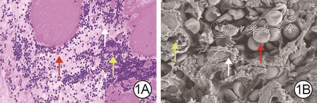
1 A-PRF的形貌特征。1A.A-PRF富含纤维蛋白、白细胞和血小板 苏木精-伊红×40;1B.A-PRF由细小的纤维蛋白组成松散排列的大网状结构,间质中散在分布着正常或变形的白细胞和血小板 扫描电子显微镜×3 000
注:A-PRF为改良型富血小板纤维蛋白;白色箭头指示纤维蛋白,黄色箭头指示白细胞,红色箭头指示血小板

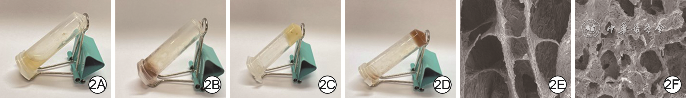
2 壳聚糖水凝胶和含15 g/L A-PRF的复合水凝胶的形态(在不同温度下)及微观形貌。2A、2B.分别为壳聚糖水凝胶及复合水凝胶,在25 ℃时均为液态;2C、2D.分别为壳聚糖水凝胶及复合水凝胶,在37 ℃水浴10 min后均为凝胶状态;2E、2F.分别为凝胶状态下的壳聚糖水凝胶及复合水凝胶的微观形貌,均为多孔网状结构,但复合水凝胶网状结构更加致密、均匀 扫描电子显微镜×3 000
注:复合水凝胶为改良型富血小板纤维蛋白(A-PRF)/壳聚糖温敏水凝胶

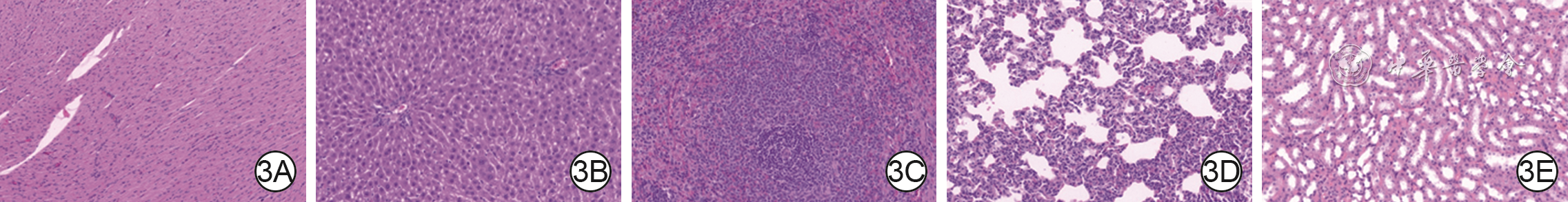
3 其中1个创面滴加含100 g/L A-PRF的复合水凝胶溶液的全层皮肤缺损糖尿病大鼠伤后14 d主要脏器情况 苏木精-伊红×200。3A、3B、3C、3D、3E.分别为心、肝、脾、肺、肾的情况,均未观察到明显的炎症、出血或坏死
注:复合水凝胶为改良型富血小板纤维蛋白(A-PRF)/壳聚糖温敏水凝胶溶液

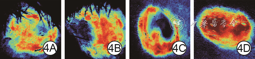
4 4组全层皮肤缺损糖尿病大鼠创面伤后10 d血流灌注情况。4A、4B、4C、4D.分别为空白组、阳性对照组、壳聚糖水凝胶组、15 g/L复合水凝胶组创面血流灌注情况,图4D血流灌注量明显多于图4A、4B、4C
注:空白组创面不进行药物干预,于阳性对照组、壳聚糖水凝胶组创面分别滴加重组人粒细胞巨噬细胞刺激因子凝胶、壳聚糖水凝胶溶液,15 g/L复合水凝胶组创面滴加含15 g/L改良型富血小板纤维蛋白(A-PRF)的A-PRF/壳聚糖温敏水凝胶溶液;黑色表示无血流灌注,绿色表示血流灌注少,红色表示血流灌注充足

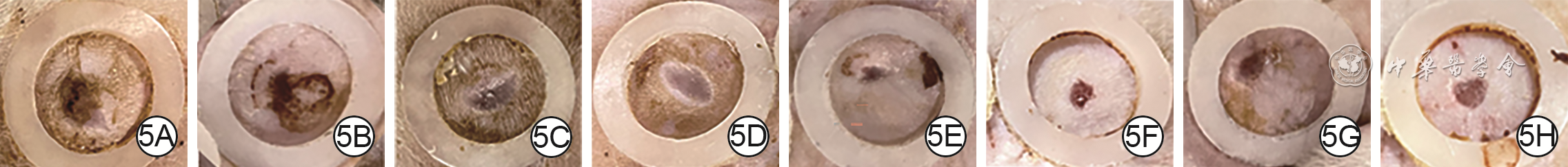
5 8组全层皮肤缺损糖尿病大鼠创面伤后14 d愈合情况。5A、5B、5C、5D、5E、5F、5G、5H.分别为空白组、壳聚糖水凝胶组、阳性对照组及10、15、20、50、100 g/L复合水凝胶组,图5E、5F、5G、5H创面面积均明显小于图5A、5B、5C、5D
注:空白组创面不进行药物干预,于阳性对照组、壳聚糖水凝胶组创面分别滴加重组人粒细胞巨噬细胞刺激因子凝胶、壳聚糖水凝胶溶液,于5个复合水凝胶组创面滴加含相应质量浓度改良型富血小板纤维蛋白(A-PRF)的A-PRF/壳聚糖温敏水凝胶溶液;创面外的白色圆环为防收缩环

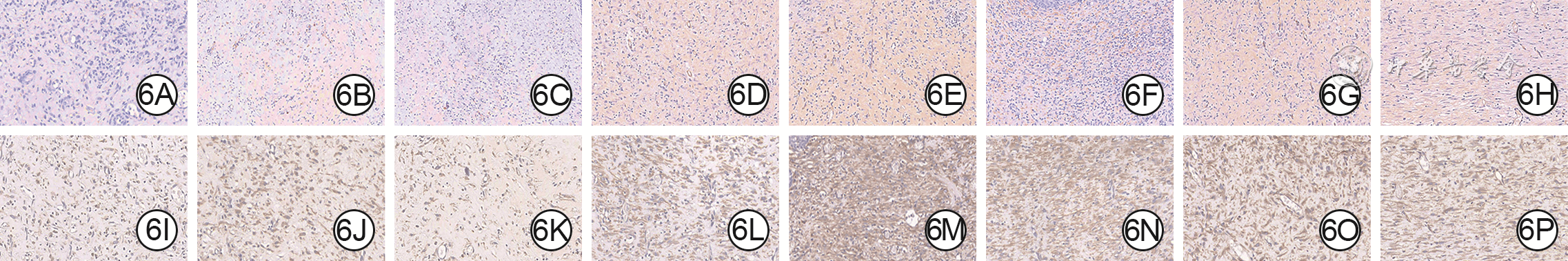
6 8组全层皮肤缺损糖尿病大鼠创面伤后14 d组织中CD31、VEGFA阳性表达情况 二氨基联苯胺-苏木精×400。6A、6B、6C、6D、6E、6F、6G、6H.分别为空白组、阳性对照组、壳聚糖水凝胶组及10、15、20、50、100 g/L复合水凝胶组创面CD31阳性表达情况,图6E、6F、6G、6H CD31阳性表达高于图6A、6B、6C、6D;6I、6J、6K、6L、6M、6N、6O、6P.分别为空白组、阳性对照组、壳聚糖水凝胶组及10、15、20、50、100 g/L 复合水凝胶组创面VEGFA阳性表达情况,图6M、6N、6O、6P VEGFA阳性表达高于图6I、6J、6K、6L
注:CD31、血管内皮生长因子A(VEGFA)阳性表达均为棕色;空白组创面不进行药物干预,于阳性对照组、壳聚糖水凝胶组创面分别滴加重组人粒细胞巨噬细胞刺激因子凝胶、壳聚糖水凝胶溶液,于5个复合水凝胶组创面滴加含相应质量浓度改良型富血小板纤维蛋白(A-PRF)的A-PRF/壳聚糖温敏水凝胶溶液

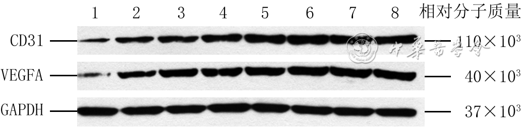
7 蛋白质印迹法检测的8组全层皮肤缺损糖尿病大鼠伤后14 d创面组织中CD31与VEGFA蛋白表达
注:1、2、3、4、5、6、7、8分别为不进行药物干预的空白组、滴加重组人粒细胞巨噬细胞刺激因子凝胶的阳性对照组、滴加壳聚糖水凝胶溶液的壳聚糖水凝胶组、滴加含相应质量浓度改良型富血小板纤维蛋白(A-PRF)的10、15、20、50、100 g/L A-PRF/壳聚糖温敏水凝胶组;VEGFA为血管内皮生长因子A,GAPDH为3-磷酸甘油醛脱氢酶
表1 8组全层皮肤缺损糖尿病大鼠创面伤后各时间点愈合率比较(%,
表1. Comparison of healing rates in 8 groups of wounds in diabetic rats with full-thickness skin defects at each time point after injury
组别 样本数 7 d 14 d 空白组 6 26.0±8.9 75.0±1.8 阳性对照组 6 45.8±3.2a 79.1±1.9a 壳聚糖水凝胶组 6 49.8±3.7a 77.2±1.7a 10 g/L 复合水凝胶组 6 51.2±2.9a 82.3±1.3a 15 g/L 复合水凝胶组 6 68.5±2.4abcd 89.6±1.9abcd 20 g/L复合水凝胶组 6 68.8±1.5abcd 89.8±1.3abcd 50 g/L 复合水凝胶组 6 72.7±2.1abcd 87.3±1.1abcd 100 g/L 复合水凝胶组 6 75.0±3.7abcd 87.9±1.3abcd F值 82.63 66.78 P值 <0.001 <0.001 注:空白组创面不进行药物干预,于阳性对照组、壳聚糖水凝胶组创面分别滴加重组人粒细胞巨噬细胞刺激因子凝胶、壳聚糖水凝胶溶液,于5个复合水凝胶组创面滴加含相应质量浓度改良型富血小板纤维蛋白(A-PRF)的A-PRF/壳聚糖温敏水凝胶溶液;与空白组比较,aP<0.05;与阳性对照组比较,bP<0.05;与壳聚糖水凝胶组比较,cP<0.05;与10 g/L复合水凝胶组比较,dP<0.05  下载: 导出CSV
表2 8组全层皮肤缺损糖尿病大鼠伤后14 d 创面组织中CD31、VEGFA阳性面积百分比比较(%,
表2. Comparison of positive area percentages of CD31 and VEGFA in wound tissue of 8 groups in diabetic rats with full-thickness skin defects on 14 d after injury
组别 样本量 CD31 VEGFA 空白组 6 26.1±1.6 25.2±2.0 阳性对照组 6 31.5±3.4a 32.5±1.2a 壳聚糖水凝胶组 6 28.9±1.9 30.7±1.7a 10 g/L 复合水凝胶组 6 29.2±1.6 38.7±2.1abc 15 g/L 复合水凝胶组 6 39.3±2.4abcd 46.1±2.4abcd 20 g/L复合水凝胶组 6 41.1±2.1abcd 42.8±1.2abcd 50 g/L 复合水凝胶组 6 40.3±1.6abcd 45.3±1.3abcd 100 g/L 复合水凝胶组 6 40.4±1.2abcd 45.8±2.3abcd F值 57.57 108.80 P值 <0.001 <0.001 注:VEGFA为血管内皮生长因子A;空白组创面不进行药物干预,于阳性对照组、壳聚糖水凝胶组创面分别滴加重组人粒细胞巨噬细胞刺激因子凝胶、壳聚糖水凝胶溶液,于5个复合水凝胶组创面滴加含相应质量浓度改良型富血小板纤维蛋白(A-PRF)的A-PRF/壳聚糖温敏水凝胶溶液;与空白组相比,aP<0.05;与阳性对照组相比,bP<0.05;与壳聚糖水凝胶组相比,cP<0.05;与10 g/L 复合水凝胶组相比,dP<0.05
下载: 导出CSV
表3 8组全层皮肤缺损糖尿病大鼠创面伤后14 d组织中CD31和VEGFA蛋白表达比较(
表3. Comparison of the protein expressions of CD31 and VEGFA in wound tissue of 8 groups in diabetic rats with full-thickness skin defects on 14 d after injury
组别 样本数 CD31 VEGFA 空白组 4 0.23±0.05 0.147±0.033 阳性对照组 4 0.53±0.07a 0.379±0.053a 壳聚糖水凝胶组 4 0.62±0.06a 0.480±0.024a 10 g/L 复合水凝胶组 4 0.71±0.05a 0.555±0.031ab 15 g/L 复合水凝胶组 4 0.96±0.05abcd 0.800±0.061abcd 20 g/L复合水凝胶组 4 1.06±0.04abcd 0.852±0.040abcd 50 g/L 复合水凝胶组 4 1.05±0.07abcd 0.866±0.050abcd 100 g/L 复合水凝胶组 4 1.06±0.07abcd 0.830±0.065abcd F值 92.89 126.80 P值 <0.001 <0.001 注:VEGFA为血管内皮生长因子A;空白组创面不进行药物干预,于阳性对照组、壳聚糖水凝胶组创面分别滴加重组人粒细胞巨噬细胞刺激因子凝胶、壳聚糖水凝胶溶液,于5个复合水凝胶组创面滴加含相应质量浓度改良型富血小板纤维蛋白(A-PRF)的A-PRF/壳聚糖温敏水凝胶溶液;与空白组相比,aP<0.05;与阳性对照组相比,bP<0.05;与壳聚糖水凝胶组相比,cP<0.05;与10 g/L 复合水凝胶组相比,dP<0.05
下载: 导出CSV
表4 8组全层皮肤缺损糖尿病大鼠创面伤后14 d组织中CD31和VEGFA mRNA表达比较(
表4. Comparison of the mRNA expressions of CD31 and VEGFA in wound tissue of 8 groups in diabetic rats with full-thickness skin defects on 14 d after injury
组别 样本数 CD31 VEGFA 空白组 4 0.99±0.05 0.993±0.023 阳性对照组 4 2.11±0.09a 1.556±0.012a 壳聚糖水凝胶组 4 1.63±0.05ab 1.429±0.044ab 10 g/L 复合水凝胶组 4 2.34±0.04abc 1.904±0.035abc 15 g/L 复合水凝胶组 4 2.93±0.04abcd 2.446±0.062abcd 20 g/L复合水凝胶组 4 2.93±0.05abcd 2.393±0.048abcd 50 g/L 复合水凝胶组 4 2.96±0.04abcd 2.436±0.039abcd 100 g/L 复合水凝胶组 4 3.01±0.03abcd 2.437±0.017abcd F值 697.00 852.70 P值 <0.001 <0.001 注:VEGFA为血管内皮生长因子A;空白组创面不进行药物干预,于阳性对照组、壳聚糖水凝胶组创面分别滴加重组人粒细胞巨噬细胞刺激因子凝胶、壳聚糖水凝胶溶液,于5个复合水凝胶组创面滴加含相应质量浓度改良型富血小板纤维蛋白(A-PRF)的A-PRF/壳聚糖温敏水凝胶溶液;与空白组相比,aP<0.05;与阳性对照组相比,bP<0.05;与壳聚糖水凝胶组相比,cP<0.05;与10 g/L复合水凝胶组相比,dP<0.05
下载: 导出CSV
-

 下载:
下载:

计量
- 文章访问数: 2346
- HTML全文浏览量: 411
- PDF下载量: 42
- 被引次数: 0
