


Clinical effects of Meek skin grafting combined with platelet-rich plasma in repairing extensive deep burn wounds
-
摘要:
目的 探讨Meek皮片移植联合富血小板血浆(PRP)修复大面积深度烧伤创面的临床效果。 方法 该研究为回顾性观察性研究。2018年8月—2023年8月,空军军医大学第一附属医院收治44例符合入选标准的患者,其中男23例、女21例,年龄22~62岁,烧伤总面积30%~80%体表总面积,四肢或躯干部位为深Ⅱ~Ⅲ度烧伤。根据四肢或躯干深度烧伤部位治疗过程,将患者分为观察组(21例)和对照组(23例),观察组患者接受Meek皮片移植联合PRP同期治疗,对照组患者接受单纯Meek皮片移植。术后10 d,观测患者Meek皮片成活情况。术后14 d,观测患者术区创面愈合率。统计术后创面愈合时间、创面分泌物标本细菌培养阳性率。 结果 术后10 d,观察组患者植皮区较为干燥,Meek皮片与创基贴合紧密、色泽多红润,皮片成活率为(89±4)%;对照组患者植皮区部分Meek皮片脱落,残余创面呈散在不规则地图状,伴有不同程度脓性分泌物,皮片成活率为(79±6)%,明显低于观察组(t=6.72,P<0.05)。术后14 d,观察组中19例患者术区创面完全愈合,另2例患者残余细小创面经换药后延期1周愈合;对照组中12例患者术区创面完全愈合,6例患者术区创面经补充邮票皮移植术后愈合,5例患者术区创面经常规换药后延期愈合,创面愈合率明显低于观察组(P<0.05)。术后,观察组患者术区创面愈合时间为(13.3±1.6)d,明显短于对照组的(16.4±3.5)d,t=3.72,P<0.05;观察组和对照组患者创面分泌物标本细菌培养阳性率比较,差异无统计学意义(P>0.05)。 结论 对于大面积深度烧伤创面,采用Meek皮片移植联合自体PRP较单纯Meek皮片移植治疗,可促进皮片成活,加快皮片扩展融合,缩短创面愈合时间,从而提高治疗效果。 Abstract:Objective To investigate the clinical effects of Meek skin grafting combined with platelet-rich plasma (PRP) in repairing of extensive deep burn wounds. Methods This study was a retrospective observational study. From August 2018 to August 2023, 44 patients who met the inclusion criteria were admitted to the First Affiliated Hospital of Air Force Medical University, including 23 males and 21 females. Their age ranged from 22 to 62 years and the total burn area was 30%-80% total body surface area, the burns on limbs or torso were deep partial-thickness to full-thickness. According to the treatment process of deep burns on the limbs or torso, the patients were divided into observation group (21 cases) and control group (23 cases). patients in observation group were treated with Meek skin grafting combined with PRP at the same time, while patients in control group were treated with Meek skin grafting alone. The survival of Meek skin grafts was observed on the 10th day after operation. The wound healing rate in the operation area of patients was observed on the 14th day after operation. The postoperative wound healing time and positive rate of bacterial culture of wound secretion specimens were recorded. Results On the 10th day after operation, the skin grafting area of patients in observation group were dry, and the skin grafts adhered closely to the wound base with ruddy color, with a survival rate of (89±4)%; whereas in control group, some of Meek skin grafts fell off in the skin graft area, and the residual wounds were scattered in irregular map shape, accompanied by purulent secretions. The survival rate of skin grafts was (79±6)%, which was significantly lower than that in observation group (t=6.72, P<0.05). On the 14th day after operation, 19 patients in observation group had complete wound healing in the operation area, and the other 2 patients had small residual wounds, which healed after 1 week delay through dressing changes; in control group, the wounds in 12 patients healed completely, the wounds in 6 patients healed after supplementary stamp skin grafting, and the wounds in 5 patients healed with delay after routine dressing changes. The wound healing rate was significantly lower than that in observation group (P<0.05). After operation, the wound healing time of the operation area of patients in observation group was (13.3±1.6) days, which was significantly shorter than (16.4±3.5) days in control group (t=3.72, P<0.05); there was no statistically significant difference in the positive rate of bacterial culture of wound secretion specimens between observation group and control group after operation (P>0.05). Conclusions Compared with Meek skin grafting alone, Meek skin grafting combined with autologous PRP can promote the survival of skin grafts, accelerate the expansion and fusion of skin grafts, and shorten the wound healing time, thereby improving the therapeutic effect in the repairing of extensive deep burn wounds. -
Key words:
- Burns /
- Platelet-rich plasma /
- Extensive deep burns /
- Meek skin grafting /
- Wound repair
-
参考文献
(58) [1] QuinteroEC, MachadoJ, RoblesR. Meek micrografting history, indications, technique, physiology and experience: a review article[J]. J Wound Care, 2018,27(Suppl 2):S12-18. DOI: 10.12968/jowc.2018.27.Sup2.S12. [2] RijpmaD, ClaesK, HoeksemaH, et al. The Meek micrograft technique for burns; review on its outcomes: searching for the superior skin grafting technique[J]. Burns, 2022,48(6):1287-1300. DOI: 10.1016/j.burns.2022.05.011. [3] ZuoKJ, MedinaA, TredgetEE. Important developments in burn care[J]. Plast Reconstr Surg, 2017,139(1):120e-138e. DOI: 10.1097/PRS.0000000000002908. [4] 邓雪. 不同浓度自体富血小板血浆治疗慢性难愈性创面的临床效果研究[J]. 中国全科医学, 2021, 24(Suppl 2):S62-64. [5] 胡建武, 任继魁, 孙晶洁, 等. 自体富血小板血浆联合负压封闭引流治疗糖尿病足溃疡的临床观察[J].中华烧伤杂志,2017,33(1):46-48. DOI: 10.3760/cma.j.issn.1009-2587.2017.01.011. [6] KaoYC, LinDZ, LeeSL, et al. Assisted therapy with platelet-rich plasma for burn patients: a meta-analysis and systematic review[J]. Burns, 2021,47(5):1012-1023. DOI: 10.1016/j.burns.2020.11.005. [7] 张鹏, 原莉莉, 罗佳, 等. 严重烧伤患者Meek植皮术后皮片存活情况的影响因素及其预测价值[J].中华烧伤杂志,2021,37(3):243-249. DOI: 10.3760/cma.j.cn501120-20201127-00503. [8] 中华医学会烧伤外科学分会MEEK植皮技术中心协作组, 海军军医大学第一附属医院烧伤外科,全军烧伤研究所. MEEK微型皮片移植技术临床操作规范[J].中华烧伤杂志,2019,35(8):561-564. DOI: 10.3760/cma.j.issn.1009-2587.2019.08.001. [9] KlosováH, StětinskýJ, BryjováI, et al. Objective evaluation of the effect of autologous platelet concentrate on post-operative scarring in deep burns[J]. Burns, 2013,39(6):1263-1276. DOI: 10.1016/j.burns.2013.01.020. [10] 狄海萍, 牛希华, 李强, 等. Meek植皮在不同年龄段大面积深度烧伤患者中的应用效果[J].中华烧伤杂志,2017,33(3):156-159. DOI: 10.3760/cma.j.issn.1009-2587.2017.03.006. [11] 张高飞, 刘文军, 王迪, 等. 微粒皮和Meek微型皮片移植修复大面积深度烧伤创面临床效果的荟萃分析[J].中华烧伤杂志,2020,36(7):560-567. DOI: 10.3760/cma.j.cn501120-20190521-00249. [12] HouschyarKS, TapkingC, NietzschmannI, et al. Five years experience with meek grafting in the management of extensive burns in an adult burn center[J]. Plast Surg (Oakv), 2019,27(1):44-48. DOI: 10.1177/2292550318800331. [13] HuG, ZhangP, ChenY, et al. Efficacy of two-stage Meek micrografting in patients with severe burns[J]. J Burn Care Res, 2022,43(5):1081-1085. DOI: 10.1093/jbcr/irab241. [14] KlosováH, Němečková CrkvenjašZ, ŠtětinskýJ. Meek micrografting technique and its use in the treatment of severe burn injuries at the university hospital Ostrava burn center [J]. Acta Chir Plast, 2017, 59 (1):11-17. PMID: 28869381. [15] LumentaDB, KamolzLP, FreyM. Adult burn patients with more than 60% TBSA involved-Meek and other techniques to overcome restricted skin harvest availability--the Viennese Concept[J]. J Burn Care Res, 2009,30(2):231-242. DOI: 10.1097/BCR.0b013e318198a2d6. [16] LeeSZ, HalimAS. Superior long term functional and scar outcome of Meek micrografting compared to conventional split thickness skin grafting in the management of burns[J]. Burns, 2019,45(6):1386-1400. DOI: 10.1016/j.burns.2019.04.011. [17] MarucciaM, TedeschiP, CorraoC, et al. Meek micro-skin grafting and acellular dermal matrix in pediatric patients: a novel approach to massive extravasation injury[J]. J Clin Med, 2023,12(14)DOI: 10.3390/jcm12144587. [18] 李兴照, 蔡晨, 徐庆连, 等. 重度烧伤患儿Meek微型皮片移植失败的原因及治疗措施[J].中华烧伤杂志,2019,35(7):525-531. DOI: 10.3760/cma.j.issn.1009-2587.2019.07.009. [19] MedinaA, RiegelT, NystadD, et al. Modified Meek micrografting technique for wound coverage in extensive burn injuries[J]. J Burn Care Res, 2016,37(5):305-313. DOI: 10.1097/BCR.0000000000000244. [20] RodeH, MartinezR, PotgieterD, et al. Experience and outcomes of micrografting for major paediatric burns[J]. Burns, 2017,43(5):1103-1110. DOI: 10.1016/j.burns.2017.02.008. [21] ZhangF, LineaweaverW. Acute and sustained effects of vascular endothelial growth factor on survival of flaps and skin grafts[J]. Ann Plast Surg, 2011,66(5):581-582. DOI: 10.1097/SAP.0b013e3182057376. [22] XuP, WuY, ZhouL, et al. Platelet-rich plasma accelerates skin wound healing by promoting re-epithelialization[J/OL]. Burns Trauma, 2020,8:tkaa028[2023-11-24]. https://pubmed.ncbi.nlm.nih.gov/32821743/. DOI: 10.1093/burnst/tkaa028. [23] QariS, BaderM, FarranE, et al. Combined synergetic effect of lipoconcentrate fat grafting, nanofat transfer, platelet-rich plasma, microneedling, and CO2 fractional laser for plastic regenerative and esthetic surgery and cosmetic care[J]. Cureus, 2023,15(8):e44035. DOI: 10.7759/cureus.44035. [24] GuillibertC, CharpinC, RaffrayM, et al. Single injection of high volume of autologous pure PRP provides a significant improvement in knee osteoarthritis: a prospective routine care study [J]. Int J Mol Sci, 2019, 20(6):1327-1335.DOI: 10.3390/ijms20061327. [25] DaiZ, LouX, ShenT, et al. Combination of ablative fractional carbon dioxide laser and platelet-rich plasma treatment to improve hypertrophic scars: a retrospective clinical observational study[J/OL]. Burns Trauma, 2021,9:tkab016[2023-11-24]. https://pubmed.ncbi.nlm.nih.gov/34337088/. DOI: 10.1093/burnst/tkab016. [26] TianJ, ChengLH, CuiX, et al. Application of standardized platelet-rich plasma in elderly patients with complex wounds[J]. Wound Repair Regen, 2019,27(3):268-276. DOI: 10.1111/wrr.12702. [27] RuizA, CuestasD, GarcıaP, et al. Early intervention in scar management and cutaneous burns with autologous platelet-rich plasma[J]. J Cosmet Dermatol, 2018,17(6):1194-1199. DOI: 10.1111/jocd.12554. [28] MarckRE, GardienK, VligM, et al. Growth factor quantification of platelet-rich plasma in burn patients compared to matched healthy volunteers[J]. Int J Mol Sci, 2019,20(2):288. DOI: 10.3390/ijms20020288. [29] LeeCH, LeeCY, YouHL, et al. The growth factor content as an indicator of platelet counts in platelet-rich plasma[J]. Clin Chim Acta, 2025,564:119901. DOI: 10.1016/j.cca.2024.119901. [30] GentileP, CalabreseC, De AngelisB, et al. Impact of the different preparation methods to obtain autologous non-activated platelet-rich plasma (A-PRP) and activated platelet-rich plasma (AA-PRP) in plastic surgery: wound healing and hair regrowth evaluation [J]. Int J Mol Sci, 2020, 21(2):431-439. DOI: 10.3390/ijms21020431. [31] ImamMS, AlotaibiA, AlotaibiN, et al. Efficiency of platelet-rich plasma in the management of burn wounds: a meta-analysis[J]. Int Wound J, 2023,21(2):e14419. DOI: 10.1111/iwj.14419. [32] LiG, LiuS, TanG, et al. Clinical observation of ultraviolet therapy combined with autologous platelet-rich plasma in the treatment of chronic refractory wounds[J]. Int Wound J, 2024,21(4):e14746. DOI: 10.1111/iwj.14746. [33] 杨思思, 肖承志. 自体富血小板血浆对烧伤创面治疗影响的研究进展[J].中华烧伤杂志,2018,34(12):910-913. DOI: 10.3760/cma.j.issn.1009-2587.2018.12.017. [34] El-TaiebMA, IbrahimHM, HegazyEM, et al. Fractional erbium-YAG laser and platelet-rich plasma as single or combined treatment for atrophic acne scars: a randomized clinical trial[J]. Dermatol Ther (Heidelb), 2019,9(4):707-717. DOI: 10.1007/s13555-019-00318-1. [35] YamaguchiR, TerashimaH, YoneyamaS, et al. Effects of platelet-rich plasma on intestinal anastomotic healing in rats: PRP concentration is a key factor[J]. J Surg Res, 2012,173(2):258-266. DOI: 10.1016/j.jss.2010.10.001. [36] NiuWZ, WangPL, GeSH, et al. Effects of platelet concentrates used in alveolar ridge preservation: a systematic review [J]. Implant Dent, 2018, 27(4):498-506. DOI: 10.1097/ID.0000000000000797. [37] KimJI, BaeHC, ParkHJ, et al. Effect of storage conditions and activation on growth factor concentration in platelet-rich plasma[J]. J Orthop Res, 2020,38(4):777-784. DOI: 10.1002/jor.24520. [38] ZhangX, YaoD, ZhaoWY, et al. Engineering platelet-rich plasma based dual-network hydrogel as a bioactive wound dressing with potential clinical translational value[J]. Adv Funct Mater, 2021, 31(8):2009258. DOI: 10.1002/adfm.202009258. [39] ZhaoM, WangJ, ZhangJ, et al. Functionalizing multi-component bioink with platelet-rich plasma for customized in-situ bilayer bioprinting for wound healing[J]. Mater Today Bio, 2022,16:100334. DOI: 10.1016/j.mtbio.2022.100334. [40] WeiS, XuP, YaoZ, et al. A composite hydrogel with co-delivery of antimicrobial peptides and platelet-rich plasma to enhance healing of infected wounds in diabetes[J]. Acta Biomater, 2021,124:205-218. DOI: 10.1016/j.actbio.2021.01.046. [41] SambergM, StoneR, NatesanS, et al. Platelet rich plasma hydrogels promote in vitro and in vivo angiogenic potential of adipose-derived stem cells[J]. Acta Biomater, 2019,87:76-87. DOI: 10.1016/j.actbio.2019.01.039. [42] MarckRE, GardienKL, StekelenburgCM, et al. The application of platelet-rich plasma in the treatment of deep dermal burns: a randomized, double-blind, intra-patient controlled study[J]. Wound Repair Regen, 2016,24(4):712-720. DOI: 10.1111/wrr.12443. [43] BenchaprathanphornK, SakulaueP, SiriwatwechakulW, et al. Expansion of fibroblast cell sheets using a modified MEEK micrografting technique for wound healing applications[J]. Sci Rep, 2022,12(1):18541. DOI: 10.1038/s41598-022-21913-x. [44] 郑健生, 刘胜利, 彭晓菁, 等. 自体富血小板血浆联合Meek微型皮片修复严重烧伤患者四肢创面的效果及其机制的前瞻性研究[J].中华烧伤杂志,2021,37(8):731-737. DOI: 10.3760/cma.j.cn501120-20200427-00241. [45] GuptaS, GoilP, ThakuraniS. Autologous platelet rich plasma as a preparative for resurfacing burn wounds with split thickness skin grafts[J]. World J Plast Surg, 2020,9(1):29-32. DOI: 10.29252/wjps.9.1.29. [46] ChenJ, WanY, LinY, et al. The application of platelet-rich plasma for skin graft enrichment: a meta-analysis[J]. Int Wound J, 2020,17(6):1650-1658. DOI: 10.1111/iwj.13445. [47] LvY, YangZ, ChenZ, et al. Artificial dermis and autologous platelet-rich plasma for treatment of refractory wounds: a clinical study[J]. Int J Low Extrem Wounds, 2024,23(2):275-282. DOI: 10.1177/15347346211050710. [48] Waiker VP, ShivalingappaS. Comparison between conventional mechanical fixation and use of autologous platelet rich plasma (PRP) in wound beds prior to resurfacing with split thickness skin graft[J]. World J Plast Surg, 2015,4(1):50-59. [49] TyagiA, GuptaA, Martires IiiVI, et al. Efficacy of platelet-rich plasma in reduction of post-operative split-thickness skin graft loss and hematoma formation: a meta-analysis[J]. Cureus, 2021,13(5):e15160. DOI: 10.7759/cureus.15160. [50] ShaoS, PanR, ChenY. Autologous platelet-rich plasma for diabetic foot ulcer[J]. Trends Endocrinol Metab, 2020,31(12):885-890. DOI: 10.1016/j.tem.2020.10.003. [51] DengJ, YangM, ZhangX, et al. Efficacy and safety of autologous platelet-rich plasma for diabetic foot ulcer healing: a systematic review and meta-analysis of randomized controlled trials[J]. J Orthop Surg Res, 2023,18(1):370. DOI: 10.1186/s13018-023-03854-x. [52] ZhengW, ZhaoDL, ZhaoYQ, et al. Effectiveness of platelet rich plasma in burn wound healing: a systematic review and meta-analysis[J]. J Dermatolog Treat, 2022,33(1):131-137. DOI: 10.1080/09546634.2020.1729949. [53] García-SánchezJM, Mirabet LisV, Ruiz-VallsA, et al. Platelet rich plasma and plasma rich in growth factors for split-thickness skin graft donor site treatment in the burn patient setting: a randomized clinical trial[J]. Burns, 2022,48(7):1662-1670. DOI: 10.1016/j.burns.2021.10.001. [54] FangZ, YangX, WuG, et al. The use of autologous platelet-rich plasma gel increases wound healing and reduces scar development in split-thickness skin graft donor sites[J]. J Plast Surg Hand Surg, 2019,53(6):356-360. DOI: 10.1080/2000656X.2019.1635489. [55] ZhuangJ, ChenY, ZhengX, et al. The application of blood products in plastic surgery: a systematic review[J]. Plast Reconstr Surg Glob Open, 2024,12(7):e6005. DOI: 10.1097/GOX.0000000000006005. [56] AlserOH, GoutosI. The evidence behind the use of platelet-rich plasma (PRP) in scar management: a literature review[J]. Scars Burn Heal, 2018,4:2059513118808773. DOI: 10.1177/2059513118808773. [57] YinYL, SunH, ZhangYX, et al. More effective strategies for wound treatment using platelet rich plasma and plasma rich in growth factors[J]. Burns, 2023,49(3):738-739. DOI: 10.1016/j.burns.2022.11.018. [58] 刘鲁冰, 文辉才, 黄进军, 等. 富血小板血液制品联合生物材料在创面修复中的应用研究进展[J].中华烧伤杂志,2021,37(4):395-400. DOI: 10.3760/cma.j.cn501120-20200531-00291. -

图 1 Meek皮片移植联合富血小板血浆(PRP)治疗例1患者右下肢大面积深度火焰烧伤创面的效果。1A.对右下肢削痂后即刻;1B.对右下肢喷涂PRP后即刻;1C.右下肢行Meek皮片移植后即刻;1D.术后5 d换药时,可见右下肢术区双绉纱干燥,无明显血肿及分泌物;1E.术后10 d换药时,可见右下肢的Meek皮片基本融合;1F.术后14 d,可见右下肢创面基本愈合
注:因对患者左、右腿创面行分期手术修复,简便起见仅对右腿情况进行描述

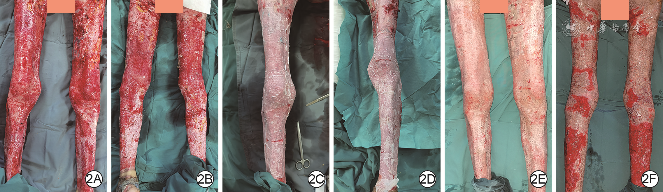
图 2 采用单纯Meek皮片移植治疗例2患者双下肢大面积深度火焰烧伤创面的效果。2A、2B.分别为双下肢创面削痂后即刻的正面观、背面观;2C、2D.分别为左、右下肢创面行Meek微型皮片移植后即刻;2E、2F.分别为术后18 d双下肢残余创面的正面观、背面观,正面的微型皮片大部分融合,背面的微型皮片部分融合
Table 1. 2组大面积深度烧伤创面患者的一般资料比较
组别 例数 性别(例) 年龄(岁, 烧伤总面积(%TBSA, 术前血红蛋白(g/L, 术前白蛋白(g/L, 女 男 观察组 21 10 11 42±10 52±13 102.0±2.7 33.2±2.0 对照组 23 11 12 41±11 50±12 102.6±4.6 33.0±1.8 t值 — 0.26 0.34 0.53 0.35 P值 >0.999 0.398 0.367 0.299 0.364 注:针对四肢或躯干深度烧伤部位,观察组患者接受Meek皮片移植联合富血小板血浆同期治疗,对照组患者接受单纯Meek皮片移植;TBSA为体表总面积;“—”表示无此项  下载: 导出CSV
Table 2. 2组大面积深度烧伤创面患者各主要观察指标的比较
组别 例数 术后10 d皮片成活率(%, 术后14 d创面完全愈合例数 创面愈合时间(d, 创面分泌物标本细菌培养阳性例数 观察组 21 89±4 19 13.3±1.6 4 对照组 23 79±6 12 16.4±3.5 5 t值 6.72 — 3.72 — P值 <0.001 0.008 0.006 0.318 注:针对四肢或躯干深度烧伤部位,观察组患者接受Meek皮片移植联合富血小板血浆同期治疗,对照组患者接受单纯Meek皮片移植;“—”表示无此项
下载: 导出CSV
-
 张婷.mp4

-

 下载:
下载:


图(3) / 表(2)
计量
- 文章访问数: 2448
- HTML全文浏览量: 1124
- PDF下载量: 55
- 被引次数: 0
