


Role and mechanism of Vγ4 T cell depletion in epidermal tissue repair after ultraviolet damage to mouse skin
-
摘要:
目的 探究清除Vγ4T细胞在小鼠皮肤紫外线损伤后表皮组织修复中的作用及其机制。 方法 该研究为实验研究。将54只6~8周龄雌性C57BL/6J野生型小鼠按照随机数字表法分为Vγ4T细胞清除组和对照组(每组27只),分别腹腔注射亚美尼亚仓鼠抗小鼠Vγ4T细胞受体(TCR)单克隆抗体200 µg、等量同型对照IgG抗体。注射后1周(之后均在此时间点取小鼠),每组取3只小鼠(之后均从这2组另取小鼠),从背部皮肤组织和腋窝、腹股沟淋巴结中分别提取真皮细胞和淋巴结细胞,行流式细胞术检测真皮细胞和淋巴结细胞中Vγ4T细胞比例;每组取5只小鼠,观察背部皮肤情况,之后行苏木精-伊红(HE)染色观察皮肤组织结构并测量表皮组织厚度;每组取5只小鼠,提取表皮细胞后行流式细胞术检测表皮细胞中树突状表皮T细胞(DETC)比例。每组取3只小鼠,分别设为Vγ4T细胞清除+5次紫外线辐射(UVR)组和对照+5次UVR组,每日1次共行5次UVR,每日照射后即刻观察背部皮肤情况;每组取5只小鼠,分别设为Vγ4T细胞清除+1次UVR组和对照+1次UVR组,均行1次UVR后即刻行HE染色后测量表皮组织厚度。每组取3只小鼠,分别设为单纯Vγ4T细胞清除组、单纯对照组;再另取3只小鼠,分别设为Vγ4T细胞清除+1次UVR组和对照+1次UVR组;均同前处理后,采用实时荧光定量反转录PCR法检测表皮组织中胰岛素样生长因子Ⅰ(IGF-Ⅰ)、角质形成细胞生长因子(KGF)、Vγ5TCR、白细胞介素15(IL-15)、IL-1β、IL-23、自然杀伤细胞2族成员D(NKG2D)、组织相容性抗原60(H60)、小鼠UL16结合蛋白样转录子1(Mult1)、维甲酸早期诱导蛋白1(Rae1)的mRNA表达。 结果 注射后1周,Vγ4T细胞清除组小鼠真皮细胞、淋巴结细胞中Vγ4T细胞比例均明显低于对照组(t值分别为27.99、13.12,P<0.05);Vγ4T细胞清除组和对照组小鼠皮肤大体情况和组织结构无明显区别,表皮组织厚度相近(P>0.05);Vγ4T细胞清除组小鼠表皮细胞中DETC比例为(3.9±0.8)%,明显高于对照组的(1.6±0.5)%(t=4.84,P<0.05)。与对照+5次UVR组相比,Vγ4T细胞清除+5次UVR组小鼠进行1次UVR后皮肤鳞屑增多,照射2次出现鳞屑样痂皮,照射3~5次鳞屑样痂皮明显增多。行UVR后即刻,Vγ4T细胞清除+1次UVR组小鼠表皮组织厚度较对照+1次UVR组明显增加(t=11.50,P<0.05)。与单纯对照组相比,单纯Vγ4T细胞清除组小鼠表皮组织中Vγ5TCR的mRNA表达明显上调(t=41.16,P<0.05),IL-23的mRNA表达明显下调(t=6.52,P<0.05);与单纯对照组相比,对照+1次UVR组小鼠表皮组织中Vγ5TCR、KGF的mRNA表达均明显上调(t值分别为15.22、13.22,P<0.05),IGF-Ⅰ、IL-23的mRNA表达均明显下调(t值分别为3.71、4.95,P<0.05);与单纯Vγ4T细胞清除组相比,Vγ4T细胞清除+1次UVR组小鼠表皮组织中IGF-Ⅰ、KGF的mRNA表达均明显上调(t值分别为11.40、18.88,P<0.05),IL-1β的mRNA表达明显下调(t=4.42,P<0.05);与对照+1次UVR组相比,Vγ4T细胞清除+1次UVR组小鼠表皮组织中Vγ5TCR、IGF-Ⅰ、KGF的mRNA表达均明显上调(t值分别为4.52、15.24、9.43,P<0.05);4组小鼠表皮组织中IL-15的mRNA表达总体相近(P>0.05)。与单纯对照组相比,单纯Vγ4T细胞清除组表皮组织中NKG2D、Rae1的mRNA表达均明显上调(t值分别为3.67、47.40,P<0.05),对照+1次UVR组小鼠表皮组织中NKG2D、Mult1、Rae1的mRNA表达均明显上调(t值分别为5.30、6.50、9.16,P<0.05);与单纯Vγ4T细胞清除组相比,Vγ4T细胞清除+1次UVR组小鼠表皮组织中NKG2D、H60、Mult1、Rae1的mRNA表达均明显下调(t值分别为4.57、4.13、4.67、27.36,P<0.05);与对照+1次UVR组相比,Vγ4T细胞清除+1次UVR组小鼠表皮组织中NKG2D、H60、Mult1、Rae1的mRNA表达均明显下调(t值分别为5.77、8.18、12.90、8.08,P<0.05)。 结论 清除Vγ4T细胞有利于DETC增殖和毒性下调,可能促进UVR后小鼠表皮损伤修复 。 Abstract:Objective To explore the role and mechanism of Vγ4 T cell depletion in epidermal tissue repair after ultraviolet damage to mouse skin. Methods The study was an experimental study. Fifty-four female C57BL/6J wild-type mice aged 6 to 8 weeks were divided into Vγ4 T cell depletion group and control group (27 mice in each group) according to the random number table, and the Armenian hamster anti-mouse Vγ4 T cell receptor (TCR) monoclonal antibody of 200 µg and an equal amount of homologous control IgG antibody were intraperitoneally injected, respectively. At one week after injection (the same time point to harvest mice below), dermal cells and lymph node cells were respectively extracted from the back skin tissue, armpit and inguinal lymph nodes of 3 mice in each group (mice in following study were all taken from these 2 groups), and the proportions of Vγ4 T cells in dermal cells and lymph node cells were detected by flow cytometry. Five mice from each group were harvested for observation of skin on the back and skin tissue structure was observed and the epidermal tissue thickness was measured after hematoxylin-eosin (HE) staining. Five mice from each group were harvested for detection of proportion of dendritic epidermal T cells (DETCs) in epidermal cells by flow cytometry after extracted. Three mice were taken from each group and recruited in Vγ4 T cell depletion+5 times ultraviolet irradiation (UVR) group and control+5 times UVR group, respectively, then UVR was administered once per day for 5 times, and the condition of skin on the back was observed immediately after daily irradiation. Five mice were taken from each group and divided into Vγ4 T cell depletion+1 UVR group and control+1 UVR group, respectively. Immediately after one UVR treatment, the epidermal tissue thickness was measured after HE staining. Three mice from each group were selected and recruited in Vγ4 T cell depletion alone group and control alone group, then 3 mice from each group rwere recruited in Vγ4 T cell depletion+1 time UVR group and control+1 time UVR group, respectively, and were treated as before. The mRNA expressions of insulin-like growth factor-Ⅰ (IGF-Ⅰ), keratinocyte growth factor (KGF), Vγ5 TCR, interleukin-15 (IL-15), IL-1β, IL-23, natural killer group 2 member D (NKG2D), histocompatibility antigen 60 (H60), mouse UL16-binding protein-like transcript 1 (Mult1), and retinoic acid early inducible protein 1 (Rae1) in the epidermal tissue were detected by real-time fluorescent quantitative reverse transcription polymerase chain reaction. Results At one week after injection, the proportions of Vγ4 T cells in dermal cells and lymph node cells of mice in Vγ4 T cell depletion group were significantly lower than those in control group (with t values of 27.99 and 13.12, respectively, P<0.05); there were no statistically significant differences in the skin general condition and tissue structure of mice between Vγ4 T cell depletion group and control group; the epidermal tissue thickness of mice between Vγ4 T cell depletion group and control group was similar (P>0.05); the proportion of DETCs in epidermal cells of mice in Vγ4 T cell depletion group was (3.9±0.8)%, which was significantly higher than (1.6±0.5)% in control group (t=4.84, P<0.05). Compared with that in control+5 times UVR group, the skin scale increased after one UVR treatment, scaly scab appeared after 2 times of irradiation, and scaly scab increased significantly after 3 to 5 times of irradiation in Vγ4 T cell depletion+5 times UVR group. Immediately after UVR treatment, the epidermal tissue thickness of mice in Vγ4 T cell depletion+1 time UVR group was significantly increased compared with that in control+1 time UVR group (t=11.50, P<0.05). Compared with those in control alone group, the mRNA expression of Vγ5 TCR in the epidermal tissue of mice in Vγ4 T cell depletion alone group was up-regulated (t=41.16, P<0.05), while the mRNA expression of IL-23 was down-regulated (t=6.52, P<0.05); compared with those in control alone group, the mRNA expressions of Vγ5 TCR and KGF in the epidermal tissue of mice in control+1 time UVR group were significantly up-regulated (with t values of 15.22 and 13.22, respectively, P<0.05), while the mRNA expressions of IGF-Ⅰ and IL-23 were significantly down-regulated (with t values of 3.71 and 4.95, respectively, P<0.05); compared with those in Vγ4 T cell depletion alone group, the mRNA expressions of IGF-Ⅰ and KGF in the epidermal tissue of mice in Vγ4 T cell depletion+1 time UVR group were significantly up-regulated (with t values of 11.40 and 18.88, respectively, P<0.05), while the mRNA expression of IL-1β was significantly down-regulated (t=4.42, P<0.05); compared with those in control+1 time UVR group, the mRNA expressions of Vγ5 TCR, IGF-Ⅰ, and KGF in the epidermal tissue of mice in Vγ4 T cell depletion+1 time UVR group were significantly up-regulated (with t values of 4.52, 15.24, and 9.43, respectively, P<0.05); the mRNA expression of IL-15 in the epidermal tissue of mice in these 4 groups was generally similar (P>0.05). Compared with those in control alone group, the mRNA expressions of NKG2D and Rae1 in the epidermal tissue of mice in Vγ4 T cell depletion alone group were significantly up-regulated (with t values of 3.67 and 47.40, respectively, P<0.05), the mRNA expressions of NKG2D, Mult1, and Rae1 in the epidermal tissue of mice in control+1 time UVR group were significantly up-regulated (with t values of 5.30, 6.50, and 9.16, respectively, P<0.05); compared with those in Vγ4 T cell depletion alone group, the mRNA expressions of NKG2D, H60, Mult1, and Rae1 in the epidermal tissue of mice in Vγ4 T cell depletion+1 time UVR group were significantly down-regulated (with t values of 4.57, 4.13, 4.67, and 27.36, respectively, P<0.05); compared with those in control group+1 time UVR group, the mRNA expressions of NKG2D, H60, Mult1, and Rae1 in the epidermal tissue of mice in Vγ4 T cell depletion+1 time UVR group were significantly down-regulated (with t values of 5.77, 8.18, 12.90, and 8.08, respectively, P<0.05). Conclusions The clearance of Vγ4 T cells is conducive to the proliferation and down-regulation of cytotoxicity of DETCs, and may promote the repair of mouse epidermal damage after UVR. -
参考文献
(31) [1] WittlichM, WesterhausenS, StrehlB, et al. The GENESIS-UV study on ultraviolet radiation exposure levels in 250 occupations to foster epidemiological and legislative efforts to combat nonmelanoma skin cancer[J]. Br J Dermatol, 2023,188(3):350-360. DOI: 10.1093/bjd/ljac093. [2] Castejón-GriñánM, CerdidoS, Sánchez-BeltránJ, et al. Melanoma-associated melanocortin 1 receptor variants confer redox signaling-dependent protection against oxidative DNA damage[J]. Redox Biol, 2024,72: 103135. DOI: 10.1016/j.redox.2024.103135. [3] SlominskiRM,ChenJY,RamanC,et al.Photo-neuro-immuno-endocrinology: how the ultraviolet radiation regulates the body, brain, and immune system[J].Proc Natl Acad Sci U S A,2024,121(14):e2308374121.DOI: 10.1073/pnas.2308374121. [4] FrascoliM, FerrajE, MiuB, et al. Skin γδ T cell inflammatory responses are hardwired in the thymus by oxysterol sensing via GPR183 and calibrated by dietary cholesterol[J]. Immunity, 2023,56(3):562-575.e6. DOI: 10.1016/j.immuni.2023.01.025. [5] WeiYX,SunGY,YangY,et al.Double-negative T cells ameliorate psoriasis by selectively inhibiting IL-17A-producing γδlow T cells[J].J Transl Med,2024,22(1):328.DOI: 10.1186/s12967-024-05132-8. [6] PeslierH,ReichartJ,BoursotC,et al.Extensive cutaneous-mucosal and muscular involvement of gamma/delta cutaneous T-cell lymphoma on 18F-FDG PET/CT[J].Clin Nucl Med,2024,49(5):e206-e207.DOI: 10.1097/RLU.0000000000005135. [7] YangYL, ZhouC, ChenQ, et al. YAP1/Piezo1 involve in the dynamic changes of lymphatic vessels in UVR-induced photoaging progress to squamous cell carcinoma[J]. J Transl Med, 2023,21(1):820. DOI: 10.1186/s12967-023-04458-z. [8] HarmonC, ZaborowskiA, MooreH, et al. γδ T cell dichotomy with opposing cytotoxic and wound healing functions in human solid tumors[J]. Nat Cancer, 2023,4(8):1122-1137. DOI: 10.1038/s43018-023-00589-w. [9] PetrovićJ,SilvaJR,BannermanCA,et al.γδ T cells modulate myeloid cell recruitment but not pain during peripheral inflammation[J].Front Immunol,2019,10:473.DOI: 10.3389/fimmu.2019.00473. [10] SonomotoK,SongR,ErikssonD,et al.High-fat-diet‐associated intestinal microbiota exacerbates psoriasis-like inflammation by enhancing systemic γδ T cell IL-17 production[J].Cell Rep,2023,42(7):112713.DOI: 10.1016/j.celrep.2023.112713. [11] LiuM, LiuZH, ChenYX, et al. Dendritic epidermal T cells secreting exosomes promote the proliferation of epidermal stem cells to enhance wound re-epithelialization[J]. Stem Cell Res Ther, 2022,13(1):121. DOI: 10.1186/s13287-022-02783-6. [12] ChenC, MengZY, RenH, et al. The molecular mechanisms supporting the homeostasis and activation of dendritic epidermal T cell and its role in promoting wound healing[J/OL]. Burns Trauma, 2021,9:tkab009[2024-01-21]. https://pubmed.ncbi.nlm.nih.gov/34212060/.DOI: 10.1093/burnst/tkab009. [13] ThelenF, WitherdenDA. Get in touch with dendritic epithelial T cells![J]. Front Immunol, 2020,11:1656. DOI: 10.3389/fimmu.2020.01656. [14] HeiligJS, TonegawaS. Diversity of murine gamma genes and expression in fetal and adult T lymphocytes[J]. Nature, 1986,322(6082):836-840. DOI: 10.1038/322836a0. [15] LiYS, WuJ, LuoGX, et al. Functions of Vγ4 T cells and dendritic epidermal T cells on skin wound healing[J]. Front Immunol, 2018,9:1099. DOI: 10.3389/fimmu.2018.01099. [16] 王珏, 张小容, 贺伟峰, 等. 树突状表皮T细胞在创面愈合中作用机制的研究进展[J].中华烧伤杂志,2021,37(3):296-300. DOI: 10.3760/cma.j.cn501120-20200226-00092. [17] LiYS, WangYP, ZhouLN, et al. Vγ4 T cells inhibit the pro-healing functions of dendritic epidermal T cells to delay skin wound closure through IL-17A[J]. Front Immunol, 2018,9:240. DOI: 10.3389/fimmu.2018.00240. [18] YangBW, LinYM, HuangYB, et al. Extracellular vesicles modulate key signalling pathways in refractory wound healing[J/OL]. Burns Trauma, 2023,11:tkad039[2024-01-21].https://pubmed.ncbi.nlm.nih.gov/38026441/.DOI: 10.1093/burnst/tkad039. [19] LiuZY,LiangGP,GuiL,et al.Weakened IL-15 production and impaired mTOR Activation alter dendritic epidermal T cell homeostasis in diabetic mice[J].Sci Rep,2017,7(1):6028.DOI: 10.1038/s41598-017-05950-5. [20] Dhillon-LaBrooyA, BrabandKL, TantawyE, et al. Inhibition of mitochondrial translation ameliorates imiquimod-induced psoriasis-like skin inflammation by targeting Vγ4+ γδ T cells[J]. J Invest Dermatol, 2024,144(4):844-854.e2. DOI: 10.1016/j.jid.2023.09.275. [21] LiYS, WangJ, WangYP, et al. IL-1β/NF-κB signaling inhibits IGF-1 production via let-7f-5p in dendritic epidermal T cells[J]. J Leukoc Biol, 2022,112(6):1677-1690. DOI: 10.1002/JLB.3MA0322-171R. [22] LiYS, HuangZG, YanRS, et al. Vγ4 γδ T cells provide an early source of IL-17A and accelerate skin graft rejection[J]. J Invest Dermatol, 2017,137(12):2513-2522. DOI: 10.1016/j.jid.2017.03.043. [23] WeiXR, LiMX, ZhengZJ, et al. Senescence in chronic wounds and potential targeted therapies[J/OL]. Burns Trauma, 2022,10:tkab045[2024-01-21]. https://pubmed.ncbi.nlm.nih.gov/35187179/.DOI: 10.1093/burnst/tkab045. [24] NitaharaA, ShimuraH, ItoA, et al. NKG2D ligation without T cell receptor engagement triggers both cytotoxicity and cytokine production in dendritic epidermal T cells[J]. J Invest Dermatol, 2006,126(5):1052-1058. DOI: 10.1038/sj.jid.5700112. [25] CunninghamTJ,TabacchiM,ElianeJP,et al.Randomized trial of calcipotriol combined with 5-fluorouracil for skin cancer precursor immunotherapy[J].J Clin Invest,2017,127(1):106-116.DOI: 10.1172/JCI89820. [26] PeterleL,SanfilippoS,BorgiaF,et al.Alopecia areata: a review of the role of oxidative stress, possible biomarkers, and potential novel therapeutic approaches[J].Antioxidants (Basel),2023,12(1):135.DOI: 10.3390/antiox12010135. [27] XiangJ, QiuMH, ZhangHY. Role of dendritic epidermal T cells in cutaneous carcinoma[J]. Front Immunol, 2020,11:1266. DOI: 10.3389/fimmu.2020.01266. [28] IbusukiA,KawaiK,YoshidaS,et al.NKG2D triggers cytotoxicity in murine epidermal γδ T cells via PI3K-dependent, Syk/ZAP70-independent signaling pathway[J].J Invest Dermatol,2014,134(2):396-404.DOI: 10.1038/jid.2013.353. [29] WangYP, BaiY, LiYS, et al. IL-15 enhances activation and IGF-1 production of dendritic epidermal T cells to promote wound healing in diabetic mice[J]. Front Immunol, 2017,8:1557. DOI: 10.3389/fimmu.2017.01557. [30] Muñoz-RuizM, LlorianM, D'AntuonoR, et al. IFN-γ-dependent interactions between tissue-intrinsic γδ T cells and tissue-infiltrating CD8 T cells limit allergic contact dermatitis[J]. J Allergy Clin Immunol, 2023,152(6):1520-1540. DOI: 10.1016/j.jaci.2023.07.015. [31] NielsenMM, Dyring-AndersenB, SchmidtJD, et al. NKG2D-dependent activation of dendritic epidermal T cells in contact hypersensitivity[J]. J Invest Dermatol, 2015,135(5):1311-1319. DOI: 10.1038/jid.2015.23. -

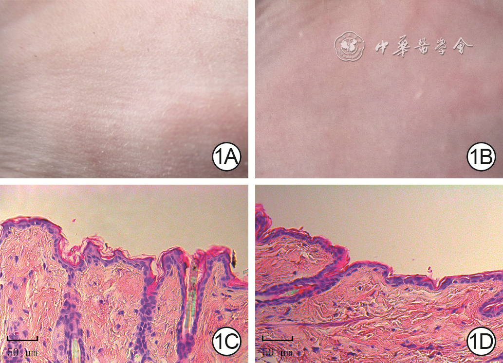
1 对照组和Vγ4T细胞清除组小鼠注射后1周皮肤大体和组织病理学情况。1A、1B.分别为对照组和Vγ4T细胞清除组皮肤大体情况,两者无明显区别;1C、1D.分别为对照组和Vγ4T细胞清除组皮肤组织病理学情况,两者表皮组织厚度、组织结构无明显区别 苏木精-伊红×40
注:对Vγ4T细胞清除组、对照组小鼠分别予以腹腔内注射亚美尼亚仓鼠抗小鼠Vγ4T细胞受体单克隆抗体、同型对照IgG抗体

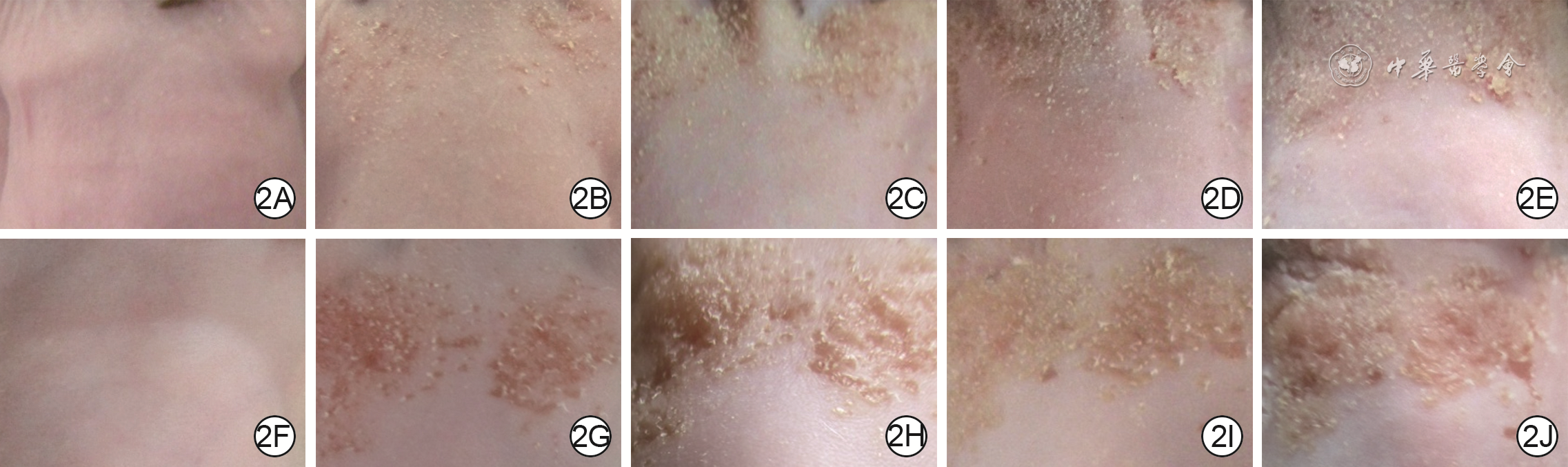
2 对照+5次UVR组和Vγ4T细胞清除+5次UVR组小鼠背部皮肤大体情况。2A、2B、2C、2D、2E.分别为对照+5次UVR组1、2、3、4、5次行UVR后即刻的皮肤大体情况,皮肤鳞屑逐渐增多;2F、2G、2H、2I、2J.分别为Vγ4T细胞清除+5次UVR组行1、2、3、4、5次UVR后即刻的皮肤大体情况,皮肤鳞屑分别较图2A、2B、2C、2D、2E明显增多
注:对Vγ4T细胞清除+5次紫外线辐射(UVR)组、对照+5次UVR组小鼠分别予以腹腔内注射亚美尼亚仓鼠抗小鼠Vγ4T细胞受体单克隆抗体、同型对照IgG抗体1周后,均另行5次UVR

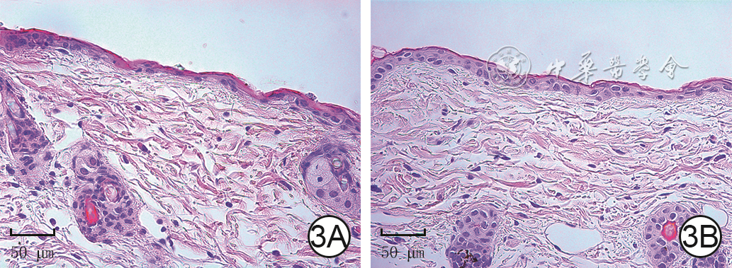
3 对照+1次UVR组和Vγ4T细胞清除+1次UVR组小鼠行UVR后即刻皮肤组织病理学情况 苏木精-伊红×40。3A、3B.分别为对照+1次UVR组和Vγ4T细胞清除+1次UVR组皮肤组织病理学情况,图3B皮肤表皮组织厚度较图3A明显增加
注:对Vγ4T细胞清除+1次紫外线辐射(UVR)组、对照+1次UVR组小鼠分别予以腹腔内注射亚美尼亚仓鼠抗小鼠Vγ4T细胞受体单克隆抗体、同型对照IgG抗体1周后,均另行1次UVR
表1 4组小鼠表皮组织中Vγ5TCR和5种细胞因子的mRNA表达比较(
表1. Comparison of the mRNA expressions of Vγ5 TCR and five cytokines in the epidermal tissue of mice in four groups
组别 样本数 Vγ5TCR IGF-Ⅰ KGF IL-15 IL-1β IL-23 单纯对照组 3 1.01±0.16 1.00±0.05 1.01±0.11 1.01±0.15 1.02±0.03 1.00±0.09 单纯Vγ4T细胞清除组 3 63.09±2.13 1.14±0.61 1.00±0.06 1.01±0.26 1.49±0.26 0.41±0.09 对照+1次UVR组 3 49.89±4.54 0.64±0.13 3.52±0.24 0.82±0.17 0.89±0.15 0.68±0.03 Vγ4T细胞清除+1次UVR组 3 66.10±2.27 9.03±0.77 6.81±0.43 0.83±0.13 0.68±0.03 0.57±0.07 t1值 41.16 0.33 0.04 — 0.31 6.52 P1值 <0.001 0.761 0.969 — 0.772 0.003 t2值 15.22 3.71 13.22 — 1.18 4.95 P2值 <0.001 0.021 <0.001 — 0.304 0.008 t3值 1.37 11.40 18.88 — 4.42 1.91 P3值 0.242 <0.001 <0.001 — 0.012 0.129 t4值 4.52 15.24 9.43 — 1.86 1.89 P4值 0.011 <0.001 <0.001 — 0.137 0.131 注:Vγ5TCR为Vγ5T细胞受体,UVR为紫外线辐射,IGF-Ⅰ为胰岛素样生长因子Ⅰ,KGF为角质形成细胞生长因子,IL为白细胞介素;对单纯Vγ4T细胞清除组、单纯对照组小鼠分别予以腹腔内注射亚美尼亚仓鼠抗小鼠Vγ4TCR单克隆抗体、同型对照IgG抗体,并于注射后1周进行检测,对对照+1次UVR组和Vγ4T细胞清除+1次UVR组小鼠分别同单纯对照组、单纯Vγ4T细胞清除组注射后1周均另行1次UVR,然后进行检测;Vγ5TCR、IGF-Ⅰ、KGF、IL-15、IL-1β、IL-23的mRNA表达的Vγ4T细胞清除主效应,F值分别为153.80、59.45、457.60、1.56、16.74、1.89,P值分别为<0.001、0.002、<0.001、0.280、0.015、0.241;UVR主效应,F值分别为479.40、2 892.00、98.50、<0.01、2.10、60.42,P值分别为<0.001、<0.001、<0.001、0.989、0.221、0.002;两者交互作用,F值分别为120.10、71.49、72.04、<0.01、9.50、16.15,P值分别为<0.001、0.001、0.001、0.976、0.037、0.016;t1值、P1值为单纯对照组与单纯Vγ4T细胞清除组比较所得;t2值、P2值为单纯对照组与对照+1次UVR组比较所得;t3值、P3值为单纯Vγ4T细胞清除组与Vγ4T细胞清除+1次UVR组比较所得,t4值、P4值为对照+1次UVR组与Vγ4T细胞清除+1次UVR组比较所得;“—”表示无此统计量值  下载: 导出CSV
表2 4组小鼠表皮组织中NKG2D及其3个配体的mRNA表达比较(
表2. Comparison of the mRNA expressions of NKG2D and the three ligands in the epidermal tissue of mice in 4 groups
组别 样本数 NKG2D H60 Mult1 Rae1 单纯对照组 3 1.03±0.25 1.04±0.29 1.05±0.32 1.02±0.18 单纯Vγ4T细胞清除组 3 1.72±0.10 0.83±0.20 1.76±0.42 19.83±0.53 对照+1次UVR组 3 2.56±0.33 1.24±0.17 2.94±0.26 57.92±8.78 Vγ4T细胞清除+1次UVR组 3 1.00±0.19 0.22±0.06 0.33±0.11 7.71±0.33 t1值 3.67 0.84 1.92 47.40 P1值 0.022 0.447 0.127 <0.001 t2值 5.30 0.87 6.50 9.16 P2值 0.006 0.435 0.003 <0.001 t3值 4.57 4.13 4.67 27.36 P3值 0.010 0.015 0.010 <0.001 t4值 5.77 8.18 12.90 8.08 P4值 0.005 0.001 <0.001 0.001 注:NKG2D为自然杀伤细胞2族成员D,UVR为紫外线辐射,H60为组织相容性抗原60,Mult1为小鼠UL16结合蛋白样转录子1,Rae1为维甲酸早期诱导蛋白1;对单纯Vγ4T细胞清除组、单纯对照组小鼠分别予以腹腔内注射亚美尼亚仓鼠抗小鼠Vγ4TCR单克隆抗体、同型对照IgG抗体,并于注射后1周进行检测,对对照+1次UVR组和Vγ4T细胞清除+1次UVR组小鼠分别同单纯对照组、单纯Vγ4T细胞清除组注射后1周均另行1次UVR,然后进行检测;NKG2D、H60、Mult1、Rae1的mRNA表达的Vγ4T细胞清除主效应,F值分别为7.75、2.49、0.95、50.65,P值分别为0.050、0.190、0.386、0.002;UVR主效应,F值分别为5.77、17.17、28.82、25.95,P值分别为0.074、0.014、0.006、0.007;两者交互作用,F值分别为58.08、10.04、47.51、120.30,P值分别为0.002、0.034、0.002、<0.001;t1值、P1值为单纯对照组与单纯Vγ4T细胞清除组比较所得;t2值、P2值为单纯对照组与对照+1次UVR组比较所得;t3值、P3值为单纯Vγ4T细胞清除组与Vγ4T细胞清除+1次UVR组比较所得,t4值、P4值为对照+1次UVR组与Vγ4T细胞清除+1次UVR组比较所得
下载: 导出CSV
-

 下载:
下载:


计量
- 文章访问数: 5543
- HTML全文浏览量: 669
- PDF下载量: 18
- 被引次数: 0
