


Effects and mechanism of electroacupuncture stimulation on the survival of multi-territory perforator flaps in rats
-
摘要:
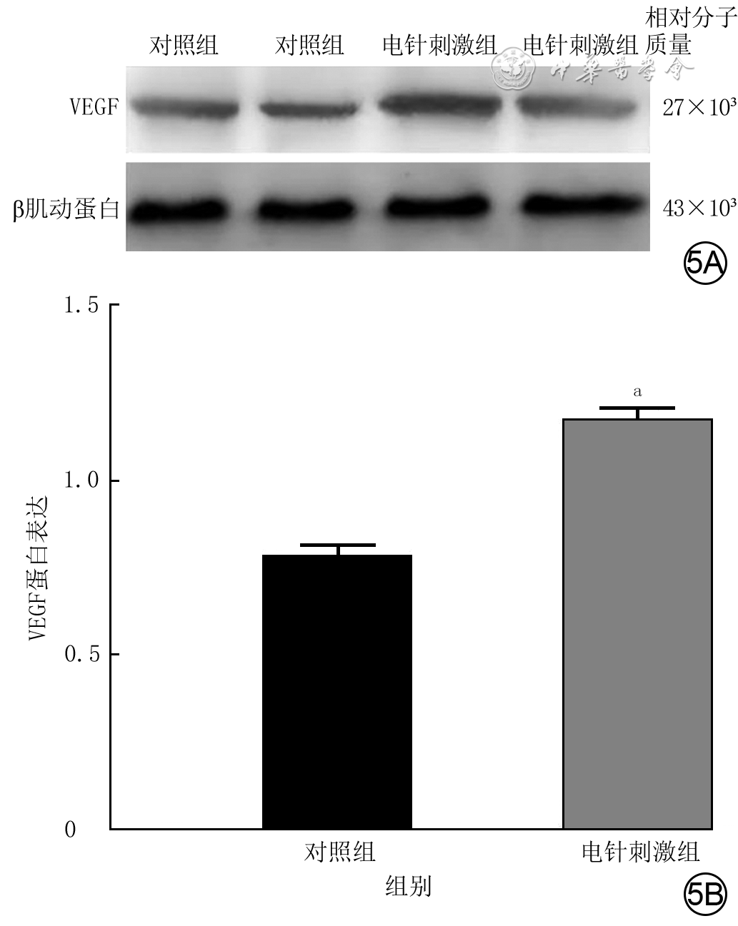
目的 探讨电针刺激对大鼠跨区穿支皮瓣成活的影响及其机制。 方法 该研究为实验研究。取30只8~10周龄雄性SD大鼠,采用随机数字表法将其分为电针刺激组和对照组,每组15只。采用多普勒血流探测仪探查2组大鼠背部旋髂深动脉、肋间后动脉、胸背动脉位置,设计并切取结扎肋间后动脉、胸背动脉后以旋髂深动脉为蒂的跨区穿支皮瓣,并将皮瓣原位回植。术前,对电针刺激组大鼠皮瓣胸背动脉与肋间后动脉所在血管体之间交界部位包含闭塞血管的区域(即闭塞区域Ⅱ)皮肤行连续7 d、每天1 h的电针刺激,对照组大鼠皮瓣不接受电针刺激。术后7 d,观察2组所有大鼠皮瓣成活情况并计算皮瓣成活率;取闭塞区域Ⅱ皮肤组织,行苏木精-伊红染色后观察微血管新生情况并计算微血管密度(样本数为3),行免疫组织化学染色观察血管内皮生长因子(VEGF)的表达与分布,采用蛋白质印迹法检测VEGF的蛋白表达(样本数为3)。 结果 术后7 d,对照组大鼠皮瓣部分发黑、坏死,电针刺激组大鼠皮瓣成活、几乎无坏死;电针刺激组大鼠皮瓣成活率为(92.1±2.1)%,显著高于对照组的(85.2±1.2)%(t=-10.95,P<0.05)。术后7 d,与对照组比较,电针刺激组大鼠皮瓣闭塞区域Ⅱ皮肤组织中新生微血管明显增多;电针刺激组大鼠皮瓣闭塞区域Ⅱ皮肤组织中微血管密度为(21.4±3.0)条/mm2,显著高于对照组的(11.5±3.7)条/mm2(t=-7.34,P<0.05)。术后7 d,与对照组比较,电针刺激组大鼠皮瓣闭塞区域Ⅱ皮肤组织中血管区VEGF的表达明显增多,VEGF蛋白表达显著增高(t=12.56,P<0.05)。 结论 电针刺激可提高大鼠跨区穿支皮瓣闭塞区域Ⅱ中VEGF的表达,通过促进该区域中血管发生形态学改变而改善皮瓣远端血供,从而提高皮瓣成活率。 Abstract:Objective To explore the effects and mechanism of electroacupuncture stimulation on the survival of multi-territory perforator flaps in rats. Methods This study was an experimental study. Thirty male Sprague-Dawley rats aged 8-10 weeks were collected and divided into electroacupuncture stimulation group and control group according to the random number table method, with 15 rats in each group. Doppler blood flow detectors were used to explore the positions of the dorsal deep circumflex artery, posterior intercostal artery, and thoracodorsal artery in the two groups of rats, and a multi-territory perforator flap was designed and resected with the dorsal deep circumflex artery as the pedicle after ligation of the posterior intercostal artery and thoracodorsal artery, and the flap was replanted in situ. Before the operation, the skin in the area containing choke vessels at the junction between the angiosomes of the thoracodorsal artery and posterior intercostal artery (i.e. the choke zone Ⅱ) in the flaps of rats in electroacupuncture stimulation group was subjected to electroacupuncture stimulation for 1 hour per day for 7 consecutive days, while the flaps of rats in control group received no electroacupuncture stimulation. Seven days after the operation, the survival status of the flaps of all rats in the two groups was observed and the flap survival rate was calculated; the skin tissue from the choke zone Ⅱ was collected and stained with hematoxylin-eosin to observe the microvascular neogenesis and calculate the microvessel density (with sample number of 3). Immunohistochemical staining was performed to observe the expression and distribution of vascular endothelial growth factor (VEGF), and Western blotting was used to detect the protein expression of VEGF (with sample number of 3). Results Seven days after the operation, the flaps of rats in control group were partially blackened and necrotic, while those in electroacupuncture stimulation group survived with almost no necrosis. The flap survival rate of rats in electroacupuncture stimulation group was (92.1±2.1)%, which was significantly higher than (85.2±1.2)% in control group (t=-10.95, P<0.05). Seven days after the operation, compared with those in control group, the number of new microvessels in the skin tissue in the choke zone Ⅱ of the flaps in rats of electroacupuncture stimulation group increased significantly. The microvessel density in the skin tissue in the choke zone Ⅱ of the flaps in rats of electroacupuncture stimulation group was (21.4±3.0) vessels/mm², which was significantly higher than (11.5±3.7) vessels/mm² in control group (t=-7.34, P<0.05). Seven days after the operation, compared with those in control group, the expression of VEGF in the vascular area of the skin tissue in the choke zone Ⅱ of the flaps in rats of electroacupuncture stimulation group was significantly increased, and the protein expression of VEGF was significantly increased (t=12.56, P<0.05). Conclusions Electroacupuncture stimulation can increase the expression of VEGF in choke zone Ⅱ of the multi-territory perforator flaps in rats, improve the blood supply at the distal end of flaps through promoting morphological changes of blood vessels in this zone, thus increasing the survival rates of flaps. -
参考文献
(40) [1] 杨亮,周荣,巨积辉,等.游离双侧股前外侧内增压型串联皮瓣修复足踝部大面积创面的临床疗效[J].中华烧伤与创面修复杂志,2025,41(1):61-69.DOI: 10.3760/cma.j.cn501225-20240508-00164. [2] WangY,ZhangX,ZhouM,et al.Irisin pre-treatment promotes multi-territory perforator flap survival in rats: an experimental study[J].Injury,2020,51(11):2442-2448.DOI: 10.1016/j.injury.2020.07.058. [3] 孙向东,李成志,悟拉木·艾力,等.股前外侧穿支皮瓣移植修复头面颈部皮肤恶性肿瘤根治性切除后创面的临床效果[J].中华烧伤与创面修复杂志,2025,41(1):28-35.DOI: 10.3760/cma.j.cn501225-20240920-00341. [4] KouniavskiE,EgoziD,WolfY.Techniques and innovations in flap engineering: a review[J].Plast Reconstr Surg Glob Open,2022,10(9):e4523.DOI: 10.1097/GOX.0000000000004523. [5] 邝依敏,黄昕,蒙旭昌,等.瞬时受体电位香草酸亚型4特异性激活剂对人血管内皮细胞功能和大鼠穿支皮瓣血供的影响及其机制[J].中华烧伤与创面修复杂志,2022,38(5):434-446.DOI: 10.3760/cma.j.cn501120-20210419-00138. [6] WagnerT,HummelinkS,UlrichD.Past, present and future in plastic flap surgery: from surgeon to bioengineer driven progress. A personal view[J].J Tissue Viability,2022,31(4):800-803.DOI: 10.1016/j.jtv.2022.06.007. [7] AfroogheA,DamavandiAR,AhmadiE,et al.The current state of knowledge on how to improve skin flap survival: a review[J].J Plast Reconstr Aesthet Surg,2023,82:48-57.DOI: 10.1016/j.bjps.2023.04.021. [8] LuoZ,WuP,QingL,et al.The hemodynamic and molecular mechanism study on the choke vessels in the multi-territory perforator flap transforming into true anastomosis[J].Gene,2019,687:99-108.DOI: 10.1016/j.gene.2018.11.019. [9] 许浩娜,谢子悦,曲芳汶,等.提高皮瓣成活率预处理方法的研究进展[J].中国美容整形外科杂志,2024,35(4):247-250,后插3.DOI: 10.3969/j.issn.1673-7040.2024.04.015. [10] CormackGC,LambertyBG.A classification of fascio-cutaneous flaps according to their patterns of vascularisation[J].Br J Plast Surg,1984,37(1):80-87.DOI: 10.1016/0007-1226(84)90049-3. [11] TaylorGI,CorlettRJ,DharSC,et al.The anatomical (angiosome) and clinical territories of cutaneous perforating arteries: development of the concept and designing safe flaps[J].Plast Reconstr Surg,2011,127(4):1447-1459.DOI: 10.1097/PRS.0b013e318208d21b. [12] 赵海福,方柏荣. choke血管血流动力学及血管重构的研究进展[J]. 中华整形外科杂志,2022,38(1):109-114. DOI: 10.3760/cma.j.cn114453-20200214-00043. [13] XiS,ZhaoJ,KangR,et al.The effects of angiosome morphology on choke vessels and flap necrosis in a rat multiterritory perforator flap[J].Ann Plast Surg,2023,91(4):479-484.DOI: 10.1097/SAP.0000000000003643. [14] 霍磊,谭琪,高加智,等.穿支皮瓣中Choke vessels的相关研究与展望[J].中国矫形外科杂志,2024,32(14):1309-1313.DOI: 10.20184/j.cnki.Issn1005-8478.11016A. [15] WuZJ,IbrahimMM,SergesketterAR,et al.The influence of topical vasodilator-induced pharmacologic delay on cutaneous flap viability and vascular remodeling[J].Plast Reconstr Surg,2022,149(3):629-637.DOI: 10.1097/PRS.0000000000008829. [16] ChenC,LiX,HuY,et al.Electrical stimulation promoting the angiogenesis in diabetic rat perforator flap through attenuating oxidative stress-mediated inflammation and apoptosis[J].PeerJ,2024,12:e16856.DOI: 10.7717/peerj.16856. [17] MiyamotoS,MinabeT,HariiK.Effect of recipient arterial blood inflow on free flap survival area[J].Plast Reconstr Surg,2008,121(2):505-513.DOI: 10.1097/01.prs.0000299185.32881.55. [18] 林丁盛,郑鑫,李志杰,等.水蛭素对大鼠随意型皮瓣存活的影响[J].中华整形外科杂志,2011,27(1):35-39.DOI: 10.3760/cma.j.issn.1009-4598.2011.01.010. [19] ZhuY,YooH,MinS,et al.Efficacy of superficial inferior epigastric vein superdrainage in free TRAM and DIEP flap: an indocyanine green angiography study of 68 cases[J].J Plast Reconstr Aesthet Surg,2023,84:385-391.DOI: 10.1016/j.bjps.2023.06.006. [20] WangX,PanJ,XiaoD,et al.Comparison of arterial supercharging and venous superdrainage on improvement of survival of the extended perforator flap in rats[J].Microsurgery,2020,40(8):874-880.DOI: 10.1002/micr.30660. [21] BrownCA,GhareebPA.Early experience with external negative pressure delay in free anterolateral thigh perforator flap reconstruction[J].Plast Reconstr Surg Glob Open,2021,9(6):e3606.DOI: 10.1097/GOX.0000000000003606. [22] FangF,LiuM,XiaoJ,et al.Arterial supercharging is more beneficial to flap survival due to quadruple dilation of venules[J].J Surg Res,2020,247:490-498.DOI: 10.1016/j.jss.2019.09.056. [23] JiJ,ChenD,NiJ,et al.Research advances in vascular remodeling in choke vessels of perforator flap: a systematic review[J].Ann Plast Surg,2024,93(2):268-275.DOI: 10.1097/SAP.0000000000003980. [24] TaylorGI,CorlettRJ,AshtonMW.The functional angiosome: clinical implications of the anatomical concept[J].Plast Reconstr Surg,2017,140(4):721-733.DOI: 10.1097/PRS.0000000000003694. [25] SergesketterAR,CasonRW,IbrahimMM,et al.Perioperative treatment with a prolyl hydroxylase inhibitor reduces necrosis in a rat ischemic skin flap model[J].Plast Reconstr Surg,2019,143(4):769e-779e.DOI: 10.1097/PRS.0000000000005441. [26] TakakuM,TomitaS,KurobeH,et al.Systemic preconditioning by a prolyl hydroxylase inhibitor promotes prevention of skin flap necrosis via HIF-1-induced bone marrow-derived cells[J].PLoS One,2012,7(8):e42964.DOI: 10.1371/journal.pone.0042964. [27] TaoX,PanX,RuiY,et al.Effects of pharmacological delay with roxadustat on multi-territory perforator flap survival in rats[J].Open Med (Wars),2023,18(1):20230762.DOI: 10.1515/med-2023-0762. [28] BlackCK,KothaVS,FanKL,et al.Pedicled and free tissue transfers[J].Clin Podiatr Med Surg,2019,36(3):441-455.DOI: 10.1016/j.cpm.2019.03.002. [29] YildizK,MirapogluSL,KilicU,et al.Effectiveness of different surgical flap delay methods and their systemic toxicities[J].J Craniofac Surg,2021,32(5):1946-1950.DOI: 10.1097/SCS.0000000000007444. [30] HamiltonK,WolfswinkelEM,WeathersWM,et al.The delay phenomenon: a compilation of knowledge across specialties[J].Craniomaxillofac Trauma Reconstr,2014,7(2):112-118.DOI: 10.1055/s-0034-1371355. [31] MachadoAF,LiebanoRE,FurtadoF,et al.Effect of high- and low-frequency transcutaneous electrical nerve stimulation on angiogenesis and myofibroblast proliferation in acute excisional wounds in rat skin[J].Adv Skin Wound Care,2016,29(8):357-363.DOI: 10.1097/01.ASW.0000488721.83423.f3. [32] DoğanF,Özyazganİ.Flap preconditioning by electrical stimulation as an alternative to surgical delay: experimental study[J].Ann Plast Surg,2015,75(5):560-564.DOI: 10.1097/SAP.0000000000000162. [33] ErdemM,TiftikciogluY,TatarBE,et al.The effect of botulinum toxin on flap viability of the posterior thigh perforator flap in rats[J].J Surg Res,2021,261:85-94.DOI: 10.1016/j.jss.2020.12.025. [34] ChenM,LiX,JiangZ,et al. Visualizing the pharmacologic preconditioning effect of botulinum toxin type A by infrared thermography in a rat pedicled perforator island flap model[J].Plast Reconstr Surg,2019,144(6):1016e-1024e.DOI: 10.1097/PRS.0000000000006251. [35] MehtaS,RolphR,CorneliusV,et al.Local heat preconditioning in skin sparing mastectomy: a pilot study[J].J Plast Reconstr Aesthet Surg,2013,66(12):1676-1682.DOI: 10.1016/j.bjps.2013.07.034. [36] KlebanS,BaynosaRC.The effect of hyperbaric oxygen on compromised grafts and flaps[J].Undersea Hyperb Med,2020,47(4):635-648.DOI: 10.22462/10.12.2020.13. [37] DoganR,Metin GulerE,KocyigitA,et al.Comparison of the efficacy of multiple antioxidant and hyperbaric oxygen treatments in the prevention of ischemia and necrosis of local random McFarlane skin flap[J].J Tissue Viability,2021,30(2):196-206.DOI: 10.1016/j.jtv.2021.02.008. [38] 苏凯奇,袁洁,凡勇福,等.电针治疗缺血性脑卒中后神经血管再生的分子机制研究进展[J].中国老年学杂志,2023,43(11):2803-2807.DOI: 10.3969/j.issn.1005-9202.2023.11.063. [39] KannoS,OdaN,AbeM,et al.Establishment of a simple and practical procedure applicable to therapeutic angiogenesis[J].Circulation,1999,99(20):2682-2687.DOI: 10.1161/01.cir.99.20.2682. [40] JeongGJ,OhJY,KimYJ,et al.Therapeutic angiogenesis via solar cell-facilitated electrical stimulation[J].ACS Appl Mater Interfaces,2017,9(44):38344-38355.DOI: 10.1021/acsami.7b13322. -

图 1 电针刺激组大鼠背部跨区穿支皮瓣制备过程。1A.电针刺激胸背动脉与肋间后动脉所在血管体之间交界部位包含闭塞血管的区域(即闭塞区域Ⅱ)中;1B.掀开皮瓣可见肋间后动脉、胸背动脉、旋髂深动脉及其穿支之间的吻合区解剖学供区、血流动力学供区、潜在供区及2个闭塞区域;1C.将肋间后动脉与胸背动脉结扎剪断,保留旋髂深动脉供应解剖学供区、血流动力学供区、潜在供区;1D.皮瓣制备完成原位回植并缝合后即刻

图 2 2组大鼠跨区穿支皮瓣原位回植术后7 d成活情况。2A.对照组皮瓣部分发黑、坏死;2B.电针刺激组皮瓣成活、未见明显坏死
注:术前,对电针刺激组大鼠皮瓣胸背动脉与肋间后动脉所在血管体之间交界部位包含闭塞血管的区域(即闭塞区域Ⅱ)皮肤行连续7 d、每天1 h的电针刺激,对照组大鼠皮瓣不接受电针刺激

图 3 2组大鼠跨区穿支皮瓣原位回植术后7 d 闭塞区域Ⅱ皮肤组织中新生微血管情况 苏木精-伊红×200。3A、3B.分别为对照组、电针刺激组,图3B中新生微血管较图3A明显增多且更清晰、完整
注:术前,对电针刺激组大鼠皮瓣胸背动脉与肋间后动脉所在血管体之间交界部位包含闭塞血管的区域(即闭塞区域Ⅱ)皮肤行连续7 d、每天1 h的电针刺激,对照组大鼠皮瓣不接受电针刺激;箭头指示新生微血管

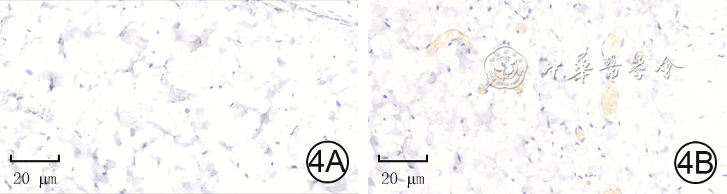
图 4 2组大鼠跨区穿支皮瓣原位回植术后7 d闭塞区域Ⅱ皮肤组织中VEGF的表达与分布 二氨基联苯胺-苏木精×400。4A.对照组血管区VEGF的表达少;4B.电针刺激组血管区VEGF的表达明显多于图4A
注:术前,对电针刺激组大鼠皮瓣胸背动脉与肋间后动脉所在血管体之间交界部位包含闭塞血管的区域(即闭塞区域Ⅱ)皮肤行连续7 d、每天1 h的电针刺激,对照组大鼠皮瓣不接受电针刺激;VEGF为血管内皮生长因子,阳性表达为黄色
-

 下载:
下载:


计量
- 文章访问数: 530
- HTML全文浏览量: 272
- PDF下载量: 3
- 被引次数: 0
