


-
摘要:
目的 探讨视黄酸对小鼠放射性皮肤损伤(RSI)的作用及其机制。 方法 该研究为实验研究。取HaCaT细胞,分为常规培养的对照组及损伤组、治疗组和拮抗组,后3组细胞均接受10 Gy X射线辐射,后2组细胞在辐射前均接受12 h的视黄酸预处理,最后1组细胞在辐射前另加入聚肌胞苷酸处理1 h。取3组辐射后24 h细胞及对照组相应时间点细胞,使用流式细胞仪检测细胞中活性氧水平,采用蛋白质印迹法检测细胞中白细胞介素6(IL-6)、肿瘤坏死因子α(TNF-α)、Toll样受体3(TLR3)、核因子κB的蛋白表达,样本数均为3。取24只6周龄雌性BALB/c小鼠,按随机数字表法分为对照组及损伤组、治疗组和拮抗组(每组6只),后3组小鼠右下肢均接受35 Gy电子线辐射造成RSI,后2组小鼠伤后0(即刻)、7、14、21、28、35、42 d均接受视黄酸处理且最后1组小鼠在这些时间点另接受聚肌胞苷酸处理,对照组小鼠模拟致假伤。伤后42 d,计算创面愈合率,采用激光散斑血流仪检测致伤处皮肤(即创面)组织血流灌注情况(以血流指数表示);取创面组织,行苏木精-伊红染色后计数炎症细胞并测量表皮厚度,行免疫组织化学染色检测IL-6和TNF-α表达,行免疫荧光染色检测TLR3表达,采用蛋白质印迹法检测TLR3和核因子κB的蛋白表达。 结果 损伤组细胞辐射后24 h活性氧水平及IL-6、TNF-α、TLR3与核因子κB的蛋白表达均显著高于对照组相应时间点(P<0.05),治疗组细胞辐射后24 h活性氧水平及IL-6、TLR3与核因子κB的蛋白表达均显著低于损伤组与拮抗组(P<0.05)。伤后42 d,对照组、损伤组、治疗组、拮抗组小鼠创面愈合率分别为(100.4±2.7)%、(77.5±2.5)%、(89.8±3.2)%、(70.1±4.8)%,治疗组小鼠创面愈合率显著高于损伤组与拮抗组(P值均<0.05)。伤后42 d,治疗组小鼠创面组织血流指数显著低于损伤组与拮抗组(P值均<0.05)。伤后42 d,与对照组比较,损伤组小鼠创面组织中炎症细胞数量显著增多且表皮厚度显著增厚(P<0.05);与治疗组比较,损伤组与拮抗组小鼠创面组织中炎症细胞数量显著增多且表皮厚度显著增厚(P<0.05)。伤后42 d,损伤组小鼠创面组织中IL-6、TNF-α、TLR3表达水平及TLR3、核因子κB的蛋白表达均显著高于对照组(P<0.05),治疗组小鼠创面组织中IL-6、TNF-α、TLR3表达水平及TLR3、核因子κB的蛋白表达均显著低于损伤组与拮抗组(P<0.05)。 结论 视黄酸通过抑制TLR3/核因子κB信号通路及其下游炎症因子的表达,显著减少辐射引起的细胞损伤,促进小鼠RSI修复。 Abstract:Objective To investigate the effects and mechanism of retinoic acid on radiation-induced skin injury (RSI) in mice. Methods This study was an experimental research. HaCaT cells were obtained and divided into control group (routinely cultured), injury group, treatment group, and antagonism group. The cells in the latter three groups were all exposed to 10 Gy X-ray radiation. The cells in the latter two groups were pretreated with retinoic acid for 12 h before radiation, and the cells in the last group were pre-treated with polyinosinic-polycytidylic acid for 1 h before radiation. Cells from the three irradiated groups at 24 h after radiation and cells from control group at the corresponding time point were collected to detect the reactive oxygen species (ROS) level in the cells by flow cytometry, and measure the protein expressions of interleukin-6 (IL-6), tumor necrosis factor-α (TNF-α), Toll-like receptor 3 (TLR3), and nuclear factor-κB (NF-κB) in the cells by Western blotting, with the sample number being 3. Twenty-four 6-week-old female BALB/c mice were obtained and divided into control group, injury group, treatment group, and antagonism group according to the random number table method (with 6 mice in each group). The right lower limbs of mice in the latter three groups were all exposed to 35 Gy electron beam radiation to induce RSI. Mice in the latter two groups were treated with retinoic acid at 0 (immediately), 7, 14, 21, 28, 35, and 42 days after injury, while mice in the last group were pre-treated with polyinosinic-polycytidylic acid at these time points. Mice in control group were simulated with sham injury. At 42 days after injury, the wound healing rate was calculated, and blood perfusion (denoted as blood flow index) in the skin tissue at the injury site (i.e. wound tissue) was detected by laser speckle flowmetry; wound tissue was collected, the hematoxylin-eosin staining was performed to count inflammatory cells and measure epidermal thickness, the immunohistochemical staining was performed to detect the expression of IL-6 and TNF-α, the immunofluorescence staining was performed to detect the expression of TLR3, and Western blotting was performed to detect the protein expressions of TLR3 and NF-κB. Results At 24 h after radiation, the ROS level and protein expressions of IL-6, TNF-α, TLR3, and NF-κB of cells in injury group were significantly higher than those in control group at the corresponding time point (P<0.05). The ROS level and protein expressions of IL-6, TLR3, and NF-κB of cells in treatment group were significantly lower than those in both injury group and antagonism group at 24 hours after radiation (P<0.05). At 42 days after injury, the wound healing rates of mice in control group, injury group, treatment group, and antagonism group were (100.4±2.7)%, (77.5±2.5)%, (89.8±3.2)%, and (70.1±4.8)%, respectively. The wound healing rate of mice in treatment group was significantly higher than that in injury group and antagonism group (both P values <0.05). At 42 days after injury, the blood flow index in the wound tissue of mice in treatment group was significantly lower than that in injury group and antagonism group (both P values <0.05). At 42 days after injury, compared with those in control group, the number of inflammatory cells in the wound tissue of mice in injury group was significantly increased, and the epidermal thickness significantly thickened (P<0.05); compared with those in treatment group, the number of inflammatory cells in the wound tissue of mice in injury group and antagonism group was significantly increased, and the epidermal thickness significantly thickened (P<0.05). At 42 days after injury, the expression levels of IL-6, TNF-α, and TLR3, as well as the protein expressions of TLR3 and NF-κB in the wound tissue of mice in injury group were significantly higher than those in control group (P<0.05), while the expression levels of IL-6, TNF-α, and TLR3, as well as the protein expressions of TLR3 and NF-κB in the wound tissue of mice in treatment group were significantly lower than those in injury group and antagonism group (P<0.05). Conclusions Retinoic acid significantly reduces radiation-induced cell injury and promotes the repair of RSI in mice by inhibiting the TLR3/NF-κB signaling pathway and the expression of downstream inflammatory factors. -
Key words:
- Radiation injuries /
- Radiation, ionizing /
- Skin ulcer /
- Toll-like receptor 3 /
- NF-kappa B /
- Retinoic acid /
- Wound healing /
- Inflammatory cytokines
-
参考文献
(40) [1] YangX,RenH,GuoX,et al.Radiation-induced skin injury: pathogenesis, treatment, and management[J].Aging (Albany NY),2020,12(22):23379-23393.DOI: 10.18632/aging.103932. [2] ShenJ,JiaoW,YangJ,et al.In situ photocrosslinkable hydrogel treats radiation-induced skin injury by ROS elimination and inflammation regulation[J].Biomaterials,2025,314:122891.DOI: 10.1016/j.biomaterials.2024.122891. [3] KiangJG,SmithJT,CannonG,et al.Ghrelin, a novel therapy, corrects cytokine and NF-κB-AKT-MAPK network and mitigates intestinal injury induced by combined radiation and skin-wound trauma[J].Cell Biosci,2020,10:63.DOI: 10.1186/s13578-020-00425-z. [4] WangY,GaoJ,SunL,et al.Jia-Wei-Si-Miao-Yong-An Fang stimulates the healing of acute radiation-induced cutaneous wounds through MAPK/ERK pathway[J].J Ethnopharmacol,2023,306:116180.DOI: 10.1016/j.jep.2023.116180. [5] 陈彦,程卓,马乐,等.放射性皮肤损伤鼠类创面中衰老细胞的数量与类型及功能异质性分析[J].中华烧伤与创面修复杂志,2025,41(6):577-587.DOI: 10.3760/cma.j.cn501225-20240604-00209. [6] 中国老年医学学会烧创伤分会,中华医学会组织修复与再生分会,中国康复医学会再生医学与康复专业委员会,等.放射性皮肤损伤的诊断和治疗专家共识(2024版)[J].中华烧伤与创面修复杂志,2024,40(8):701-712.DOI: 10.3760/cma.j.cn501225-20240126-00033. [7] StaceySK,McEleneyM.Topical corticosteroids: choice and application[J].Am Fam Physician,2021,103(6):337-343. [8] NiX,HuG,CaiX.The success and the challenge of all-trans retinoic acid in the treatment of cancer[J].Crit Rev Food Sci Nutr,2019,59(Suppl 1):S71-S80.DOI: 10.1080/10408398.2018.1509201. [9] WuD,KhanFA,ZhangK,et al.Retinoic acid signaling in development and differentiation commitment and its regulatory topology[J].Chem Biol Interact,2024,387:110773.DOI: 10.1016/j.cbi.2023.110773. [10] GhyselinckNB,DuesterG.Retinoic acid signaling pathways[J].Development,2019,146(13):dev167502.DOI: 10.1242/dev.167502. [11] PousoMR,CairraoE.Effect of retinoic acid on the neurovascular unit: a review[J].Brain Res Bull,2022,184:34-45.DOI: 10.1016/j.brainresbull.2022.03.011. [12] SzymańskiŁ,SkopekR,PalusińskaM,et al.Retinoic acid and its derivatives in skin[J].Cells,2020,9(12):2660.DOI: 10.3390/cells9122660. [13] SeyaT,TakedaY,MatsumotoM.A Toll-like receptor 3 (TLR3) agonist ARNAX for therapeutic immunotherapy[J].Adv Drug Deliv Rev,2019,147:37-43.DOI: 10.1016/j.addr.2019.07.008. [14] ChenY,LinJ,ZhaoY,et al.Toll-like receptor 3 (TLR3) regulation mechanisms and roles in antiviral innate immune responses[J].J Zhejiang Univ Sci B,2021,22(8):609-632.DOI: 10.1631/jzus.B2000808. [15] TakemuraN,KawasakiT,KunisawaJ,et al.Blockade of TLR3 protects mice from lethal radiation-induced gastrointestinal syndrome[J].Nat Commun,2014,5:3492.DOI: 10.1038/ncomms4492. [16] PuJ,ChenD,TianG,et al.All-trans retinoic acid attenuates transmissible gastroenteritis virus-induced inflammation in IPEC-J2 cells via suppressing the RLRs/NF-κB signaling pathway[J].Front Immunol,2022,13:734171.DOI: 10.3389/fimmu.2022.734171. [17] 黄从书,朱贵花,刘桂芳,等.60Co γ射线对HaCaT细胞损伤的模型建立及其机制[J].辐射研究与辐射工艺学报,2021,39(4):38-44.DOI: 10.11889/j.1000-3436.2021.rrj.39.040304. [18] ChenE,ChenC,NiuZ,et al.Poly(I:C) preconditioning protects the heart against myocardial ischemia/reperfusion injury through TLR3/PI3K/Akt-dependent pathway[J].Signal Transduct Target Ther,2020,5(1):216.DOI: 10.1038/s41392-020-00257-w. [19] MaL,ChenY,GongQ,et al.Cold atmospheric plasma alleviates radiation-induced skin injury by suppressing inflammation and promoting repair[J].Free Radic Biol Med,2023,204:184-194.DOI: 10.1016/j.freeradbiomed.2023.05.002. [20] TuW,TangS,YanT,et al.Integrative multi-omic analysis of radiation-induced skin injury reveals the alteration of fatty acid metabolism in early response of ionizing radiation[J].J Dermatol Sci,2022,108(3):178-186.DOI: 10.1016/j.jdermsci.2023.01.001. [21] 张进勇,谢宗能,孔平,等.散斑血流灌注成像在医学中的应用[J].激光与光电子学进展,2022,59(22):43-54.DOI: 10.3788/LOP202259.2200003. [22] LennickeC,CocheméHM.Redox metabolism: ROS as specific molecular regulators of cell signaling and function[J].Mol Cell,2021,81(18):3691-3707.DOI: 10.1016/j.molcel.2021.08.018. [23] SiesH,BelousovVV,ChandelNS,et al.Defining roles of specific reactive oxygen species (ROS) in cell biology and physiology[J].Nat Rev Mol Cell Biol,2022,23(7):499-515.DOI: 10.1038/s41580-022-00456-z. [24] AnX,YuW,LiuJ,et al.Oxidative cell death in cancer: mechanisms and therapeutic opportunities[J].Cell Death Dis,2024,15(8):556.DOI: 10.1038/s41419-024-06939-5. [25] ChenS,LiQ,ShiH,et al.New insights into the role of mitochondrial dynamics in oxidative stress-induced diseases[J].Biomed Pharmacother,2024,178:117084.DOI: 10.1016/j.biopha.2024.117084. [26] WangB,WangY,ZhangJ,et al.ROS-induced lipid peroxidation modulates cell death outcome: mechanisms behind apoptosis, autophagy, and ferroptosis[J].Arch Toxicol,2023,97(6):1439-1451.DOI: 10.1007/s00204-023-03476-6. [27] SchofieldJH,SchaferZT.Mitochondrial reactive oxygen species and mitophagy: a complex and nuanced relationship[J].Antioxid Redox Signal,2021,34(7):517-530.DOI: 10.1089/ars.2020.8058. [28] HuntM,TorresM,Bachar-WikstromE,et al.Cellular and molecular roles of reactive oxygen species in wound healing[J].Commun Biol,2024,7(1):1534.DOI: 10.1038/s42003-024-07219-w. [29] DongY,WangZ.ROS-scavenging materials for skin wound healing: advancements and applications[J].Front Bioeng Biotechnol,2023,11:1304835.DOI: 10.3389/fbioe.2023.1304835. [30] MoreiraHR,MarquesAP.Vascularization in skin wound healing: where do we stand and where do we go?[J].Curr Opin Biotechnol,2022,73:253-262.DOI: 10.1016/j.copbio.2021.08.019. [31] PeñaOA,MartinP.Cellular and molecular mechanisms of skin wound healing[J].Nat Rev Mol Cell Biol,2024,25(8):599-616.DOI: 10.1038/s41580-024-00715-1. [32] DaiS,WenY,LuoP,et al.Therapeutic implications of exosomes in the treatment of radiation injury[J/OL].Burns Trauma,2022,10:tkab043[2024-04-20].https://pubmed.ncbi.nlm.nih.gov/35071650/.DOI: 10.1093/burnst/tkab043. [33] ChenW,WangY,ZhengJ,et al.Characterization of cellular senescence in radiation ulcers and therapeutic effects of mesenchymal stem cell-derived conditioned medium[J/OL].Burns Trauma,2023,11:tkad001[2024-04-20].https://pubmed.ncbi.nlm.nih.gov/37188110/.DOI: 10.1093/burnst/tkad001. [34] KawczakP,FeszakI,BrzezińskiP,et al.Structure-activity relationships and therapeutic applications of retinoids in view of potential benefits from drug repurposing process[J].Biomedicines,2024,12(5):1059.DOI: 10.3390/biomedicines12051059. [35] FerreiraR,NapoliJ,EnverT,et al.Advances and challenges in retinoid delivery systems in regenerative and therapeutic medicine[J].Nat Commun,2020,11(1):4265.DOI: 10.1038/s41467-020-18042-2. [36] GattuS,BangYJ,PendseM,et al.Epithelial retinoic acid receptor β regulates serum amyloid A expression and vitamin A-dependent intestinal immunity[J].Proc Natl Acad Sci U S A,2019,116(22):10911-10916.DOI: 10.1073/pnas.1812069116. [37] SakaniwaK,FujimuraA,ShibataT,et al.TLR3 forms a laterally aligned multimeric complex along double-stranded RNA for efficient signal transduction[J].Nat Commun,2023,14(1):164.DOI: 10.1038/s41467-023-35844-2. [38] KimD,GarzaLA.Hypothesis: wound-induced TLR3 activation stimulates endogenous retinoic acid synthesis and signalling during regeneration[J].Exp Dermatol,2019,28(4):450-452.DOI: 10.1111/exd.13931. [39] LimCS,JangYH,LeeGY,et al.TLR3 forms a highly organized cluster when bound to a poly(I:C) RNA ligand[J].Nat Commun,2022,13(1):6876.DOI: 10.1038/s41467-022-34602-0. [40] LavudiK,NuguriSM,OlversonZ,et al.Targeting the retinoic acid signaling pathway as a modern precision therapy against cancers[J].Front Cell Dev Biol,2023,11:1254612.DOI: 10.3389/fcell.2023.1254612. -

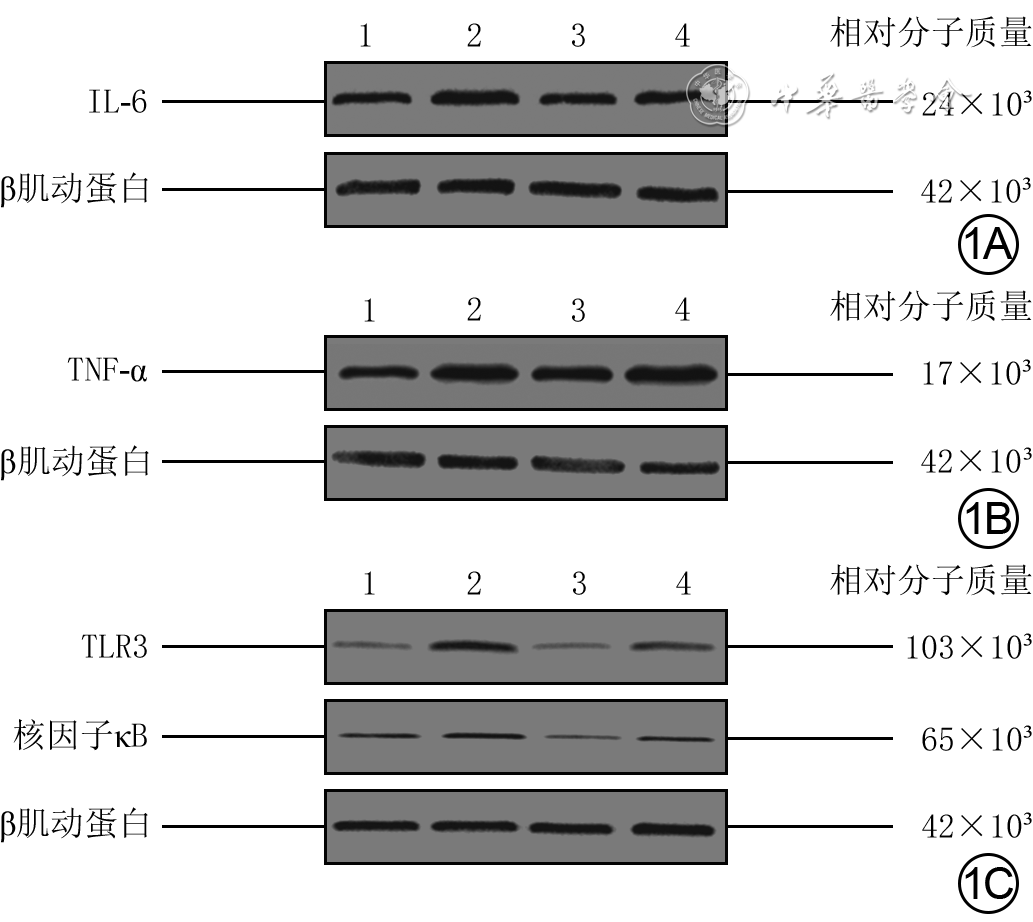
图 1 采用蛋白质印迹法检测的3组放射性损伤HaCaT细胞辐射后24 h及对照组HaCaT细胞相应时间点炎症因子蛋白表达。1A.IL-6;1B.TNF-α;1C.TLR3与核因子κB
注:条带上方1、2、3、4分别指示细胞常规培养的对照组、细胞仅接受10 Gy X射线辐射的损伤组、细胞同前辐射并接受12 h视黄酸预处理的治疗组、细胞同前辐射及预处理并在辐射前加入聚肌胞苷酸处理1 h的拮抗组;IL-6为白细胞介素6,TNF-α为肿瘤坏死因子α,TLR3为Toll样受体3

图 2 4组放射性皮肤损伤小鼠伤后42 d致伤处皮肤组织血流灌注情况。2A、2B、2C、2D.分别为对照组、损伤组、治疗组、拮抗组,图2A与2C血流灌注量较低,明显低于图2B与2D
注:对照组小鼠模拟致假伤,损伤组小鼠仅接受35 Gy电子线辐射,治疗组小鼠同前辐射并于伤后0(即刻)、7、14、21、28、35、42 d均接受视黄酸处理,拮抗组小鼠同前辐射及处理并于前述各时间点另接受聚肌胞苷酸处理;颜色越红表示血流灌注量越大,颜色越蓝表示血流灌注量越小

图 3 4组放射性皮肤损伤小鼠伤后42 d致伤处皮肤组织中IL-6和TNF-α表达 二氨基联苯胺-苏木精×100。3A、3B、3C、3D.分别为对照组、损伤组、治疗组、拮抗组IL-6表达(棕色)情况,图3A与3C中IL-6表达较低,明显低于图3B与3D;3E、3F、3G、3H.分别为对照组、损伤组、治疗组、拮抗组TNF-α表达(棕色)情况,图3E与3G中TNF-α表达较低,明显低于图3F与3H
注:对照组小鼠模拟致假伤,损伤组小鼠仅接受35 Gy电子线辐射,治疗组小鼠同前辐射并于伤后0(即刻)、7、14、21、28、35、42 d均接受视黄酸处理,拮抗组小鼠同前辐射及处理并于前述各时间点另接受聚肌胞苷酸处理;IL-6为白细胞介素6,TNF-α为肿瘤坏死因子α

图 4 4组放射性皮肤损伤小鼠伤后42 d致伤处皮肤组织中TLR3表达 Alexa Fluor 488-4',6-二脒基-2-苯基吲哚×100。4A、4B、4C、4D.分别为对照组、损伤组、治疗组、拮抗组,图4A与4C中TLR3表达较低,明显低于图4B与4D
注:对照组小鼠模拟致假伤,损伤组小鼠仅接受35 Gy电子线辐射,治疗组小鼠同前辐射并于伤后0(即刻)、7、14、21、28、35、42 d均接受视黄酸处理,拮抗组小鼠同前辐射及处理并于前述各时间点另接受聚肌胞苷酸处理;Toll样受体3(TLR3)阳性表达呈绿色,细胞核阳性表达呈蓝色

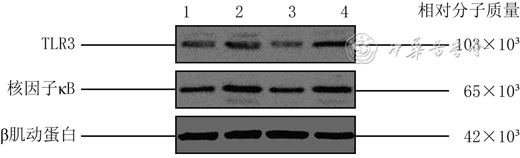
图 5 采用蛋白质印迹法检测的4组放射性皮肤损伤小鼠伤后42 d致伤处皮肤组织中TLR3与核因子κB的蛋白表达
注:条带上方1、2、3、4分别指示对照组、损伤组、治疗组、拮抗组;对照组小鼠模拟致假伤,损伤组小鼠仅接受35 Gy电子线辐射,治疗组小鼠同前辐射并于伤后0(即刻)、7、14、21、28、35、42 d均接受视黄酸处理,拮抗组小鼠同前辐射及处理并于前述各时间点另接受聚肌胞苷酸处理;TLR3为Toll样受体3
组别 样本数 白细胞介素6 肿瘤坏死因子α Toll样受体3 核因子κB 对照组 3 0.91 0.83 0.88 0.946 损伤组 3 1.69 2.47 3.23 1.363 治疗组 3 0.78 2.24 1.02 0.588 拮抗组 3 1.12 3.42 2.14 0.986 F值 55.85 5.18 54.75 23.09 P值 <0.001 0.026 <0.001 <0.001 P1值 <0.001 0.039 <0.001 0.002 P2值 <0.001 0.738 <0.001 <0.001 P3值 0.002 0.114 0.001 0.003 注:对照组细胞常规培养,损伤组细胞仅接受10 Gy X射线辐射,治疗组细胞同前辐射并接受12 h视黄酸预处理,拮抗组细胞同前辐射及预处理并在辐射前加入聚肌胞苷酸处理1 h;F值、P值为组间各指标总体比较所得;P1值、P2值、P3值分别为对照组与损伤组、损伤组与治疗组、治疗组与拮抗组各指标比较所得  下载: 导出CSV
Table 2. 4组放射性皮肤损伤小鼠伤后各时间点致伤处皮肤组织损伤程度评分比较(分,
组别 样本数 7 d 14 d 21 d 28 d 35 d 42 d 对照组 6 0 0 0 0 0 0 损伤组 6 0 1.08 1.79 2.25 2.00 2.04 治疗组 6 0 0 0.71 0.95 0.87 0.25 拮抗组 6 0 1.29 2.62 2.79 2.62 2.45 P1值 — <0.001 <0.001 <0.001 <0.001 <0.001 P2值 — <0.001 <0.001 <0.001 <0.001 <0.001 P3值 — <0.001 <0.001 <0.001 <0.001 <0.001 注:对照组小鼠模拟致假伤,损伤组小鼠仅接受35 Gy电子线辐射,治疗组小鼠同前辐射并于伤后0(即刻)、7、14、21、28、35、42 d均接受视黄酸处理,拮抗组小鼠同前辐射及处理并于前述各时间点另接受聚肌胞苷酸处理;处理因素主效应,F=963.74,P<0.001;时间因素主效应,F=257.83,P<0.001;两者交互作用,F=51.42,P<0.001;P1值、P2值、P3值分别为对照组与损伤组、损伤组与治疗组、治疗组与拮抗组各时间点比较所得;“—”表示无此项
下载: 导出CSV
-

 下载:
下载:


计量
- 文章访问数: 1142
- HTML全文浏览量: 482
- PDF下载量: 15
- 被引次数: 0
