


Analysis of the number, type, and functional heterogeneity of senescent cells in the radiation-induced skin wounds in mice
-
摘要:
目的 探讨放射性皮肤损伤鼠类创面中衰老细胞的数量与类型以及功能异质性。 方法 该研究为实验研究。取40只6~8周龄雄性可示踪衰老细胞的p16启动子驱动番茄红蛋白和白喉毒素受体(p16DTR/Tom)转基因小鼠,按随机数字表法分为35 Gy组和50 Gy组(每组20只),分别对小鼠右后肢皮肤施加35、50 Gy X射线照射建立3、4度放射性皮肤损伤模型,照射前和照射后10、20、30 d,检测2组小鼠创面组织中衰老细胞阳性面积百分比;照射后10 d,采用免疫荧光法观察50 Gy组小鼠创面组织中内皮细胞、单核巨噬细胞、角质形成细胞(KC)、成纤维细胞(Fb)与衰老细胞共定位情况。取9只6~8周龄雄性p16DTR/Tom转基因小鼠,按随机数字表法分为不进行任何处理的未照射组和同前处理的35 Gy组与50 Gy组(每组3只),取35 Gy组和50 Gy组小鼠照射后10 d创面组织及未照射组小鼠相应时间点正常皮肤组织,采用流式细胞术检测KC、Fb、内皮细胞、单核巨噬细胞的衰老百分比。对公开获取的健康大鼠正常皮肤组织(设为对照组)和经30 Gy电子线照射后7、14 d大鼠创面混合组织(设为辐照组)单细胞转录组测序数据进行生物信息学分析,对2组细胞进行衰老评定筛选衰老细胞,分析辐照组各类衰老细胞衰老相关分泌表型(SASP)表达谱之间的相关性,筛选出辐照组衰老细胞相较于对照组相应正常细胞差异表达显著的差异表达基因(DEG)进行基因本体论(GO)富集分析。 结果 50 Gy组小鼠照射后20、30 d创面组织中衰老细胞阳性面积百分比均显著高于35 Gy组(t值分别为-5.56、-5.48,P < 0.05);50 Gy组小鼠照射后10 d创面组织中可见内皮细胞、单核巨噬细胞、KC、Fb与衰老细胞共定位;50 Gy组小鼠照射后10 d创面组织中KC、Fb、内皮细胞、单核巨噬细胞的衰老百分比分别为(21.07±9.49)%、(16.10±3.27)%、(16.90±5.29)%、(34.13±8.76)%,显著高于未照射组小鼠相应时间点正常皮肤组织中的(3.58±1.13)%、(4.13±0.19)%、(3.86±1.28)%、(10.14±4.95)%,P值均 < 0.05。生物信息学分析显示,辐照组大鼠创面组织中内皮细胞、Fb、KC、巨噬细胞、单核细胞和施万细胞衰老评分均显著高于对照组大鼠正常皮肤组织(Z值分别为-8.71、-9.58、-7.19、-8.82、-6.66、-2.70,P < 0.05),即筛选出6种衰老细胞。辐照组大鼠创面组织中单核细胞与巨噬细胞SASP表达谱之间显著相关(r=0.83,P < 0.05),其余各类衰老细胞SASP表达谱之间的相关性均无统计学意义(P > 0.05)。GO富集分析显示,与对照组相应正常细胞比较,辐照组大鼠创面组织中6种衰老细胞显著上调的DEG均显著富集于对凋亡信号通路的调控,多种衰老细胞显著上调的DEG显著富集于髓系细胞分化通路、显著下调的DEG显著富集于细胞分裂相关通路,P值均 < 0.05。 结论 放射性皮肤损伤鼠类创面中衰老细胞数量上调,且衰老细胞的积累呈辐射剂量和时间依赖性;内皮细胞、Fb、KC、单核巨噬细胞等多种类型细胞可发生衰老,各类衰老细胞之间功能和SASP表达谱存在明显差异。 Abstract:Objective To investigate the number, type, and functional heterogeneity of senescent cells in the radiation-induced skin wounds in mice. Methods The study was an experimental study. Forty male p16-diphtheria toxin receptor-tdTomato (p16DTR/Tom) transgenic mice aged 6-8 weeks, which could be used to trace senescent cells, were divided into 35 Gy group and 50 Gy group (with 20 mice in each group) according to the random number table method, and 35 or 50 Gy X-ray irradiation was applied to the skin of the right hind limb of the mice to establish 3 or 4 degree of radiation-induced skin injury model, respectively. The positive area percentage of senescent cells in the wound tissue of mice in two groups was detected before irradiation and at 10, 20, and 30 d after irradiation; at 10 d after irradiation, the co-localization of endothelial cells (ECs), mononuclear macrophages (MMs), keratinocytes (KCs), fibroblasts (Fbs) and senescent cells in the wound tissue of mice in 50 Gy group was observed by immunofluorescence method. Nine male p16DTR/Tom transgenic mice aged 6-8 weeks were divided into unirradiated group without any treatment and 35 Gy group and 50 Gy group with the same treatment as above (with 3 mice in each group) according to the random number table method. The wound tissue of mice in 35 Gy group and 50 Gy group at 10 d after irradiation and the normal skin tissue of mice in unirradiated group at the corresponding time point was taken, and the senescence percentages of KCs, Fbs, ECs, and MMs were detected by flow cytometry. Bioinformatics analysis was performed on publicly available single-cell transcriptome sequencing data from normal skin tissue of healthy rats (setting as control group) and mixed wound tissue of rats for 7 and 14 d after irradiation with 30 Gy electron beams (setting as irradiated group), and the two groups of cells were subjected to senescence assessment to screen for senescent cells, the correlation between the expression profiles of senescence-associated secretory phenotypes (SASPs) of various types of senescent cells in irradiated group was analyzed, and the differentially expressed genes (DEGs) with significantly differential expression between the senescent cells in irradiated group and the corresponding normal cells in control group were screened for gene ontology (GO) enrichment analysis. Results The positive area percentage of senescent cells in the wound tissue of mice in 50 Gy group was significantly higher than that in 35 Gy group at 20 and 30 d after irradiation (with t values of -5.56 and -5.48, respectively, P < 0.05). ECs, MMs, KCs, and Fbs co-localized with senescent cells in the wound tissue of mice in 50 Gy group at 10 d after irradiation. The senescence percentages of KCs, Fbs, ECs, and MMs in the wound tissue of mice in 50 Gy group at 10 d after irradiation were (21.07±9.49)%, (16.10±3.27)%, (16.90±5.29)%, and (34.13±8.76)%, respectively, which were significantly higher than (3.58±1.13)%, (4.13±0.19)%, (3.86±1.28)%, and (10.14±4.95)% in the normal skin tissue of mice in unirradiated group at the corresponding time point, with P values all < 0.05. Bioinformatics analysis showed that the senescence scores of ECs, Fbs, KCs, macrophages, monocytes, and Schwann cells in the wound tissue of rats in irradiated group were significantly higher than those in the normal skin tissue of rats in control group (with Z values of -8.71, -9.58, -7.19, -8.82, -6.66, and -2.70, respectively, P < 0.05), i.e., 6 types of senescent cells were screened. The SASPs expression profiles of monocytes and macrophages in the wound tissue of rats in irradiated group were significantly correlated (r=0.83, P < 0.05), but there was no statistically significant correlation between the SASPs expression profiles of the remaining types of senescent cells (P > 0.05). GO enrichment analysis showed that, compared with the corresponding normal cells in control group, the significantly up-regulated DEGs of the 6 types of senescent cells in the wound tissue of rats in irradiated group were significantly enriched in the regulation of apoptosis signaling pathway, the significantly up-regulated DEGs of multiple senescent cells were significantly enriched in the myeloid cell differentiation pathway, and the significantly down-regulated DEGs of multiple senescent cells were significantly enriched in the cell division-related pathway, with P values all < 0.05. Conclusions The number of senescent cells in the wounds of mice with radiation-induced skin injury is up-regulated, and the accumulation of senescent cells is radiation dose- and time-dependent; multiple types of cells including ECs, Fbs, KCs, and MMs can undergo senescence, and there are obvious differences in the function and SASP expression profiles among various types of senescent cells. 本文亮点(1) 揭示了放射性皮肤损伤鼠类创面中衰老细胞积累的剂量与时间依赖性。(2) 阐明了皮肤放射性损伤后发生衰老的细胞功能的改变及衰老相关分泌表型表达谱的差异。 -
参考文献
(40) [1] Iddins CJ, DiCarlo AL, Ervin MD, et al. Cutaneous and local radiation injuries[J]. J Radiol Prot, 2022, 42(1): 011001. DOI: 10.1088/1361-6498/ac241a. [2] Yang X, Ren H, Guo X, et al. Radiation-induced skin injury: pathogenesis, treatment, and management[J]. Aging (Albany NY), 2020, 12(22): 23379-23393. DOI: 10.18632/aging.103932. [3] Davan-Wetton CSA, Pessolano E, Perretti M, et al. Senescence under appraisal: hopes and challenges revisited[J]. Cell Mol Life Sci, 2021, 78(7): 3333-3354. DOI: 10.1007/s00018-020-03746-x. [4] Adjemian S, Oltean T, Martens S, et al. Ionizing radiation results in a mixture of cellular outcomes including mitotic catastrophe, senescence, methuosis, and iron-dependent cell death[J]. Cell Death Dis, 2020, 11(11): 1003. DOI: 10.1038/s41419-020-03209-y. [5] Demaria M, Ohtani N, Youssef SA, et al. An essential role for senescent cells in optimal wound healing through secretion of PDGF-AA[J]. Dev Cell, 2014, 31(6): 722-733. DOI: 10.1016/j.devcel.2014.11.012. [6] Wei X, Li M, Zheng Z, et al. Senescence in chronic wounds and potential targeted therapies[J/OL]. Burns Trauma, 2022, 10: tkab045[2024-06-04]. https://pubmed.ncbi.nlm.nih.gov/35187179/ . DOI:10.1093/burnst/tkab045 .[7] Admasu TD, Kim K, Rae M, et al. Selective ablation of primary and paracrine senescent cells by targeting iron dyshomeostasis[J]. Cell Rep, 2023, 42(2): 112058. DOI: 10.1016/j.celrep.2023.112058. [8] Huang W, Hickson LJ, Eirin A, et al. Cellular senescence: the good, the bad and the unknown[J]. Nat Rev Nephrol, 2022, 18(10): 611-627. DOI: 10.1038/s41581-022-00601-z. [9] Chen Y, Ma L, Cheng Z, et al. Senescent fibroblast facilitates re-epithelization and collagen deposition in radiation-induced skin injury through IL-33-mediated macrophage polarization[J]. J Transl Med, 2024, 22(1): 176. DOI: 10.1186/s12967-024-04972-8. [10] Iwakawa M, Noda S, Ohta T, et al. Different radiation susceptibility among five strains of mice detected by a skin reaction[J]. J Radiat Res, 2003, 44(1): 7-13. DOI: 10.1269/jrr.44.7. [11] Rodgers KE, Tan A, Kim L, et al. Development of a guinea pig cutaneous radiation injury model using low penetrating X-rays[J]. Int J Radiat Biol, 2016, 92(8): 434-443. DOI: 10.1080/09553002.2016.1186302. [12] Tu W, Tang S, Yan T, et al. Integrative multi-omic analysis of radiation-induced skin injury reveals the alteration of fatty acid metabolism in early response of ionizing radiation[J]. J Dermatol Sci, 2022, 108(3): 178-186. DOI: 10.1016/j.jdermsci.2023.01.001. [13] Stuart T, Butler A, Hoffman P, et al. Comprehensive integration of single-cell data[J]. Cell, 2019, 177(7): 1888-1902.e21. DOI: 10.1016/j.cell.2019.05.031. [14] Wu T, Hu E, Xu S, et al. clusterProfiler 4.0: a universal enrichment tool for interpreting omics data[J]. Innovation (Camb), 2021, 2(3): 100141. DOI: 10.1016/j.xinn.2021.100141. [15] 中国老年医学学会烧创伤分会, 中华医学会组织修复与再生分会, 中国康复医学会再生医学与康复专业委员会, 等. 放射性皮肤损伤的诊断和治疗专家共识(2024版)[J]. 中华烧伤与创面修复杂志, 2024, 40(8): 701-712. DOI: 10.3760/cma.j.cn501225-20240126-00033. [16] Dai S, Wen Y, Luo P, et al. Therapeutic implications of exosomes in the treatment of radiation injury[J/OL]. Burns Trauma, 2022, 10: tkab043[2024-06-04]. https://pubmed.ncbi.nlm.nih.gov/35071650/ . DOI:10.1093/burnst/tkab043 .[17] Huayllani MT, Ruiz-Garcia H, Boczar D, et al. Adipose-derived stem cells therapy for radiation-induced skin injury[J]. Ann Plast Surg, 2021, 87(6): 639-649. DOI: 10.1097/SAP.0000000000003039. [18] 夏成德, 杨阳. 放射性皮肤溃疡的治疗和预防策略[J]. 中华烧伤与创面修复杂志, 2024, 40(8): 719-724. DOI: 10.3760/cma.j.cn501225-20240415-00134. [19] DiCarlo AL, Bandremer AC, Hollingsworth BA, et al. Cutaneous radiation injuries: models, assessment and treatments[J]. Radiat Res, 2020, 194(3): 315-344. DOI: 10.1667/RADE-20-00120.1. [20] Chen W, Wang Y, Zheng J, et al. Characterization of cellular senescence in radiation ulcers and therapeutic effects of mesenchymal stem cell-derived conditioned medium[J/OL]. Burns Trauma, 2023, 11: tkad001[2024-06-04]. https://pubmed.ncbi.nlm.nih.gov/37188110/ . DOI:10.1093/burnst/tkad001 .[21] Wang H, Wang Z, Huang Y, et al. Senolytics (DQ) mitigates radiation ulcers by removing senescent cells[J]. Front Oncol, 2020, 9: 1576. DOI: 10.3389/fonc.2019.01576. [22] Haston S, Gonzalez-Gualda E, Morsli S, et al. Clearance of senescent macrophages ameliorates tumorigenesis in KRAS-driven lung cancer[J]. Cancer Cell, 2023, 41(7): 1242-1260.e6. DOI: 10.1016/j.ccell.2023.05.004. [23] Moiseeva V, Cisneros A, Cobos AC, et al. Context-dependent roles of cellular senescence in normal, aged, and disease states[J]. FEBS J, 2023, 290(5): 1161-1185. DOI: 10.1111/febs.16573. [24] Wang W, Luo J, Sheng W, et al. Proteomic profiling of radiation-induced skin fibrosis in rats: targeting the ubiquitin-proteasome system[J]. Int J Radiat Oncol Biol Phys, 2016, 95(2): 751-760. DOI: 10.1016/j.ijrobp.2016.01.021. [25] Campbell RA, Docherty MH, Ferenbach DA, et al. The role of ageing and parenchymal senescence on macrophage function and fibrosis[J]. Front Immunol, 2021, 12: 700790. DOI: 10.3389/fimmu.2021.700790. [26] Nguyen HQ, To NH, Zadigue P, et al. Ionizing radiation-induced cellular senescence promotes tissue fibrosis after radiotherapy. A review[J]. Crit Rev Oncol Hematol, 2018, 129: 13-26. DOI: 10.1016/j.critrevonc.2018.06.012. [27] Paramos-de-Carvalho D, Jacinto A, Saúde L. The right time for senescence[J]. Elife, 2021, 10: e72449. DOI: 10.7554/eLife.72449. [28] Kirschner K, Rattanavirotkul N, Quince MF, et al. Functional heterogeneity in senescence[J]. Biochem Soc Trans, 2020, 48(3): 765-773. DOI: 10.1042/BST20190109. [29] Wilkinson HN, Hardman MJ. Cellular senescence in acute and chronic wound repair[J]. Cold Spring Harb Perspect Biol, 2022, 14(11): a041221. DOI: 10.1101/cshperspect.a041221. [30] Alessio N, Acar MB, Squillaro T, et al. Progression of irradiated mesenchymal stromal cells from early to late senescence: changes in SASP composition and anti-tumour properties[J]. Cell Prolif, 2023, 56(6): e13401. DOI: 10.1111/cpr.13401. [31] Cohn RL, Gasek NS, Kuchel GA, et al. The heterogeneity of cellular senescence: insights at the single-cell level[J]. Trends Cell Biol, 2023, 33(1): 9-17. DOI: 10.1016/j.tcb.2022.04.011. [32] Wu F, Zhang Z, Wang M, et al. Cellular atlas of senescent lineages in radiation-or immunotherapy-induced lung injury by single-cell RNA-sequencing analysis[J]. Int J Radiat Oncol Biol Phys, 2023, 116(5): 1175-1189. DOI: 10.1016/j.ijrobp.2023.02.005. [33] Yan T, Yang P, Bai H, et al. Single-cell RNA-Seq analysis of molecular changes during radiation-induced skin injury: the involvement of Nur77[J]. Theranostics, 2024, 14(15): 5809-5825. DOI: 10.7150/thno.100417. [34] Guerrero-Juarez CF, Dedhia PH, Jin S, et al. Single-cell analysis reveals fibroblast heterogeneity and myeloid-derived adipocyte progenitors in murine skin wounds[J]. Nat Commun, 2019, 10(1): 650. DOI: 10.1038/s41467-018-08247-x. [35] Paldor M, Levkovitch-Siany O, Eidelshtein D, et al. Single-cell transcriptomics reveals a senescence-associated IL-6/CCR6 axis driving radiodermatitis[J]. EMBO Mol Med, 2022, 14(8): e15653. DOI: 10.15252/emmm.202115653. [36] Lucas V, Cavadas C, Aveleira CA. Cellular senescence: from mechanisms to current biomarkers and senotherapies[J]. Pharmacol Rev, 2023, 75(4): 675-713. DOI: 10.1124/pharmrev.122.000622. [37] Muñoz-Espín D, Cañamero M, Maraver A, et al. Programmed cell senescence during mammalian embryonic development[J]. Cell, 2013, 155(5): 1104-1118. DOI: 10.1016/j.cell.2013.10.019. [38] Zhang J, Yu H, Man MQ, et al. Aging in the dermis: fibroblast senescence and its significance[J]. Aging Cell, 2024, 23(2): e14054. DOI: 10.1111/acel.14054. [39] Sharpless NE, Sherr CJ. Forging a signature of in vivo senescence[J]. Nat Rev Cancer, 2015, 15(7): 397-408. DOI: 10.1038/nrc3960. [40] Gorgoulis V, Adams PD, Alimonti A, et al. Cellular senescence: defining a path forward[J]. Cell, 2019, 179(4): 813-827. DOI: 10.1016/j.cell.2019.10.005. -

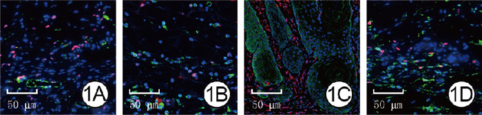
图 1 50 Gy组放射性皮肤损伤小鼠照射后10 d创面组织中内皮细胞、单核巨噬细胞、KC、Fb均与衰老细胞存在共定位 番茄红蛋白-Alexa Fluor 647-4',6-二脒基-2-苯基吲哚×400。1A.内皮细胞;1B.单核巨噬细胞;1C.KC;1D.Fb
注:50 Gy组小鼠右后肢皮肤接受50 Gy X射线照射;内皮细胞、单核巨噬细胞、角质形成细胞(KC)、成纤维细胞(Fb)阳性染色均为绿色,衰老细胞阳性染色为红色,细胞核阳性染色为蓝色
Figure 1. Endothelial cells, mononuclear macrophages, KCs, and Fbs showed co-localization with senescent cells in the wound tissue of mice with radiation-induced skin injury in 50 Gy group at 10 d after irradiation

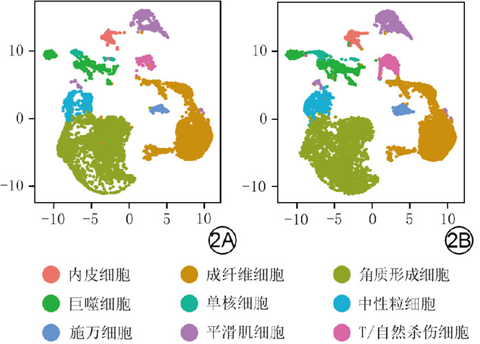
图 2 对照组大鼠正常皮肤组织和辐照组大鼠创面组织中细胞类型的二维流形近似和投影云图。2A.对照组;2B.辐照组
注:对照组大鼠不进行任何处理,辐照组大鼠创面组织为采用30 Gy电子线照射后7、14 d的混合组织
Figure 2. Two-dimensional streaming approximation and projection cloud of cell types in the normal skin tissue of rats in control group and the wound tissue of rats in irradiated group

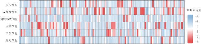
图 3 辐照组大鼠创面组织中衰老细胞SASP基因表达热图
注:辐照组大鼠创面组织为经30 Gy电子线照射后7、14 d的混合组织;衰老相关分泌表型(SASP)基因从左至右依次为激活素A受体1B型、血管生成素1、血管生成素配体4、双调蛋白、酪氨酸蛋白激酶受体、脑表达X-连锁3、骨形态发生蛋白2(Bmp2)、Bmp6、补体3、趋化因子配体1(Ccl1)、Ccl2、Ccl20、Ccl24、Ccl26、Ccl3、Ccl4、Ccl5、Ccl7、分化群55、分化群9、集落刺激因子1(Csf1)、Csf2、Csf2受体亚基β、连环蛋白β1、组织蛋白酶b、CXC基序趋化因子配体1(Cxcl1)、Cxcl10、Cxcl12、Cxcl16、Cxcl2、Cxcl3、CXC基序趋化因子受体2、Dickkopf相关蛋白1、内皮素1、表皮生长因子、表皮生长因子受体、上皮调节素、内皮细胞特异性分子1、E26转化特异性相关序列2、Fas细胞表面死亡受体、成纤维细胞生长因子1(Fgf1)、Fgf2、Fgf7、生长分化因子15、三磷酸鸟苷结合蛋白、胶质细胞成熟因子γ、肝细胞生长因子、高迁移率族蛋白B1、细胞间黏附分子1、胰岛素样生长因子1、胰岛素样生长因子结合蛋白1(Igfbp1)、Igfbp2、Igfbp3、Igfbp4、Igfbp5、Igfbp6、Igfbp7、白细胞介素10(Il10)、Il13、Il15、Il18、Il1a、Il1b、Il2、Il6、Il7、Il33、抑制素α亚基、含IQ基序的鸟苷三磷酸酶激活蛋白2、整合素亚基a2、肌醇三磷酸3-激酶A、Jun原癌基因、受体酪氨酸激酶配体、淋巴细胞胞质蛋白1、基质金属蛋白酶1(Mmp1)、Mmp10、Mmp12、Mmp13、Mmp14、Mmp2、Mmp3、Mmp9、核小体组装蛋白14、神经调节蛋白1、胎盘生长因子、纤溶酶原激活因子t(Plat)、Plau、Plau受体、聚嘧啶束结合蛋白1、前列腺素E受体2、谷胱甘肽依赖性前列腺素E合酶、核糖体蛋白S6激酶A5、分泌型载体膜蛋白4、选择素P配体、信号蛋白3F、serpin家族E成员1(Serpine1)、Serpine2、分泌型磷蛋白1、spexin激素、组织金属蛋白酶抑制物金属肽酶抑制因子2、肿瘤坏死因子(Tnf)、Tnf受体超家族成员1A、Tnf受体超家族成员1B、微管蛋白γ复合体相关蛋白2、血管内皮生长因子a(Vegfa)、Vegfc、神经生长因子诱导型、Wnt家族成员16
Figure 3. Heat map of SASP gene expression in senescent cells of rat wound tissue in irradiated group

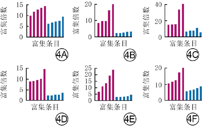
图 4 辐照组大鼠创面组织中衰老细胞相较于对照组大鼠正常皮肤组织相应正常细胞差异表达显著的DEG的基因本体论富集分析结果。4A、4B、4C、4D、4E、4F.分别为内皮细胞、成纤维细胞、角质形成细胞、单核细胞、巨噬细胞、施万细胞
注:红色指示显著上调的差异表达基因(DEG),蓝色指示显著下调的DEG;辐照组大鼠创面组织为经30 Gy电子线照射后7、14 d的混合组织,对照组大鼠不进行任何处理;图 4A横坐标从左至右对应富集条目为内在凋亡信号通路、蛋白酶体介导的泛素依赖性蛋白质分解代谢过程、细胞对肽激素刺激的反应、细胞因子介导的信号通路、有丝分裂细胞周期、角质形成细胞分化、表皮细胞分化、核染色体分离、皮肤发育、细胞分裂,图 4B横坐标从左至右对应富集条目为细胞对化学应激的反应、对缺氧的反应、肌动蛋白丝组织、创面愈合、髓系细胞分化、染色体分离、参与有丝分裂的微管细胞骨架组织、有丝分裂纺锤体组织、细胞分裂、主轴组织,图 4C横坐标从左至右对应富集条目为还原型烟酰胺腺嘌呤二核苷酸脱氢酶复合物组装、线粒体呼吸链复合体Ⅰ组装、细胞呼吸、前体代谢产物和能量的产生、蛋白质在细胞器中定位的建立、有丝分裂、参与有丝分裂的微管细胞骨架组织、细胞分裂、核染色体分离、染色体分离,图 4D横坐标从左至右对应富集条目为防御反应的正向调节、凋亡信号通路的调控、核糖核蛋白复合体生物发生、细胞对生物刺激的反应、无膜细胞器组装、内皮细胞迁移、对内质网应激的反应、类固醇代谢过程、细胞内运输的正调控、炎症反应的正调控,图 4E横坐标从左至右对应富集条目为细胞因子介导的信号通路、炎症反应的调节、内在凋亡信号通路、凋亡信号通路的调控、防御反应的正向调节、Wnt的细胞-细胞信号传导、Wnt信号通路、经典Wnt信号通路的调控、Wnt信号通路的调控、乳腺发育,图 4F横坐标从左至右对应富集条目为核糖核蛋白复合体生物发生、蛋白质-RNA复合物组织、内在凋亡信号通路、凋亡信号通路的调控、细胞对化学应激的反应、细胞-基质黏附的调节、核糖体RNA代谢过程、核糖体RNA处理、核糖体生物发生、核糖核蛋白复合体生物发生
Figure 4. Results of gene ontology enrichment analysis of DEGs with significantly differential expressions of senescent cells in the wound tissue of rats in irradiated group in comparison with the corresponding normal cells in the normal skin tissue of rats in control group
表 1 2组放射性皮肤损伤小鼠照射后各时间点损伤情况大体评分比较(分,x ± s)
Table 1. Comparison of the gross scores of injuries in 2 groups of mice with radiation-induced skin injury at each time point after irradiation
组别 样本数 5 d 10 d 15 d 20 d 25 d 30 d 35 Gy组 5 0.80±0.27 1.10±0.14 1.50±0.31 0.85±0.14 0.60±0.14 0.60±0.22 50 Gy组 5 1.00±0.31 2.35±0.65 2.80±0.45 2.70±0.45 1.10±0.14 0.95±0.21 t值 -1.09 -4.20 -5.36 -8.84 -5.77 -2.56 P值 0.308 0.003 < 0.001 < 0.001 < 0.001 0.034 注:35 Gy组和50 Gy组小鼠右后肢皮肤分别接受35、50 Gy X射线照射;处理因素主效应,F=258.28,P=0.002;时间因素主效应,F=75.76,P=0.011;两者交互作用,F=15.49,P=0.024  下载: 导出CSV
表 2 2组放射性皮肤损伤小鼠各时间点创面组织中衰老细胞阳性面积百分比比较(%,x ± s)
Table 2. Comparison of the positive area percentage of senescent cells in the wound tissue of 2 groups of mice with radiation-induced skin injury at each time point
组别 样本数 照射前 照射后10 d 照射后20 d 照射后30 d 35 Gy组 5 0.8±0.3 4.0±1.3 3.1±1.2 1.5±0.6 50 Gy组 5 0.9±0.3 5.6±1.7 11.2±3.0 5.5±1.7 t值 -0.26 -1.67 -5.56 -5.48 P值 0.803 0.135 0.002 0.002 P1值 — < 0.001 0.005 0.696 P2值 — 0.006 < 0.001 0.007 注:35 Gy组和50 Gy组小鼠右后肢皮肤分别接受35、50 Gy X射线照射;处理因素主效应,F=98.73,P=0.001;时间因素主效应,F=35.91,P=0.012;两者交互作用,F=9.44,P=0.048;t值、P值为2组间不同时间点比较所得;P1值、P2值分别为35 Gy组、50 Gy组照射后各时间点与组内照射前比较所得;“—”表示无此项
下载: 导出CSV
表 3 2组放射性皮肤损伤小鼠照射后10 d创面组织及未照射组小鼠相应时间点正常皮肤组织中主要细胞的衰老百分比比较(%,x ± s)
Table 3. Comparison of senescence percentages of predominant cells in the wound tissue of 2 groups of mice with radiation-induced skin injury at 10 d after irradiation and the normal skin tissue of mice in unirradiated group at the corresponding time point
组别 样本数 角质形成细胞 成纤维细胞 内皮细胞 单核巨噬细胞 35 Gy组 3 7.88±1.07 7.58±3.43 11.93±6.87 13.73±1.25 50 Gy组 3 21.07±9.49 16.10±3.27 16.90±5.29 34.13±8.76 未照射组 3 3.58±1.13 4.13±0.19 3.86±1.28 10.14±4.95 F值 10.10 4.61 5.29 20.50 P值 < 0.001 0.020 0.012 < 0.001 P1值 0.632 0.338 0.205 0.744 P2值 0.020 0.004 0.045 0.006 注:未照射组小鼠不进行任何处理,35 Gy组和50 Gy组小鼠右后肢皮肤分别接受35、50 Gy X射线照射;处理因素主效应,F=35.66,P=0.001;细胞类型主效应,F=7.64,P=0.002;两者交互作用,F=1.61,P=0.237;F值、P值为组间各细胞总体比较所得;P1值、P2值分别为35 Gy组、50 Gy组与未照射组间各细胞比较所得
下载: 导出CSV
表 4 对照组大鼠正常皮肤组织和辐照组大鼠创面组织中各类细胞衰老评分比较[分,M(Q1,Q3)]
Table 4. Comparison of senescence scores of various types of cells in the normal skin tissue of rats in control group and the wound tissue of rats in irradiated group
组别 内皮细胞 成纤维细胞 角质形成细胞 巨噬细胞 单核细胞 中性粒细胞 施万细胞 平滑肌细胞 T/自然杀伤细胞 对照组 0(-0.4,0.5) -0.3(-0.5,0) 0.2(-0.1,0.5) 0(-0.3,0.4) -0.4(-0.5,0.1) 0(-0.2,0.2) -0.5(-0.6,0) 0.2(-0.1,0.4) -0.5(-0.6,-0.4) 辐照组 0.6(0.3,0.8) 0.4(0.1,0.7) 0.4(0,0.6) 0.4(-0.3,0.4) 0.4(-0.4,0.7) -0.2(-0.4,0.2) 0.4(-0.1,0.7) 0.1(-0.2,0.5) -0.5(-0.6,0.1) Z值 -8.71 -9.58 -7.19 -8.82 -6.66 -0.82 -2.70 -1.71 -0.68 P值 < 0.001 < 0.001 < 0.001 < 0.001 < 0.001 0.411 0.007 0.086 0.498 注:对照组大鼠不进行任何处理,辐照组大鼠创面组织为经30 Gy电子线照射后7、14 d的混合组织;对照组表中从左至右各细胞样本数依次为205、3 514、4 019、405、100、512、134、560、97,辐照组表中从左至右各细胞样本数依次为537、6 856、2 895、1 122、213、539、289、1 000、567
下载: 导出CSV
-

 下载:
下载:

计量
- 文章访问数: 756
- HTML全文浏览量: 292
- PDF下载量: 14
- 被引次数: 0
