


Proteomics analysis of the effect and mechanism of ADSCs on full-thickness skin defects in diabetic rats
-
摘要:
目的 基于蛋白质组学分析,探讨脂肪间充质干细胞(ADSC)对糖尿病大鼠全层皮肤缺损的作用及其机制。 方法 该研究为自身对照设计实验研究。取4只8~10周龄雄性SD大鼠,从其附睾脂肪组织中提取ADSC并成功鉴定,然后取第3代ADSC用于下述实验。取24只4~6周龄雄性SD大鼠并成功构建2型糖尿病模型,选取其中体重为350~400 g的16只糖尿病大鼠,在其背部脊柱两侧同一水平位置各制作1个全层皮肤缺损创面。采用随机数字表法,将每只大鼠的2个创面分别纳入实验组与对照组(每组16个创面),伤后即刻,于创面周围及基底组织分别多点注射含ADSC的细胞悬液和磷酸盐缓冲液。计算大鼠伤后7、10、14 d创面愈合率。大鼠伤后7 d,取创面组织并提取蛋白质,采用四维数据非依赖采集非标记定量蛋白质谱技术行定量蛋白质组学分析及生物信息学分析,筛选2组创面组织中的差异表达蛋白(DEP)。然后通过蛋白质-蛋白质相互作用网络筛选出关键DEP,并应用基因本体论(GO)行功能注释及富集分析、京都基因和基因组百科全书(KEGG)行通路富集分析,同时进一步筛选目标DEP。取大鼠伤后7 d创面组织,采用蛋白质印迹法检测胸腺细胞分化抗原-1(Thy-1)和G蛋白偶联受体177/Wnt配体分泌介质(GPR177/Wls)的蛋白表达量。 结果 大鼠伤后7、10、14 d,实验组创面愈合率分别为(66±16)%、(83±8)%、(93±4)%,均显著高于对照组的(30±8)%、(62±6)%、(77±8)%,t值分别为-4.41、-7.46、-6.65,P<0.05。大鼠伤后7 d,相较于对照组,从实验组创面组织中共筛选出474个DEP(P<0.05)。进一步筛选出224个关键DEP,其中78个DEP显著上调,146个DEP显著下调。GO功能注释分析显示,显著上调和显著下调的DEP主要在细胞过程和生物调控的条件下影响蛋白质的表达,与细胞解剖实体和含蛋白质复合物有关,与生物分子之间的特异性结合和催化活性有关。GO功能富集分析显示,首要的DEP(显著上调)显著富集于Wnt-蛋白结合。KEGG通路富集分析显示,显著上调和显著下调的DEP富集通路包括淀粉和蔗糖代谢、核苷酸代谢、p53信号通路、细胞外基质-受体相互作用等。共筛选出4个目标蛋白:Thy-1、GPR177/Wls、FCH结构域蛋白2、线粒体核糖体蛋白L21,前两者为显著上调DEP,后两者为显著下调DEP。大鼠伤后7 d,实验组创面组织中GPR177/Wls和Thy-1的蛋白表达量分别为0.93±0.07、0.96±0.05,均显著高于对照组的0.39±0.07、0.36±0.12(t值分别为11.61、9.41,P<0.05)。 结论 基于蛋白质组学分析,揭示了大鼠ADSC能通过上调GPR177/Wls和Thy-1的蛋白表达量促进糖尿病大鼠全层皮肤缺损创面愈合。 Abstract:Objective To explore the effect and mechanism of adipose-derived mesenchymal stem cells (ADSCs) on full-thickness skin defects in diabetic rats using proteomics analysis. Methods This study was a self-control design experimental study. Four 8 to 10 weeks old male Sprague-Dawley rats were selected, and ADSCs were extracted from their epididymal adipose tissue and successfully identified. The third passage of ADSCs were used for the following experiments. Twenty-four 4 to 6 weeks old male Sprague-Dawley rats were selected and the type 2 diabetes model was successfully established. Among them, 16 diabetic rats weighing 350 to 400 g were chosen, and a full-thickness skin defect wound was created on each side of the spine on their backs at the same level. Using the random number table method, the two wounds of each rat were included in experimental group and control group (with 16 wounds in each group), and the cell suspensions containing ADSCs and phosphate buffered saline were injected at multiple points around and at the base of the wounds immediately after injury, respectively. The wound healing rates were calculated at day 7, 10, and 14 after injury in rats. At day 7 after injury in rats, wound tissue was collected and proteins were extracted. Four-dimensional data-independent acquisition label-free quantitative proteomics technology was used for quantitative proteomics analysis and bioinformatics analysis to screen differentially expressed proteins (DEPs) in the two groups of wound tissue. Then, key DEPs were screened through protein-protein interaction networks, gene ontology (GO) was used for functional annotation and enrichment analysis, and Kyoto encyclopedia of genes and genomes (KEGG) was used for pathway enrichment analysis to further screen target DEPs. The wound tissue of rats 7 days after injury was collected, and Western blotting was used to detect the protein expression of thymocyte differentiation antigen 1 (Thy-1) and G protein-coupled receptor 177/Wnt ligand secretion mediator (GPR177/Wls). Results At day 7, 10, and 14 after injury in rats, the wound healing rates in experimental group were (66±16)%, (83±8)%, and (93±4)%, respectively, which were significantly higher than (30±8)%, (62±6)%, and (77±8)% in control group (with t values of -4.41, -7.46, and -6.65, respectively, P<0.05). At day 7 after injury in rats, compared with those in control group, a total of 474 DEPs were screened from the wound tissue in experimental group (P<0.05). A total of 224 key DEPs were further screened out, among which 78 DEPs were significantly upregulated and 146 DEPs were significantly downregulated. GO functional annotation analysis showed that the most significantly upregulated and downregulated DEPs mainly affected protein expression under cellular processes and biological regulation conditions, and were related to cell anatomical entities and protein-containing complexes, as well as specific binding and catalytic activity between biomolecules. GO functional enrichment analysis showed that the most significantly upregulated DEPs were significantly enriched in Wnt-protein binding. KEGG pathway enrichment analysis showed that the significantly upregulated and downregulated DEPs were enriched in pathways such as starch and sucrose metabolism, nucleotide metabolism, p53 signaling pathway, and extracellular matrix -receptor interaction, and so on. A total of 4 target proteins were screened out, including Thy-1, GPR177/Wls, Fer/CIP4 homology domain only 2, and mitochondrial ribosomal protein L21, with the first two being significantly upregulated DEPs while the latter two being significantly downregulated DEPs. At day 7 after injury in rats, the protein expressions of GPR177/Wls and Thy-1 in the wound tissue in experimental group were 0.93±0.07 and 0.96±0.05, respectively, which were significantly higher than 0.39±0.07 and 0.36±0.12 in control group (with t values of 11.61 and 9.41, respectively, P<0.05). Conclusions Based on proteomics analysis, it was revealed that ADSCs from rats can promote the wound healing of full-thickness skin defects in diabetic rats by upregulating the protein expression of GPR177/Wls and Thy-1. -
参考文献
(40) [1] NitzanO, EliasM, ChazanB, et al. Urinary tract infections in patients with type 2 diabetes mellitus: review of prevalence, diagnosis, and management[J]. Diabetes Metab Syndr Obes, 2015,8:129-136. DOI: 10.2147/DMSO.S51792. [2] AschnerP, KarurangaS, JamesS, et al. The International Diabetes Federation's guide for diabetes epidemiological studies[J]. Diabetes Res Clin Pract, 2021,172:108630. DOI: 10.1016/j.diabres.2020.108630. [3] 中国老年2型糖尿病防治临床指南编写组,中国老年医学学会老年内分泌代谢分会,中国老年保健医学研究会老年内分泌与代谢分会,等 .中国老年2型糖尿病防治临床指南(2022年版)[J|.中华内科杂志,2022,61(1):12-50. DOI: 10.3760/cma.j.cn112138-20211027-00751. [4] LiuH, LiZ, ZhaoY, et al. Novel diabetic foot wound dressing based on multifunctional hydrogels with extensive temperature-tolerant, durable, adhesive, and intrinsic antibacterial properties[J]. ACS Appl Mater Interfaces, 2021,13(23):26770-26781. DOI: 10.1021/acsami.1c05514. [5] SchaperNC, van NettenJJ, ApelqvistJ, et al. Practical guidelines on the prevention and management of diabetes-related foot disease (IWGDF 2023 update)[J]. Diabetes Metab Res Rev, 2024,40(3):e3657. DOI: 10.1002/dmrr.3657. [6] JiangY, WangX, XiaL, et al. A cohort study of diabetic patients and diabetic foot ulceration patients in China[J]. Wound Repair Regen, 2015,23(2):222-230. DOI: 10.1111/wrr.12263. [7] ChoH, BlatchleyMR, DuhEJ, et al. Acellular and cellular approaches to improve diabetic wound healing[J]. Adv Drug Deliv Rev, 2019,146:267-288. DOI: 10.1016/j.addr.2018.07.019. [8] ArmstrongDG, SwerdlowMA, ArmstrongAA, et al. Five year mortality and direct costs of care for people with diabetic foot complications are comparable to cancer[J]. J Foot Ankle Res, 2020,13(1):16. DOI: 10.1186/s13047-020-00383-2. [9] KongD, ZhuangX, WangD, et al. Umbilical cord mesenchymal stem cell transfusion ameliorated hyperglycemia in patients with type 2 diabetes mellitus[J]. Clin Lab, 2014,60(12):1969-1976. DOI: 10.7754/clin.lab.2014.140305. [10] LeiL, ZhangX, MaoY, et al. Statin therapy and bone marrow CD34+ cell frequency in type 2 diabetes mellitus: a cross-sectional study[J]. Int J Cardiol, 2014,175(1):214-216. DOI: 10.1016/j.ijcard.2014.04.248. [11] QiuX, LiuJ, ZhengC, et al. Exosomes released from educated mesenchymal stem cells accelerate cutaneous wound healing via promoting angiogenesis[J]. Cell Prolif, 2020,53(8):e12830. DOI: 10.1111/cpr.12830. [12] YangJ, ChenZ, PanD, et al. Umbilical cord-derived mesenchymal stem cell-derived exosomes combined pluronic F127 hydrogel promote chronic diabetic wound healing and complete skin regeneration[J]. Int J Nanomedicine, 2020,15:5911-5926. DOI: 10.2147/IJN.S249129. [13] MaT, FuB, YangX, et al. Adipose mesenchymal stem cell-derived exosomes promote cell proliferation, migration, and inhibit cell apoptosis via Wnt/β-catenin signaling in cutaneous wound healing[J]. J Cell Biochem, 2019,120(6):10847-10854. DOI: 10.1002/jcb.28376. [14] XuF, XiangQ, HuangJ, et al. Exosomal miR-423-5p mediates the proangiogenic activity of human adipose-derived stem cells by targeting Sufu[J]. Stem Cell Res Ther, 2019,10(1):106. DOI: 10.1186/s13287-019-1196-y. [15] MaT, SunJ, ZhaoZ, et al. A brief review: adipose-derived stem cells and their therapeutic potential in cardiovascular diseases[J]. Stem Cell Res Ther, 2017,8(1):124. DOI: 10.1186/s13287-017-0585-3. [16] SavageN. Proteomics: high-protein research[J]. Nature, 2015,527(7576):S6-S7. DOI: 10.1038/527S6a. [17] WangN, ZhuF, ChenL, et al. Proteomics, metabolomics and metagenomics for type 2 diabetes and its complications[J]. Life Sci, 2018,212:194-202. DOI: 10.1016/j.lfs.2018.09.035. [18] WangX, GuH, QinD, et al. Exosomal miR-223 contributes to mesenchymal stem cell-elicited cardioprotection in polymicrobial sepsis[J]. Sci Rep, 2015,5:13721. DOI: 10.1038/srep13721. [19] FriedensteinAJ, ChailakhjanRK, LalykinaKS. The development of fibroblast colonies in monolayer cultures of guinea-pig bone marrow and spleen cells[J]. Cell Tissue Kinet, 1970,3(4):393-403. DOI: 10.1111/j.1365-2184.1970.tb00347.x. [20] CaplanAI. Mesenchymal stem cells: time to change the name![J]. Stem Cells Transl Med, 2017,6(6):1445-1451. DOI: 10.1002/sctm.17-0051. [21] ZukPA, ZhuM, AshjianP, et al. Human adipose tissue is a source of multipotent stem cells[J]. Mol Biol Cell, 2002,13(12):4279-4295. DOI: 10.1091/mbc.e02-02-0105. [22] GentileP, GarcovichS. Advances in regenerative stem cell therapy in androgenic alopecia and hair loss: Wnt pathway, growth-factor, and mesenchymal stem cell signaling impact analysis on cell growth and hair follicle development[J]. Cells, 2019, 8(5):466. DOI: 10.3390/cells8050466. [23] 白晓智,陶克,刘洋,等. 人脂肪间充质干细胞外泌体对脓毒症小鼠急性肺损伤的影响及其机制[J]. 中华烧伤与创面修复杂志,2024,40(12):1132-1142. DOI: 10.3760/cma.j.cn501225-20240927-00355. [24] MaziniL, RochetteL, AdmouB, et al. Hopes and limits of adipose-derived stem cells (ADSCs) and mesenchymal stem cells (MSCs) in wound healing[J]. Int J Mol Sci, 2020,21(4):1306. DOI: 10.3390/ijms21041306. [25] BariE, SilvestreDD, MastracciL, et al. GMP-compliant sponge-like dressing containing MSC lyo-secretome: proteomic network of healing in a murine wound model[J]. Eur J Pharm Biopharm, 2020,155:37-48. DOI: 10.1016/j.ejpb.2020.08.003. [26] 姜敏敏. MSC与成纤维细胞的增殖及分化在创面愈合中的作用及其机制研究[D/OL].重庆:第三军医大学,2015[2024-06-17]. https://wap.cnki.net/touch/web/Dissertation/Article/1016042800.nh.html. https://wap.cnki.net/touch/web/Dissertation/Article/1016042800.nh.html [27] OuyangX, HanX, ChenZ, et al. Correction: MSC-derived exosomes ameliorate erectile dysfunction by alleviation of corpus cavernosum smooth muscle apoptosis in a rat model of cavernous nerve injury[J]. Stem Cell Res Ther, 2022,13(1):508. DOI: 10.1186/s13287-022-03190-7. [28] YuB, ShaoH, SuC, et al. Exosomes derived from MSCs ameliorate retinal laser injury partially by inhibition of MCP-1[J]. Sci Rep, 2016,6:34562. DOI: 10.1038/srep34562. [29] LorenowiczMJ, KorswagenHC. Sailing with the Wnt: charting the Wnt processing and secretion route[J]. Exp Cell Res, 2009,315(16):2683-2689. DOI: 10.1016/j.yexcr.2009.06.015. [30] NusseR, CleversH. Wnt/β-catenin signaling, disease, and emerging therapeutic modalities[J]. Cell, 2017,169(6):985-999. DOI: 10.1016/j.cell.2017.05.016. [31] KorenE, FeldmanA, YusupovaM, et al. Thy1 marks a distinct population of slow-cycling stem cells in the mouse epidermis[J]. Nat Commun, 2022,13(1):4628. DOI: 10.1038/s41467-022-31629-1. [32] AnZ, SabalicM, BloomquistRF, et al. A quiescent cell population replenishes mesenchymal stem cells to drive accelerated growth in mouse incisors[J]. Nat Commun, 2018,9(1):378. DOI: 10.1038/s41467-017-02785-6. [33] GargettCE. Identification and characterisation of human endometrial stem/progenitor cells[J]. Aust N Z J Obstet Gynaecol, 2006,46(3):250-253. DOI: 10.1111/j.1479-828X.2006.00582.x. [34] YovchevMI, GrozdanovPN, ZhouH, et al. Identification of adult hepatic progenitor cells capable of repopulating injured rat liver[J]. Hepatology, 2008,47(2):636-647. DOI: 10.1002/hep.22047. [35] SedovE, KorenE, ChopraS, et al. THY1-mediated mechanisms converge to drive YAP activation in skin homeostasis and repair[J]. Nat Cell Biol, 2022,24(7):1049-1063. DOI: 10.1038/s41556-022-00944-6. [36] Mendoza-ReinosoV, BeverdamA. Epidermal YAP activity drives canonical WNT16/β-catenin signaling to promote keratinocyte proliferation in vitro and in the murine skin[J]. Stem Cell Res, 2018,29:15-23. DOI: 10.1016/j.scr.2018.03.005. [37] NygaardR, YuJ, KimJ, et al. Structural basis of WLS/Evi-mediated Wnt transport and secretion[J]. Cell, 2021,184(1):194-206.e14. DOI: 10.1016/j.cell.2020.11.038. [38] HausmannG, BänzigerC, BaslerK. Helping Wingless take flight: how WNT proteins are secreted[J]. Nat Rev Mol Cell Biol, 2007,8(4):331-336. DOI: 10.1038/nrm2141. [39] MichauxG, BorgneRL. Sorting, recycling and WNT signaling: Wntless and retromer functions[J]. Med Sci (Paris), 2009,25(6/7):617-621. DOI: 10.1051/medsci/2009256-7617. [40] Franch-MarroX, WendlerF, GuidatoS, et al. Wingless secretion requires endosome-to-Golgi retrieval of Wntless/Evi/Sprinter by the retromer complex[J]. Nat Cell Biol, 2008,10(2):170-177. DOI: 10.1038/ncb1678. -

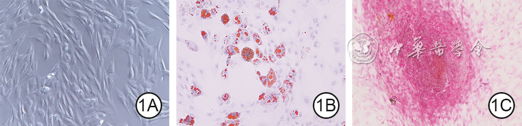
图 1 大鼠脂肪间充质干细胞的形态及其分化能力鉴定 倒置显微镜 ×200。1A.原代细胞培养7 d后呈旋涡状排列;1B.成脂诱导培养21 d后细胞内可见红色脂滴 油红O;1C.成骨诱导培养21 d后细胞内可见红色或橘红色沉淀物 茜素红S

图 2 糖尿病大鼠伤后各时间点2组全层皮肤缺损创面的愈合情况。2A、2B、2C、2D、2E.分别为大鼠伤后0(即刻)、3、7、10、14 d对照组创面;2F、2G、2H、2I、2J.分别为大鼠伤后0、3、7、10、14 d实验组创面,图2H、2I、2J的创面面积分别明显小于图2C、2D、2E
注:大鼠实验组、对照组创面分别注射含大鼠脂肪间充质干细胞的细胞悬液和等量磷酸盐缓冲液

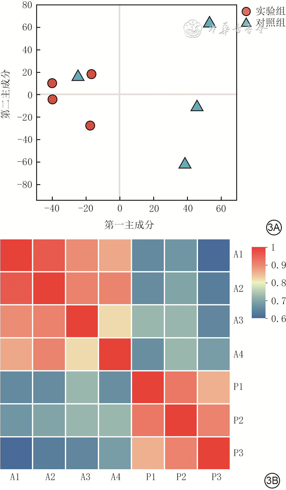
图 3 糖尿病大鼠伤后7 d的2组全层皮肤缺损创面的蛋白质样本的相关性分析。3A.主成分分析;3B.热图
注:大鼠实验组、对照组创面分别注射含大鼠脂肪间充质干细胞的细胞悬液和等量磷酸盐缓冲液;图3A中的横坐标代表数据中方差最大的方向,其单独解释了约31.90%的数据变异,是降维后的核心指标;纵坐标代表数据中方差次大的方向,其对原始数据总方差的解释贡献率为28.00%;各个样本点之间的距离代表了样本间的相似程度,距离越短表明样本间相似性越高;图3B中的纵坐标和横坐标上的A1、A2、A3、A4和P1、P2、P3分别指实验组的4个样本和对照组的3个样本;不同颜色代表样本间相关系数的大小,红色越深代表2个样本间相关性越大,蓝色越深代表2个样本间相关性越小

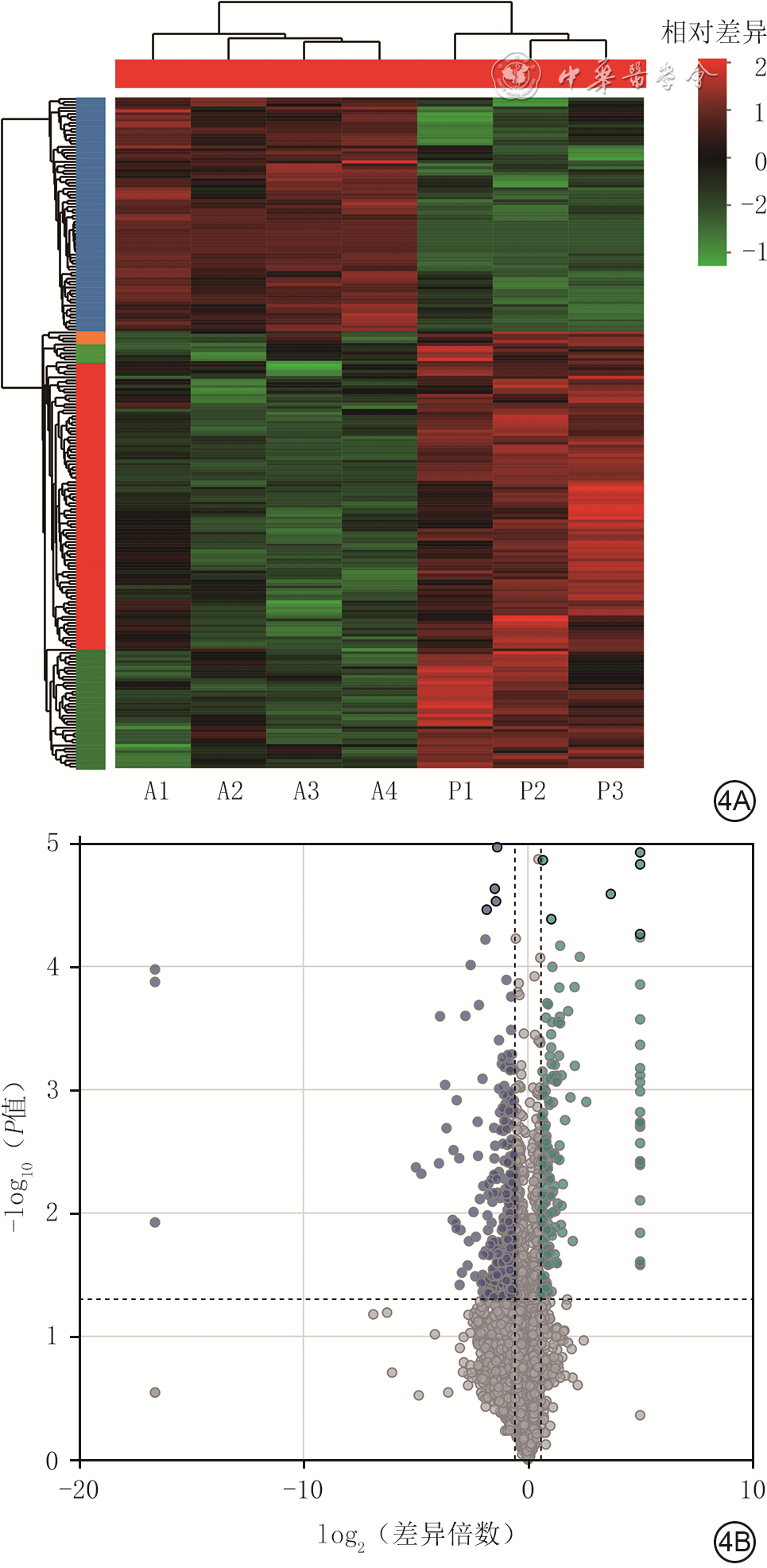
图 4 糖尿病大鼠伤后7 d的2组全层皮肤缺损创面组织中差异表达蛋白的聚类热图和火山图。4A.聚类热图;4B.火山图
注:大鼠实验组、对照组创面分别注射含大鼠脂肪间充质干细胞的细胞悬液和等量磷酸盐缓冲液;图4A中横坐标上的A1、A2、A3、A4和P1、P2、P3分别指实验组的4个样本和对照组的3个样本;上方为样本聚类的树状图,2个样本分支之间的距离越近则其蛋白质的表达模式越接近;左侧为蛋白质聚类的树状图,2个蛋白质分支之间的距离越近则其表达量越接近;图4B中绿色点为实验组较对照组显著上调的蛋白,蓝色点为实验组较对照组显著下调的蛋白,灰色点为实验组较对照组无显著变化的蛋白

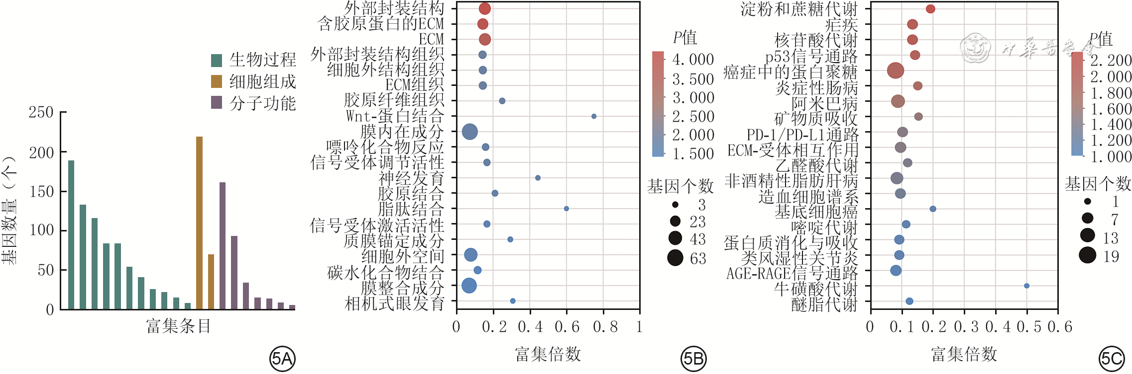
图 5 糖尿病大鼠伤后7 d的2组全层皮肤缺损创面组织中的差异表达蛋白的GO功能注释及富集分析和KEGG通路富集分析。5A.GO功能注释分析;5B.GO功能富集分析(样本数为4);5C.KEGG通路富集分析(样本数为4)
注:大鼠实验组、对照组创面分别注射含大鼠脂肪间充质干细胞的细胞悬液和等量磷酸盐缓冲液;图5A中横坐标从左至右对应富集条目为细胞过程、生物调控、代谢过程、对刺激的反应、发育过程、定位、多细胞生物过程、免疫系统过程、涉及相互作用的生物过程、生殖过程、信号转导,细胞解剖实体、含蛋白质复合物,特异性结合、催化活性、分子功能调节活性、转运蛋白活性、结构分子活性、分子转导活性、分子接头活性;ECM为细胞外基质,PD-1为程序性死亡受体1,PD-L1为程序性死亡配体1,AGE-RAGE为晚期糖基化终末产物-晚期糖基化终末产物受体,GO为基因本体论,KEGG为京都基因和基因组百科全书

图 6 蛋白质印迹法检测的糖尿病大鼠伤后7 d的2组全层皮肤缺损创面组织中Thy-1和GPR177/Wls的蛋白表达
注:大鼠实验组、对照组创面分别注射含大鼠脂肪间充质干细胞的细胞悬液和等量磷酸盐缓冲液;1-1、2-1、3-1为实验组的3个样本;1-2、2-2、3-2为对照组的3个样本;Thy-1为胸腺细胞分化抗原-1,GPR177/Wls为G蛋白偶联受体177/Wnt配体分泌介质
Table 1. 糖尿病大鼠2组全层皮肤缺损创面伤后各时间点的愈合率比较(%,
组别 样本数 3 d 7 d 10 d 14 d 对照组 8 18±7 30±8 62±6 77±8 实验组 8 30±11 66±16 83±8 93±4 t值 -2.21 -4.41 -7.46 -6.65 P值 0.063 <0.001 <0.001 0.003 注:大鼠实验组、对照组创面分别注射含大鼠脂肪间充质干细胞的细胞悬液和等量磷酸盐缓冲液;处理因素主效应,F=40.21,P<0.001;时间因素主效应,F=176.34,P<0.001;二者交互作用,F=8.17,P<0.001  下载: 导出CSV
-

 下载:
下载:


计量
- 文章访问数: 887
- HTML全文浏览量: 703
- PDF下载量: 18
- 被引次数: 0
