


Influence and mechanism of extracellular vesicles derived from human dermal papilla cells on skin fibrosis in mice
-
摘要:
目的 探究人真皮毛乳头细胞(hDPC)来源的细胞外囊泡(hDPC-EV)对小鼠皮肤纤维化的影响及其机制。 方法 该研究为实验研究。收集2024年9月于兰州大学第二医院行毛发移植手术的2例分别为25、40岁的男性患者的100个废弃的毛囊单位,提取原代hDPC并成功鉴定。取第3~5代hDPC,培养后提取hDPC-EV并成功鉴定。采用实时荧光定量反转录PCR(RT-PCR)法检测hDPC和hDPC-EV中微小RNA-182-5p(miRNA-182-5p)的表达(样本数为4)。取30只6周龄雄性C57BL/6J小鼠,皮内注射博来霉素4周制作小鼠皮肤纤维化模型。采用随机数字表法(后续分组方法同此)选取6只造模后的小鼠,另取6只健康未处理6周龄雄性C57BL/6J小鼠,采用蛋白质印迹法检测小鼠正常皮肤组织与纤维化皮肤组织中转化生长因子β1(TGF-β1)的蛋白表达(样本数为3)。将剩余24只造模后的小鼠分为磷酸盐缓冲液(PBS)+miRNA模拟物对照组、细胞外囊泡(EV)+miRNA模拟物对照组、EV+miRNA抑制剂组、miRNA模拟物组(每组6只),分别于注射与组名相对应的试剂2周后,采用蛋白质印迹法检测纤维化皮肤组织中α-平滑肌肌动蛋白(α-SMA)和Ⅰ型胶原的蛋白表达(样本数为3),采用实时荧光定量RT-PCR法检测纤维化皮肤组织中miRNA-182-5p的表达和TGF-β1的mRNA表达(样本数为4)。取人增生性瘢痕成纤维细胞(HSF),分为miRNA-182-5p模拟物+野生型-TGF-β1组、miRNA-182-5p对照+野生型-TGF-β1组、miRNA-182-5p模拟物+突变型-TGF-β1组、miRNA-182-5p对照+突变型-TGF-β1组并转染相应质粒培养36 h后,行双荧光素酶报告基因实验检测miRNA-182-5p与TGF-β1的相互作用,以相对荧光素酶活性表示(样本数为5)。 结果 hDPC-EV中miRNA-182-5p的表达明显高于hDPC(t=5.48,P < 0.05)。与小鼠正常皮肤组织比较,小鼠纤维化皮肤组织中TGF-β1的蛋白表达升高。处理2周后,与PBS+miRNA模拟物对照组比较,EV+miRNA模拟物对照组小鼠纤维化皮肤组织中α-SMA、Ⅰ型胶原的蛋白表达均明显降低(P < 0.05);与EV+miRNA模拟物对照组比较,EV+miRNA抑制剂组小鼠纤维化皮肤组织中α-SMA、Ⅰ型胶原的蛋白表达均明显升高(P < 0.05);与EV+miRNA抑制剂组比较,miRNA模拟物组小鼠纤维化皮肤组织中α-SMA、Ⅰ型胶原的蛋白表达均明显降低(P < 0.05)。处理2周后,与EV+miRNA模拟物对照组比较,PBS+miRNA模拟物对照组和EV+miRNA抑制剂组小鼠纤维化皮肤组织中miRNA-182-5p表达均明显降低(P < 0.05)而TGF-β1的mRNA表达均明显升高(P < 0.05);与EV+miRNA抑制剂组比较,PBS+miRNA模拟物对照组小鼠纤维化皮肤组织中miRNA-182-5p表达明显升高(P < 0.05),miRNA模拟物组小鼠纤维化皮肤组织中miRNA-182-5p表达明显升高(P < 0.05)而TGF-β1的mRNA表达明显降低(P < 0.05)。培养36 h后,miRNA-182-5p模拟物+野生型-TGF-β1组HSF的相对荧光素酶活性为0.594±0.019,明显低于miRNA-182-5p对照+野生型-TGF-β1组的1.000±0.153(t=5.87,P < 0.05);miRNA-182-5p模拟物+突变型-TGF-β1组HSF的相对荧光素酶活性为0.911±0.085,与miRNA-182-5p对照+突变型-TGF-β1组的0.934±0.027比较,差异无统计学意义(P > 0.05)。表明miRNA-182-5p可靶向调控TGF-β1。 结论 hDPC-EV可通过递送miRNA-182-5p靶向抑制TGF-β1信号通路来减轻博来霉素诱导的小鼠皮肤纤维化。 Abstract:Objective To explore the influence and mechanism of extracellular vesicles (EVs) derived from human dermal papilla cells (hDPCs), i. e. hDPC-EVs on skin fibrosis in mice. Methods This study was an experimental research. One hundred discarded hair follicle units from 2 male patients aged 25 years and 40 years who underwent hair transplantation surgery at the Second Hospital of Lanzhou University in September 2024 were collected, and primary hDPCs were extracted and successfully identified. After hDPCs of passage 3 to 5 were taken and cultured, the hDPC-EVs were extracted and successfully identified. The expression of microRNA-182-5p (miRNA-182-5p) in hDPCs and hDPC-EVs was detected by real-time fluorescence quantitative reverse transcription polymerase chain reaction (RT-PCR, n=4). Thirty 6-week-old male C57BL/6J mice were taken and injected intradermal bleomycin for 4 weeks to establish mouse skin fibrosis models. Six mice after modeling were selected according to the random number table method (the same grouping method applied hereafter), and another 6 healthy untreated 6-week-old male C57BL/6J mice were taken. The protein expression of transforming growth factor β1 (TGF-β1) in normal skin tissue and fibrotic skin tissue of mice was detected by Western blotting (n=3). The remaining 24 mice after modeling were divided into phosphate buffered solution (PBS)+miRNA mimic control group, EV+miRNA mimic control group, EV+miRNA inhibitor group, and miRNA mimic group (n=6). Two weeks after injection of the reagents corresponding to the group names, the protein expressions of α-smooth muscle actin (α-SMA) and type Ⅰ collagen in fibrotic skin tissue was detected by Western blotting (n=3), and the expression of miRNA-182-5p and the mRNA expression of TGF-β1 in fibrotic skin tissue was detected by real-time fluorescence quantitative RT-PCR (n=4). Human hypertrophic scar fibroblasts (HSFs) were taken and divided into miRNA-182-5p mimic+wild-type TGF-β1 group, miRNA-182-5p control+wild-type TGF-β1 group, miRNA-182-5p mimic+mutant-type TGF-β1 group, and miRNA-182-5p control+mutant-type TGF-β1 group. Cells in each group were transfected with the corresponding plasmids and cultured for 36 h. Double luciferase reporter gene assay was performed to detect the interaction between miRNA-182-5p and TGF-β1 (denoted as relative luciferase activity, n=5). Results The expression of miRNA-182-5p in hDPC-EVs was significantly higher than that in hDPCs (t=5.48, P < 0.05). Compared with that in normal skin tissue of mice, the protein expression of TGF-β1 was increased in fibrotic skin tissue of mice. After 2 weeks of treatment, compared with those in PBS+miRNA mimic control group, the protein expressions of α-SMA and type Ⅰ collagen in the fibrotic skin tissue of mice in EV+miRNA mimic control group were significantly decreased (P < 0.05); compared with those in EV+miRNA mimic control group, the protein expressions of α-SMA and type Ⅰ collagen in the fibrotic skin tissue of mice in EV+miRNA inhibitor group were significantly increased (P < 0.05); compared with those in EV+miRNA inhibitor group, the protein expressions of α-SMA and type Ⅰ collagen in the fibrotic skin tissue of mice in miRNA mimic group were significantly decreased (P < 0.05). After 2 weeks of treatment, compared with those in EV+miRNA mimic control group, the expression of miRNA-182-5p in the fibrotic skin tissue of mice in PBS+miRNA mimic control group and EV+miRNA inhibitor group was significantly decreased (P < 0.05), while the mRNA expression of TGF-β1 was significantly increased (P < 0.05). Compared with those in EV+miRNA inhibitor group, the expression of miRNA-182-5p in fibrotic skin tissue of mice in PBS+miRNA mimic control was significantly increased (P < 0.05); the expression of miRNA-182-5p in the fibrotic skin tissue of mice was significantly increased (P < 0.05), while the mRNA expression of TGF-β1 was significantly decreased in miRNA mimic group (P < 0.05). After 36 h of culture, the relative luciferase activity of HSFs in miRNA-182-5p mimic+wild-type TGF-β1 group was 0.594±0.019, which was significantly lower than 1.000±0.153 in miRNA-182-5p control+wild-type TGF-β1 group (t=5.87, P < 0.05); the relative luciferase activity of HSFs in miRNA-182-5p mimic+mutant-type TGF-β1 group was 0.911±0.085, which has no statistically significant difference with 0.934±0.027 of miRNA-182-5p control+mutant-type TGF-β1 group (P > 0.05), indicating that miRNA-182-5p could exerted targeted regulation of TGF-β1. Conclusions hDPC-EVs alleviate bleomycin-induced skin fibrosis in mice by delivering miRNA-182-5p to inhibit the TGF-β1 signal pathway. -
Key words:
- Skin /
- Cicatrix /
- Fibrosis /
- Extracellular vesicles /
- Transforming growth factor beta1 /
- MicroRNAs /
- Dermal papilla cells
本文亮点(1) 揭示人真皮毛乳头细胞来源的细胞外囊泡(hDPC-EV)通过递送微小RNA-182-5p靶向抑制转化生长因子β1信号通路,减轻博来霉素诱导的小鼠皮肤纤维化。(2) 证实hDPC-EV中的微小RNA-182-5p可抑制皮肤纤维化标志物的表达,为毛囊来源干细胞在体应用提供新靶点。 -
参考文献
(40) [1] Edwards J. Hypertrophic scar management[J]. Br J Nurs, 2022, 31(20): S24-S31. DOI: 10.12968/bjon.2022.31.20.S24. [2] Jung BK, Roh TS, Roh H, et al. Effect of mortalin on scar formation in human dermal fibroblasts and a rat incisional scar model[J]. Int J Mol Sci, 2022, 23(14): 7918. DOI: 10.3390/ijms23147918. [3] 王运帷, 罗亮, 曹鹏, 等. 真皮毛乳头细胞分离培养技术的研究进展[J/CD]. 中华损伤与修复杂志(电子版), 2022, 17(6): 520-523. DOI: 10.3877/cma.j.issn.1673-9450.2022.06.010 .[4] Wang YW, Shen K, Sun YL, et al. Extracellular vesicles from 3D cultured dermal papilla cells improve wound healing via Krüppel-like factor 4/vascular endothelial growth factor A -driven angiogenesis[J/OL]. Burns Trauma, 2023, 11: tkad034[2024-09-25]. https://pubmed.ncbi.nlm.nih.gov/37908562/ . DOI:10.1093/burnst/tkad034 .[5] 王运帷, 张浩, 曹鹏, 等. 小鼠真皮毛乳头细胞外囊泡对人增生性瘢痕成纤维细胞的影响及其机制[J]. 中华烧伤与创面修复杂志, 2024, 40(3): 258-265. DOI: 10.3760/cma.j.cn501225-20231107-00185. [6] Wang J, Barr MM, Wehman AM. Extracellular vesicles[J]. Genetics, 2024, 227(4): iyae088. DOI: 10.1093/genetics/iyae088. [7] Ramírez-Hernández AA, Velázquez-Enríquez JM, Santos-Álvarez JC, et al. The role of extracellular vesicles in idiopathic pulmonary fibrosis progression: an approach on their therapeutics potential[J]. Cells, 2022, 11(4): 630. DOI: 10.3390/cells11040630. [8] Xiao T, Meng W, Jin Z, et al. miR-182-5p promotes hepatocyte-stellate cell crosstalk to facilitate liver regeneration[J]. Commun Biol, 2022, 5(1): 771. DOI: 10.1038/s42003-022-03714-0. [9] 曹鹏, 王运帷, 官浩, 等. 机械张力对兔耳增生性瘢痕的形成及转化生长因子β1/Smad信号通路的影响[J]. 中华烧伤与创面修复杂志, 2022, 38(12): 1162-1169. DOI: 10.3760/cma.j.cn501120-20211213-00412. [10] Ahuja S, Zaheer S. Multifaceted TGF-β signaling, a master regulator: from bench-to-bedside, intricacies, and complexities[J]. Cell Biol Int, 2024, 48(2): 87-127. DOI: 10.1002/cbin.12097. [11] Topouzi H, Logan NJ, Williams G, et al. Methods for the isolation and 3D culture of dermal papilla cells from human hair follicles[J]. Exp Dermatol, 2017, 26(6): 491-496. DOI: 10.1111/exd.13368. [12] 王运帷. 新型三维真皮毛乳头细胞外囊泡对皮肤创面愈合的作用与机制研究[D]. 西安: 空军军医大学, 2023. [13] Xu C, Zhang H, Yang C, et al. miR-125b-5p delivered by adipose-derived stem cell exosomes alleviates hypertrophic scarring by suppressing Smad2[J/OL]. Burns Trauma, 2024, 12: tkad064[2024-09-25]. https://pubmed.ncbi.nlm.nih.gov/38765787/ . DOI:10.1093/burnst/tkad064 .[14] Zheng X, Huang M, Xing L, et al. The circRNA circSEPT9 mediated by E2F1 and EIF4A3 facilitates the carcinogenesis and development of triple-negative breast cancer[J]. Mol Cancer, 2020, 19(1): 73. DOI: 10.1186/s12943-020-01183-9. [15] Atiyeh BS. Nonsurgical management of hypertrophic scars: evidence-based therapies, standard practices, and emerging methods[J]. Aesthetic Plast Surg, 2020, 44(4): 1320-1344. DOI: 10.1007/s00266-020-01820-0. [16] Yuan B, Upton Z, Leavesley D, et al. Vascular and collagen target: a rational approach to hypertrophic scar management[J]. Adv Wound Care (New Rochelle), 2023, 12(1): 38-55. DOI: 10.1089/wound.2020.1348. [17] Hameedi SG, Saulsbery A, Olutoye OO. The pathophysiology and management of pathologic scarring-a contemporary review[J]. Adv Wound Care (New Rochelle), 2025, 14(1): 48-64. DOI: 10.1089/wound.2023.0185. [18] 周圳滔, 赵沁园, 赵钧, 等. 毛囊及相关干细胞在创面无瘢痕愈合中的研究进展[J]. 中国修复重建外科杂志, 2021, 35(2): 241-245. DOI: 10.7507/1002-1892.202005086. [19] Rippa AL, Kalabusheva EP, Vorotelyak EA. Regeneration of dermis: scarring and cells involved[J]. Cells, 2019, 8(6): 607. DOI: 10.3390/cells8060607. [20] Qi SH, Liu P, Xie JL, et al. Experimental study on repairing of nude mice skin defects with composite skin consisting of xenogeneic dermis and epidermal stem cells and hair follicle dermal papilla cells[J]. Burns, 2008, 34(3): 385-392. DOI: 10.1016/j.burns.2007.04.003. [21] Leirós GJ, Kusinsky AG, Drago H, et al. Dermal papilla cells improve the wound healing process and generate hair bud-like structures in grafted skin substitutes using hair follicle stem cells[J]. Stem Cells Transl Med, 2014, 3(10): 1209-1219. DOI: 10.5966/sctm.2013-0217. [22] 石傲, 王运帷, 康宇晨, 等. 水凝胶促进创面血管化的研究进展[J]. 中华烧伤与创面修复杂志, 2025, 41(3): 295-300. DOI: 10.3760/cma.j.cn501225-20240521-00193. [23] Yin S, Zhou S, Ren D, et al. Mesenchymal stem cell-derived exosomes attenuate epithelial-mesenchymal transition of HK-2 cells[J]. Tissue Eng Part A, 2022, 28(13/14): 651-659. DOI: 10.1089/ten.TEA.2021.0190. [24] Cecchin R, Troyer Z, Witwer K, et al. Extracellular vesicles: the next generation in gene therapy delivery[J]. Mol Ther, 2023, 31(5): 1225-1230. DOI: 10.1016/j.ymthe.2023.01.021. [25] Zhang X, Zhang H, Gu J, et al. Engineered extracellular vesicles for cancer therapy[J]. Adv Mater, 2021, 33(14): e2005709. DOI: 10.1002/adma.202005709. [26] Egal E, Kamdem SD, Yoshigi M, et al. EphB2 receptor promotes dermal fibrosis in systemic sclerosis[J]. Arthritis Rheumatol, 2024, 76(8): 1303-1316. DOI: 10.1002/art.42858. [27] 李超. 曲安奈德联合5-氟尿嘧啶对博来霉素诱导小鼠增生性瘢痕的实验研究[D]. 延吉: 延边大学, 2022. [28] 周思政. 皮肤创伤愈合和增生性瘢痕动物模型的研究进展[J]. 组织工程与重建外科杂志, 2018, 14(1): 48-52. DOI: 10.3969/j.issn.1673-0364.2018.01.013. [29] Wang J, Zhao M, Zhang H, et al. KLF4 alleviates hypertrophic scar fibrosis by directly activating BMP4 transcription[J]. Int J Biol Sci, 2022, 18(8): 3324-3336. DOI: 10.7150/ijbs.71167. [30] Li Y, Zhang J, Shi J, et al. Correction to: exosomes derived from human adipose mesenchymal stem cells attenuate hypertrophic scar fibrosis by miR-192-5p/IL-17RA/Smad axis[J]. Stem Cell Res Ther, 2021, 12(1): 490. DOI: 10.1186/s13287-021-02568-3. [31] 贺伟峰, 吴军, 罗高兴, 等. 增生性瘢痕组织和正常皮肤来源成纤维细胞的差异蛋白质组学研究[G]. 漳州: 第十届全国烧伤救治专题研讨会暨福建省第八次烧伤外科学术研讨会, 2013. [32] Wu B, Feng J, Guo J, et al. ADSCs-derived exosomes ameliorate hepatic fibrosis by suppressing stellate cell activation and remodeling hepatocellular glutamine synthetase-mediated glutamine and ammonia homeostasis [J]. Stem Cell Res Ther, 2022, 13(1): 494. DOI: 10.1186/s13287-022-03049-x. [33] Hwangbo C, Tae N, Lee S, et al. Syntenin regulates TGF-β1-induced Smad activation and the epithelial-to-mesenchymal transition by inhibiting caveolin-mediated TGF-β type Ⅰ receptor internalization[J]. Oncogene, 2016, 35(3): 389-401. DOI: 10.1038/onc.2015.100. [34] Zheng Z, Zhang XL, Dang C, et al. Fibromodulin is essential for fetal-type scarless cutaneous wound healing[J]. Am J Pathol, 2016, 186(11): 2824-2832. DOI: 10.1016/j.ajpath.2016.07.023. [35] Chen L, Li J, Li Q, et al. Non-coding RNAs: the new insight on hypertrophic scar[J]. J Cell Biochem, 2017, 118(8): 1965-1968. DOI: 10.1002/jcb.25873. [36] Yu F, Liu Z, Feng J, et al. Hyaluronic acid modified extracellular vesicles targeting hepatic stellate cells to attenuate hepatic fibrosis[J]. Eur J Pharm Sci, 2024, 198: 106783. DOI: 10.1016/j.ejps.2024.106783. [37] Nosalski R, Siedlinski M, Denby L, et al. T-cell-derived miRNA-214 mediates perivascular fibrosis in hypertension [J]. Circ Res, 2020, 126(8): 988-1003. DOI: 10.1161/CIRCRESAHA.119.315428. [38] Zou A, Liu P, Liu T, et al. Long non-coding RNA HOXA11-AS contributes to the formation of keloid by relieving the inhibition of miR-182-5p on ZNF217[J]. Burns, 2023, 49(5): 1157-1169. DOI: 10.1016/j.burns.2022.07.010. [39] Xu Q, Miao Y, Ren J, et al. Silencing of Nesprin-2 inhibits the differentiation of myofibroblasts from fibroblasts induced by mechanical stretch[J]. Int Wound J, 2022, 19(5): 978-986. DOI: 10.1111/iwj.13694. [40] Chen Y, Zhang Q, Zhou Y, et al. Inhibition of miR-182-5p attenuates pulmonary fibrosis via TGF-β/Smad pathway[J]. Hum Exp Toxicol, 2020, 39(5): 683-695. DOI: 10.1177/0960327119895549. -

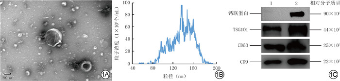
图 1 hDPC-EV的鉴定。1A.hDPC-EV为盘状囊泡结构 透射电子显微镜×40 000;1B.hDPC-EV平均粒径为142.91 nm;1C.hDPC-EV的囊泡阳性标志物TSG101、CD63、CD9阳性,阴性标志物钙联蛋白阴性
注:hDPC-EV为人真皮毛乳头细胞来源的细胞外囊泡,TSG101为肿瘤易感基因101;条带上方1、2分别指示hDPC-EV、人真皮毛乳头细胞
Figure 1. Identification of hDPC-EVs

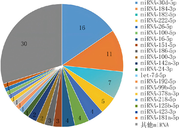
图 2 高通量测序检测的人真皮毛乳头细胞来源的细胞外囊泡中miRNA的丰度(%)
注:miRNA为微小RNA;其他miRNA为丰度 < 1%的miRNA
Figure 2. The abundance of miRNAs in extracellular vesicles derived from human dermal papilla cells detected by high-throughput sequencing

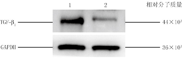
图 3 蛋白质印迹法检测的小鼠正常皮肤组织和纤维化皮肤组织中TGF-β1的蛋白表达
注:TGF-β1为转化生长因子β1,GAPDH为3-磷酸甘油醛脱氢酶;条带上方1、2分别指示行博来霉素注射4周的小鼠的纤维化皮肤组织和未行博来霉素注射的小鼠的正常皮肤组织
Figure 3. The protein expression of TGF-β1 in normal skin tissue and fibrotic skin tissue of mice detected by Western blotting

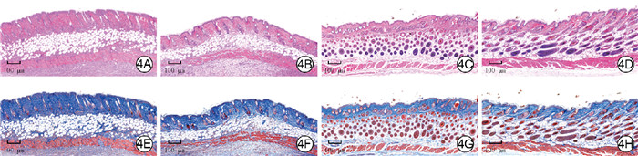
图 4 4组皮肤纤维化小鼠处理2周后纤维化皮肤组织病理学和胶原沉积情况。4A、4B、4C、4D.分别为PBS+miRNA模拟物对照组、EV+miRNA抑制剂组、EV+miRNA模拟物对照组、miRNA模拟物组小鼠纤维化皮肤组织,图4A和图4B中皮肤附属器缺乏,图4C和图4D中皮肤附属器丰富 苏木精-伊红×40;4E、4F、4G、4H.分别为PBS+miRNA模拟物对照组、EV+miRNA抑制剂组、EV+miRNA模拟物对照组、miRNA模拟物组小鼠纤维化皮肤组织,图4E和图4F中胶原大量沉积、纤维排列紊乱,图4G和图4H中胶原沉积良好、纤维排列趋于正常 Masson×40
注:对磷酸盐缓冲液(PBS)+微小RNA(miRNA)模拟物对照组、细胞外囊泡(EV)+miRNA抑制剂组、EV+miRNA模拟物对照组、miRNA模拟物组小鼠分别注射PBS+miRNA模拟物对照、人真皮毛乳头细胞来源的EV(hDPC-EV)+miRNA-182-5p抑制剂、hDPC-EV+miRNA模拟物对照、miRNA-182-5p模拟物
Figure 4. Histopathology and collagen deposition in the fibrotic skin tissue of 4 groups of mice with skin fibrosis after 2 weeks of treatment

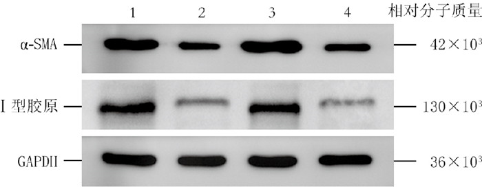
图 5 蛋白质印迹法检测的4组皮肤纤维化小鼠处理2周后纤维化皮肤组织中α-SMA和Ⅰ型胶原的蛋白表达
注:α-SMA为α-平滑肌肌动蛋白,GAPDH为3-磷酸甘油醛脱氢酶,条带上方1、2、3、4分别指示对小鼠注射磷酸盐缓冲液(PBS)+微小RNA(miRNA)模拟物对照、人真皮毛乳头细胞来源的细胞外囊泡(hDPC-EV)+miRNA模拟物对照、hDPC-EV+miRNA-182-5p抑制剂、miRNA-182-5p模拟物的PBS+miRNA模拟物对照组、细胞外囊泡(EV)+miRNA模拟物对照组、EV+miRNA抑制剂组、miRNA模拟物组
Figure 5. The protein expressions of α-SMA and type Ⅰ collagen in the fibrotic skin tissue of 4 groups of mice with skin fibrosis after 2 weeks of treatment detected by Western blotting
表 1 4组皮肤纤维化小鼠处理2周后纤维化皮肤组织中miRNA-182-5p的表达和TGF-β1的mRNA表达(x ± s)
Table 1. Expression of miRNA-182-5p and mRNA expression of TGF-β1 in the fibrotic skin tissue of 4 groups of mice with skin fibrosis after 2 weeks of treatment
组别 样本数 miRNA-182-5p TGF-β1 PBS+miRNA模拟物对照组 4 0.31±0.04 0.76±0.09 EV+miRNA模拟物对照组 4 1.19±0.11 0.29±0.05 EV+miRNA抑制剂组 4 0.13±0.06 0.88±0.08 miRNA模拟物组 4 1.30±0.05 0.23±0.03 F值 289.00 118.60 P值 < 0.001 < 0.001 P1值 < 0.001 < 0.001 P2值 < 0.001 < 0.001 P3值 0.021 0.115 P4值 < 0.001 < 0.001 P5值 0.119 0.085 注:miRNA为微小RNA,TGF-β1为转化生长因子β1;对磷酸盐缓冲液(PBS)+miRNA模拟物对照组、细胞外囊泡(EV)+miRNA模拟物对照组、EV+miRNA抑制剂组、miRNA模拟物组小鼠分别注射PBS+miRNA模拟物对照、人真皮毛乳头细胞来源的EV(hDPC-EV)+miRNA模拟物对照、hDPC-EV+miRNA-182-5p抑制剂、miRNA-182-5p模拟物;F值、P值为组间各指标总体比较所得;P1值、P2值、P3值、P4值、P5值分别为PBS+miRNA模拟物对照组与EV+miRNA模拟物对照组、EV+miRNA模拟物对照组与EV+miRNA抑制剂组、EV+miRNA抑制剂组与PBS+miRNA模拟物对照组、miRNA模拟物组与EV+miRNA抑制剂组、miRNA模拟物组与EV+miRNA模拟物对照组各指标比较所得  下载: 导出CSV
-

 下载:
下载:

计量
- 文章访问数: 972
- HTML全文浏览量: 462
- PDF下载量: 5
- 被引次数: 0
