


Effects of ultrathin inguinal flap free transplantation in repairing hand and foot wounds
-
摘要:
目的 探讨超薄腹股沟皮瓣游离移植修复手足创面的效果。 方法 该研究为病例系列研究。2021年1月—2024年3月,武汉大学同仁医院暨武汉市第三医院收治18例符合入选标准的手足部皮肤软组织缺损伴肌腱和/或骨损伤、外露的患者(男14例、女4例,年龄19~66岁),共21个创面,其中12个位于手部、9个位于足部。清创后皮肤软组织缺损面积为5.0 cm×2.5 cm~16.0 cm×5.0 cm。以纯皮穿支为中心,应用逆行解剖法获取含真皮下血管网的超薄腹股沟皮瓣,皮瓣大小为6.0 cm×3.0 cm~17.0 cm×6.5 cm。其中,15个皮瓣以旋髂浅动脉为血管蒂,5个皮瓣以旋髂浅动脉和腹壁浅动脉共干为血管蒂,1个皮瓣以旋髂浅动脉和腹壁浅动脉双血管为血管蒂。将皮瓣转移至受区创面,将血管蒂中的动静脉与受区动静脉端端吻合,然后封闭创面。减张缝合供区创面。术中,测量并记录皮瓣厚度、血管蒂中动脉吻合口径、携带的纯皮穿支数及多穿支皮瓣中相邻纯皮穿支间距离。术后,观察皮瓣成活情况、并发症发生情况及供区创面愈合情况。随访时,观察受区修复情况。末次随访时,根据总主动活动度(TAM)评估患指功能,采用美国足踝外科医师协会踝-后足评分系统(AOFAS-AHS)对患足功能进行评分,采用利克特量表评价患者对疗效的满意度。 结果 患者皮瓣厚度为0.2~0.5 cm,平均0.4 cm;血管蒂中动脉吻合口径为0.5~1.2 mm,平均0.7 mm;7个皮瓣各携带1条纯皮穿支,6个皮瓣各携带2条纯皮穿支,8个皮瓣各携带3条纯皮穿支;多穿支皮瓣中相邻纯皮穿支间距离为0.8~3.5 cm,平均1.7 cm。术后,1个皮瓣出现小范围坏死,经清创缝合后成活;其余皮瓣均顺利成活。3个患指远端存在血运障碍,行动脉再通后均成活。1个供区创面因局部张力较高愈合不良,经清创缝合后愈合;其余供区创面均顺利愈合。术后6~35个月随访时,皮瓣质地柔软、弹性良好,其中2个皮瓣因局部稍显臃肿,于术后3~5个月行修薄处理。末次随访时,10个患指的TAM评估为优、2个为良,9个患足的AOFAS-AHS评分为99~100分,18例患者对疗效均表示非常满意。 结论 超薄腹股沟皮瓣具有厚度薄、血运丰富、切取方法可靠等优点,用于修复手足烧创伤后创面,可获得理想的外形与功能效果,且术后二次修整率低,值得临床推广应用。 Abstract:Objective To explore the effects of ultrathin inguinal flap free transplantation in repairing hand and foot wounds. Methods This study was a case series study. From January 2021 to March 2024, 18 patients (14 males and 4 females, aged 19 to 66 years) with skin and soft tissue defects of hands and feet accompanied by tendon and/or bone injuries and exposure were admitted to Tongren Hospital of Wuhan University & Wuhan Third Hospital. A total of 21 wounds were involved, including 12 on hands and 9 on feet. The area of skin and soft tissue defects after debridement was 5.0 cm×2.5 cm to 16.0 cm×5.0 cm. Using the reverse dissection method, ultrathin inguinal flaps containing the subdermal vascular network were obtained with the pure skin perforator as the center. The size of flaps was 6.0 cm×3.0 cm to 17.0 cm×6.5 cm. Among them, 15 flaps were based on the superficial circumflex iliac artery as the vascular pedicle, 5 flaps were based on the common trunk of the superficial circumflex iliac artery and the superficial epigastric artery as the vascular pedicle, and 1 flap was based on the dual vessels of the superficial circumflex iliac artery and the superficial epigastric artery as the vascular pedicle. The flaps were transferred to the recipient wounds, and the arteries and veins of the vascular pedicles were end-to-end anastomosed with the recipient arteries and veins, and then the wounds were closed. The donor site wounds were sutured with tension reduction. During the operation, the thickness of flaps, the diameter of arterial anastomosis in vascular pedicle, the number of pure skin perforators carried, and the distance between adjacent pure skin perforators in multi-perforator flaps were measured and recorded. Postoperatively, the survival of the flaps, the occurrence of complications, as well as the healing of the donor site wounds were observed. During follow-up, the repair of the recipient areas was observed. At the last follow-up, the total active motion (TAM) was used to evaluate the function of the affected fingers, and the American Orthopaedic Foot and Ankle Society Ankle-Hindfoot Scale (AOFAS-AHS) was used to score the function of the affected feet. The Likert scale was used to evaluate the patients' satisfaction with the therapeutic effect. Results The thickness of flaps of patients was 0.2 to 0.5 cm, with an average of 0.4 cm; the diameter of arterial anastomosis in vascular pedicle was 0.5 to 1.2 mm, with an average of 0.7 mm; 7 flaps each carried one pure skin perforator, 6 flaps each carried two pure skin perforators, and 8 flaps each carried three pure skin perforators. The distance between adjacent pure skin perforators in multi-perforator flaps was 0.8 to 3.5 cm, with an average of 1.7 cm. Postoperatively, one flap showed small area of necrosis, which survived after debridement and suture; the remaining flaps survived smoothly. Three affected fingers had blood supply disorder at the distal end, and all survived after arterial recanalization. One donor site wound healed poorly due to high local tension and healed after debridement and suture, and the remaining donor site wounds healed smoothly. During the 6 to 35 months of follow-up, the flaps were soft and elastic. Two flaps were slightly swollen and underwent thinning treatment 3 to 5 months after surgery. At the last follow-up, the TAM of 10 affected fingers were excellent, and two were good, the AOFAS-AHS score of 9 affected feet was 99 to 100 points, and all 18 patients were very satisfied with the effects. Conclusions The ultrathin inguinal flap has the advantages of thin thickness, rich blood supply, and reliable harvesting method. Its application in repairing hand and foot wounds after burns and trauma can achieve ideal aesthetic and functional results, with a low rate of secondary revision, and it is worthy of clinical promotion and application. -
Key words:
- Burns /
- Perforator flap /
- Microsurgery /
- Arteriovenous anastomosis /
- Reconstructive surgical procedures /
- Hand /
- Foot /
- Soft tissue injury
-
参考文献
(40) [1] AltiparmakM, ChaHG, HongJP, et al. Superficial circumflex iliac artery perforator flap as a workhorse flap: systematic review and meta-analysis[J]. J Reconstr Microsurg, 2020,36(8):600-605. DOI: 10.1055/s-0040-1713666. [2] 亢向辉,赵建强,段冰,等. 游离髂腹股沟皮瓣在手足部组织缺损修复中的应用[J]. 中华手外科杂志,2023,39(6):550-552. DOI: 10.3760/cma.j.cn311653-20230604-00117. [3] BarnhillCW, GreysonMA, IorioML. Superficial circumflex iliac artery perforator flap reconstruction of the upper extremity[J]. Hand Clin, 2024,40(2):179-187. DOI: 10.1016/j.hcl.2023.10.001. [4] WangJ, ChenY, ZhouG, et al. Polydopamine-coated Antheraea pernyi (A. pernyi) silk fibroin films promote cell adhesion and wound healing in skin tissue repair[J]. ACS Appl Mater Interfaces, 2019,11(38):34736-34743. DOI: 10.1021/acsami.9b12643. [5] 陶泉.手部损伤康复[M]. 上海:上海交通大学出版社, 2006:41. [6] KitaokaHB, AlexanderIJ, AdelaarRS, et al. Clinical rating systems for the ankle-hindfoot, midfoot, hallux, and lesser toes[J]. Foot Ankle Int, 1994,15(7):349-353. DOI: 10.1177/107110079401500701. [7] SchweizerR, PedrazziN, KleinHJ, et al. Risk factors for mortality and prolonged hospitalization in electric burn injuries[J]. J Burn Care Res, 2021,42(3):505-512. DOI: 10.1093/jbcr/iraa192. [8] 王克彬,尹国瑞,刘凯,等. 游离足底内侧皮瓣修复手部掌侧软组织缺损17例[J]. 中华显微外科杂志,2024,47(3):321-324. DOI: 10.3760/cma.j.cn441206-20230926-00042. [9] 张卫东,张伟,余刚,等. 腓肠内侧动脉穿支皮瓣游离移植修复手足电烧伤创面的效果[J]. 中华烧伤与创面修复杂志,2025,41(4):386-393. DOI: 10.3760/cma.j.cn501225-20240611-00226. [10] 张丕红. 浅谈薄型穿支皮瓣切取与穿支血管探测和皮瓣血运评估[J]. 中华烧伤与创面修复杂志,2023,39(10):911-918. DOI: 10.3760/cma.j.cn501225-20230812-00047. [11] 狄海萍,邢培朋,郑军杰,等. 超薄股前外侧皮瓣一期分指修复手掌合并多指创面的疗效[J]. 中华烧伤与创面修复杂志,2023,39(9):835-841. DOI: 10.3760/cma.j.cn501225-20221129-00514. [12] 杨成兰,曾雪琴,闫雪萍,等.双侧腹壁真皮下血管网超薄皮瓣在手指皮肤软组织缺损中的应用[J].中华手外科杂志, 2025, 41(2):167-170.DOI: 10.3760/cma.j.cn311653-20240802-00209. [13] 张伟,张卫东,徐军辉,等. 分叶腹股沟皮瓣游离移植的临床应用效果[J]. 中华烧伤与创面修复杂志,2025,41(1):36-44. DOI: 10.3760/cma.j.cn501225-20241014-00384. [14] KimHB, MinJC, PakCJ, et al. Maximizing the versatility of thin flap from the groin area as a workhorse flap: the selective use of superficial circumflex iliac artery perforator (SCIP) free flap and superficial inferior epigastric artery (SIEA) free flap with precise preoperative planning[J]. J Reconstr Microsurg, 2023,39(2):148-155. DOI: 10.1055/a-1887-7274. [15] ZublerC, HaberthürD, HlushchukR, et al. The anatomical reliability of the superficial circumflex iliac artery perforator (SCIP) flap[J]. Ann Anat, 2021,234:151624. DOI: 10.1016/j.aanat.2020.151624. [16] 刘峰,张伟,谢卫国,等. 微型游离腹股沟穿支皮瓣修复手部小型创面的效果[J]. 中华烧伤与创面修复杂志,2023,39(10):933-938. DOI: 10.3760/cma.j.cn501225-20230701-00244. [17] 陈斓,张伟,谢卫国,等. 扩张髂腹股沟皮瓣游离移植整复大面积烧伤后严重瘢痕挛缩畸形的临床效果[J]. 中华烧伤与创面修复杂志,2022,38(4):321-327. DOI: 10.3760/cma.j.cn501225-20220210-00021. [18] 李辉. 带蒂真皮下血管网超薄皮瓣在手部皮肤缺损中的应用[J].实用医学杂志,1999(5):83-84. DOI: 10.3969/j.issn.1006-5725.1999.05.066. [19] 顾荣,王海文,江新民,等. “U”形修薄髂腹股沟皮瓣移植修复四肢皮肤软组织缺损[J]. 中华显微外科杂志,2016,39(4):340-343. DOI: 10.3760/cma.j.issn.1001-2036.2016.04.008. [20] 李月辉. 带蒂真皮下血管网超薄皮瓣在手烧伤后畸形修复中的应用[J].现代预防医学,2010,37(9):1757-1758. [21] NarushimaM, YamasobaT, IidaT, et al. Pure skin perforator flap for microtia and congenital aural atresia using supermicrosurgical techniques[J]. J Plast Reconstr Aesthet Surg, 2011,64(12):1580-1584. DOI: 10.1016/j.bjps.2011.07.005. [22] LeeK, ParkBY, KimE, et al. Superthin SCIP flap for reconstruction of subungual melanoma: aesthetic functional surgery[J]. Plast Reconstr Surg, 2017,140(6):1278-1289. DOI: 10.1097/PRS.0000000000003881. [23] ParkSY, LeeK. Use of the superthin superficial circumflex iliac artery perforator flap for reconstruction of lower lip defects[J]. Plast Reconstr Surg, 2024,154(3):585e-588e. DOI: 10.1097/PRS.0000000000010978. [24] KarakawaR, IidaT, YoshimatsuH, et al. Functional and aesthetic reconstruction for microtia using the combination of superficial circumflex iliac artery perforator superthin flap transfer and skin grafting[J]. Plast Reconstr Surg Glob Open, 2019,7(7):e2312. DOI: 10.1097/GOX.0000000000002312. [25] NarushimaM, YamasobaT, IidaT, et al. Supermicrosurgical reconstruction for congenital aural atresia using a pure skin perforator flap: concept and long-term results[J]. Plast Reconstr Surg, 2013,131(6):1359-1366. DOI: 10.1097/PRS.0b013e31828bd466. [26] NarushimaM, YamasobaT, IidaT, et al. Pure skin perforator flaps: the anatomical vascularity of the superthin flap[J]. Plast Reconstr Surg, 2018,142(3):351e-360e. DOI: 10.1097/PRS.0000000000004698. [27] KimuraN, SatohK. Consideration of a thin flap as an entity and clinical applications of the thin anterolateral thigh flap[J]. Plast Reconstr Surg, 1996,97(5):985-992. DOI: 10.1097/00006534-199604001-00016. [28] KimuraN. A microdissected thin tensor fasciae latae perforator flap[J]. Plast Reconstr Surg, 2002,109(1):69-77; discussion 78-80. DOI: 10.1097/00006534-200201000-00012. [29] KimSY, LeeYJ, MunGH. Anatomical understanding of target subcutaneous tissue layer for thinning procedures in thoracodorsal artery perforator, superficial circumflex iliac artery perforator, and anterolateral thigh perforator flaps[J]. Plast Reconstr Surg, 2018,142(2):521-534. DOI: 10.1097/PRS.0000000000004619. [30] 郭涛, 刘宏君, 张翘楚, 等. 浅筋膜层浅、深脂肪交界平面切取超薄游离股前外侧皮瓣修复足部软组织缺损[J].中华整形外科杂志,2024,40(9):954-962. DOI: 10.3760/cma.j.cn114453-20240108-00015. [31] 徐军辉, 张伟, 周锦秀, 等. 游离移植腹股沟皮瓣修复四肢电烧伤创面的临床效果[J].中华烧伤与创面修复杂志,2023,39(11):1030-1037. DOI: 10.3760/cma.j.cn501225-20230804-00039. [32] NarushimaM, IidaT, KajiN, et al. Superficial circumflex iliac artery pure skin perforator-based superthin flap for hand and finger reconstruction[J]. J Plast Reconstr Aesthet Surg, 2016,69(6):827-834. DOI: 10.1016/j.bjps.2016.03.005. [33] 高文华, 赵书明, 张路, 等. 吲哚菁绿血管造影技术在四肢软组织缺损修复术中对游离穿支皮瓣血液灌注监测的价值[J]. 中华创伤杂志,2023,39(10):919-924. DOI: 10.3760/cma.j.cn501098-20230522-00291. [34] 赵书明, 刘娜, 刘学亮, 等. 彩色多普勒超声辅助下超薄胸背动脉穿支皮瓣的切取方案及临床应用效果[J].中华烧伤与创面修复杂志,2024,40(3):281-288. DOI: 10.3760/cma.j.cn501225-20231012-00111. [35] 钟少开,王海文,梅雄军,等.应用超级显微外科技术微型髂腹股沟自由穿支皮瓣修复手足部皮肤软组织缺损[J].中华手外科杂志, 2025, 41(5):451-454.DOI: 10.3760/cma.j.cn311653-20231115-00080. [36] SchiltzD, LenhardJ, KleinS, et al. Do-it-yourself preoperative high-resolution ultrasound-guided flap design of the superficial circumflex iliac artery perforator flap (SCIP)[J]. J Clin Med, 2021, 10(11):2427.DOI: 10.3390/jcm10112427. [37] 张伟,陈斓,杨飞,等. 上肢毁损性电烧伤的救治方法及其临床疗效[J]. 中华烧伤与创面修复杂志,2023,39(8):731-737. DOI: 10.3760/cma.j.cn501225-20230530-00188. [38] 薛继东,狄海萍,梁琰,等. 足部微型皮瓣游离移植修复手指Ⅳ度电烧伤创面的疗效[J]. 中华烧伤与创面修复杂志,2023,39(8):724-730. DOI: 10.3760/cma.j.cn501225-20230323-00094. [39] 邢培朋,薛继东,郭海娜,等. 游离足底内侧皮瓣修复拇指深度电烧伤创面的临床效果观察[J]. 中华显微外科杂志,2023,46(5):500-504. DOI: 10.3760/cma.j.cn441206-20221123-00236. [40] 杜全红, 毕卫伟, 仲崇华, 等. 游离足底内侧动脉穿支皮瓣修复手指掌侧软组织缺损的远期随访[J].中华显微外科杂志, 2020,43(4):357-360. DOI: 10.3760/cma.j.cn441206-20200205-00036. -

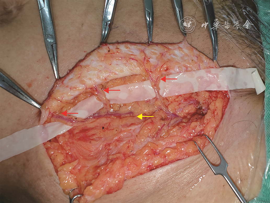
图 2 超薄腹股沟皮瓣游离移植修复例1患者左足踝挤压伤创面的效果。2A.受伤当天创面情况,呈皮革样焦痂;2B.创面清创后基底情况,可见肌腱损伤、外露;2C.设计右侧腹股沟皮瓣;2D、2E.分别为皮瓣游离后即刻的腹面、背面观;2F.将皮瓣与创面缝合后即刻;2G、2H.分别为术后7个月随访时,左足侧面、正面观,可见皮瓣存活良好,形态佳

图 3 双侧超薄腹股沟皮瓣游离移植修复例2患者双手电烧伤创面的效果。3A.术前右手电烧伤创面,示指存在凹陷性焦痂;3B.术前荧光显影显示,示指远端无血流灌注(箭头所示);3C.术中清创,切除栓塞血管、肌腱及指骨后;3D.术中设计左侧超薄腹股沟皮瓣;3E.将皮瓣游离移植修复示指创面并桥接远节动脉后即刻;3F.缝合创面后即刻,荧光显影显示皮瓣及示指远端血流灌注良好;3G.术前左中指创面存在凹陷性焦痂;3H.术中切取右侧超薄腹股沟皮瓣,携带1条纯皮穿支;3I、3J、3K、3L.分别为伤后6个月随访时,双侧腹股沟供区情况、右手掌侧面、左手背侧面观及双手屈曲形态,见供区创面愈合良好,皮瓣外形佳,功能良好
-

 下载:
下载:


图(5)
计量
- 文章访问数: 847
- HTML全文浏览量: 585
- PDF下载量: 60
- 被引次数: 0
