Wang X,Cheng W,Liu XJ,et al.Effects of microneedle injection of rat epidermal stem cells on wound healing of full-thickness skin defects in rats[J].Chin J Burns Wounds,2025,41(9):887-894.DOI: 10.3760/cma.j.cn501225-20241031-00427.
Citation:
Wang X,Cheng W,Liu XJ,et al.Effects of microneedle injection of rat epidermal stem cells on wound healing of full-thickness skin defects in rats[J].Chin J Burns Wounds,2025,41(9):887-894.DOI: 10.3760/cma.j.cn501225-20241031-00427.
Wang X,Cheng W,Liu XJ,et al.Effects of microneedle injection of rat epidermal stem cells on wound healing of full-thickness skin defects in rats[J].Chin J Burns Wounds,2025,41(9):887-894.DOI: 10.3760/cma.j.cn501225-20241031-00427.
Citation:
Wang X,Cheng W,Liu XJ,et al.Effects of microneedle injection of rat epidermal stem cells on wound healing of full-thickness skin defects in rats[J].Chin J Burns Wounds,2025,41(9):887-894.DOI: 10.3760/cma.j.cn501225-20241031-00427.
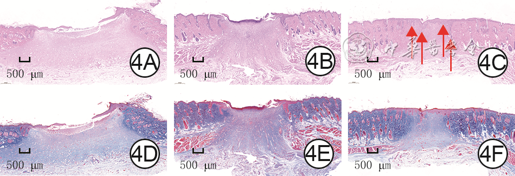
Abstract:Objective To investigate the effects of microneedle injection of rat epidermal stem cells (ESCs) on wound healing of full-thickness skin defects in rats.Methods This study was an experimental study. The hollow silicon-based microneedle injection device was developed successfully by utilizing laser-etching technology. ESCs were isolated and cultured from the skin of two 2-day-old female Sprague-Dawley rats and identified successfully by using flow cytometry and immunofluorescence method. A total of 18 eight-week-old female Sprague-Dawley rats were divided into control group, spray group, and microneedle group according to the random number table method, with 6 rats in each group. All rats were inflicted with a full-thickness skin defect wound on the dorsum. On day 0 (immediately after injury), the wounds in control group and spray group were sprayed with 0.5 mL phosphate-buffered solution and 0.5 mL rat ESCs suspension, respectively, and the wounds in microneedle group of rats were injected with 0.5 mL rat ESCs suspension through the hollow silicon-based microneedle injection device. On days 3, 7, and 14 after injury, the wound healing was observed and the wound healing rate was calculated. On day 14 after injury, the wound tissue of rats were harvested for hematoxylin and eosin staining to observe the epithelialization of wounds, for Masson staining to detect the collagen fiber content in wound tissue, and for immunohistochemical staining to detect the neovascular number in wound tissue.Results Within 14 days after injury, the wounds in three groups of rats progressively healed. On day 3 after injury, there was no statistically significant difference in the overall comparison of the wound healing rate among the three groups of rats (P>0.05). On days 7 and 14 after injury, the wound healing rates in microneedle group of rats were (77.0±4.6)% and (95.0±2.1)%, respectively, which were significantly higher than (66.5±8.6)% and (88.3±3.1)% in spray group and (44.5±5.7)% and (78.8±6.3)% in control group (P<0.05); the wound healing rates in spray group of rats were significantly higher than those in control group (P<0.05). On day 14 after injury, the degree of wound epithelialization in the microneedle group of rats was higher than that in spray group and control group, and hair follicles developed in the newly regenerated skin tissue. On day 14 after injury, the collagen fiber content in the wound tissue in microneedle group and spray group of rats was significantly higher than that in control group (P<0.05), and the collagen fiber content in the wound tissue in microneedle group of rats was significantly higher than that in spray group (P<0.05); the neovascular number in the wound tissue in microneedle group and spray group of rats was significantly more than that in control group (P<0.05), and the neovascular number in the wound tissue in microneedle group of rats was significantly more than that in spray group (P<0.05).Conclusions ESCs can promote neovascularization and collagen fiber formation in the wound tissue, thereby accelerating the wound healing and improve the quality of wound healing in rats with full-thickness skin defects. Injecting rat ESCs suspension through hollow silicon-based microneedle injection device shows better wound healing effects than using conventional approaches, making it an effective cell delivery method.
LoughDM,YangM,BlumA,et al.Transplantation of the LGR6+ epithelial stem cell into full-thickness cutaneous wounds results in enhanced healing, nascent hair follicle development, and augmentation of angiogenic analytes[J].Plast Reconstr Surg,2014,133(3):579-590.DOI: 10.1097/PRS.0000000000000075.
[3]
YangY, HuangJ, ZengA, et al. The role of the skin microbiome in wound healing[J/OL]. Burns Trauma, 2024, 12:tkad059[2024-10-31]. https://pubmed.ncbi.nlm.nih.gov/38444635/. DOI: 10.1093/burnst/tkad059.
[4]
JamesSE, BoothS, DheansaB, et al. Sprayed cultured autologous keratinocytes used alone or in combination with meshed autografts to accelerate wound closure in difficult-to-heal burns patients[J]. Burns, 2010, 36(3):e10-20. DOI: 10.1016/j.burns.2008.11.011.
[5]
JacksonCJ,TønsethKA,UtheimTP.Cultured epidermal stem cells in regenerative medicine[J].Stem Cell Res Ther,2017,8(1):155.DOI: 10.1186/s13287-017-0587-1.
[6]
ChengD,ZhuX,YanS,et al.New insights into inflammatory memory of epidermal stem cells[J].Front Immunol,2023,14:1188559.DOI: 10.3389/fimmu.2023.1188559.
[7]
YangRH,QiSH,ShuB,et al.Epidermal stem cells (ESCs) accelerate diabetic wound healing via the Notch signalling pathway[J].Biosci Rep,2016,36(4):e00364.DOI: 10.1042/BSR20160034.
LeeK,XueY,LeeJ,et al.A patch of detachable hybrid microneedle depot for localized delivery of mesenchymal stem cells in regeneration therapy[J].Adv Funct Mater,2020,30(23):2000086.DOI: 10.1002/adfm.202000086.
[11]
SumathyB,VelayudhanS.Fabrication and evaluation of a bi-layered gelatin based scaffold with arrayed micro-pits for full-thickness skin construct[J].Int J Biol Macromol,2023,251:126360.DOI: 10.1016/j.ijbiomac.2023.126360.
[12]
WeidnerN,FolkmanJ,PozzaF,et al.Tumor angiogenesis: a new significant and independent prognostic indicator in early-stage breast carcinoma[J].J Natl Cancer Inst,1992,84(24):1875-1887.DOI: 10.1093/jnci/84.24.1875.
[13]
PersaOD,KoesterJ,NiessenCM.Regulation of cell polarity and tissue architecture in epidermal aging and cancer[J].J Invest Dermatol,2021,141(4S):1017-1023.DOI: 10.1016/j.jid.2020.12.012.
[14]
PeñaOA,MartinP.Cellular and molecular mechanisms of skin wound healing[J].Nat Rev Mol Cell Biol,2024,25(8):599-616.DOI: 10.1038/s41580-024-00715-1.
[15]
HuangS,KuriP,AubertY,et al.Lgr6 marks epidermal stem cells with a nerve-dependent role in wound re-epithelialization[J].Cell Stem Cell,2021,28(9):1582-1596.e6. DOI: 10.1016/j.stem.2021.05.007.
[16]
ZhangM,HuangM,DongX,et al.Rotating cell culture system-induced injectable self-assembled microtissues with epidermal stem cells for full-thickness skin repair[J].PeerJ,2024,12:e18418.DOI: 10.7717/peerj.18418.
[17]
KangD,WangX,ChenW,et al.Epidermal stem cell-derived exosomes improve wound healing by promoting the proliferation and migration of human skin fibroblasts[J/OL].Burns Trauma,2024,12:tkae047[2024-10-31].https://pubmed.ncbi.nlm.nih.gov/39687464/. DOI: 10.1093/burnst/tkae047.
[18]
SzaboAZ,FongS,YueL,et al.The CD44+ ALDH+ population of human keratinocytes is enriched for epidermal stem cells with long-term repopulating ability[J].Stem Cells,2013,31(4):786-799.DOI: 10.1002/stem.1329.
[19]
ShiY,YangR,TuL,et al.Long non-coding RNA HOTAIR promotes burn wound healing by regulating epidermal stem cells[J].Mol Med Rep,2020,22(3):1811-1820.DOI: 10.3892/mmr.2020.11268.
HuZ,ChenY,GaoM,et al.Novel strategy for primary epithelial cell isolation: combination of hyaluronidase and collagenase I[J].Cell Prolif,2022,56(1):e13320.DOI: 10.1111/cpr.13320.
[22]
SuzukiY,YanagisawaM,YagiH,et al.Involvement of beta1-integrin up-regulation in basic fibroblast growth factor- and epidermal growth factor-induced proliferation of mouse neuroepithelial cells[J].J Biol Chem,2010,285(24):18443-18451.DOI: 10.1074/jbc.M110.114645.
[23]
JinY, LiS, YuQ, et al. Application of stem cells in regeneration medicine[J]. MedComm (2020), 2023, 4(4):e291. DOI: 10.1002/mco2.291.
[24]
HuangS,HuZ,WangP,et al.Rat epidermal stem cells promote the angiogenesis of full-thickness wounds[J].Stem Cell Res Ther,2020,11(1):344.DOI: 10.1186/s13287-020-01844-y.
[25]
KucharzewskiM,RojczykE,Wilemska-KucharzewskaK,et al.Novel trends in application of stem cells in skin wound healing[J].Eur J Pharmacol,2019,843:307-315.DOI: 10.1016/j.ejphar.2018.12.012.
[26]
LiuM,LiuZ,ChenY,et al.Dendritic epidermal T cells secreting exosomes promote the proliferation of epidermal stem cells to enhance wound re-epithelialization[J].Stem Cell Res Ther,2022,13(1):121.DOI: 10.1186/s13287-022-02783-6.
[27]
DonnellyRF,SinghTR,TunneyMM,et al.Microneedle arrays allow lower microbial penetration than hypodermic needles in vitro[J].Pharm Res,2009,26(11):2513-2522.DOI: 10.1007/s11095-009-9967-2.
CaoH,DuanL,ZhangY,et al.Current hydrogel advances in physicochemical and biological response-driven biomedical application diversity[J]. Signal Transduct Target Ther,2021,6(1):426.DOI: 10.1038/s41392-021-00830-x.
[31]
XiaoT, HeG, GengH, et al. Microneedle assisted delivery of epidermal and dermal stem cells to promote hair growth[J]. J Mater Sci, 2024, 59(19): 8427-8440.DOI: 10.1007/s10853-024-09592-7.
ZhaoY, WangM, LiangF, et al. Recent strategies for enhancing the therapeutic efficacy of stem cells in wound healing[J]. Stem Cell Res Ther, 2021, 12(1): 588. DOI: 10.1186/s13287-021-02657-3.
[34]
ZhangY,ChenZ,WuW,et al.Focused ion beam (FIB) prepared microtextured microneedle array capable of delivering drugs through punctured skin[J].Sci Rep,2024,14(1):31489. DOI: 10.1038/s41598-024-83094-z.
[35]
WuJ, LiX, YanX, et al. Fabrication of self-powered silicon microneedles for transdermal drug delivery[J]. J Phy: Conf Ser, 2024, 2884: 012004.DOI: 10.1088/1742-6596/2884/1/012004.
[36]
Pradeep NarayananS, RaghavanS. Solid silicon microneedles for drug delivery applications[J]. Int J Adv Manuf Technol, 2017, 93(1/2/3/4): 407-422.DOI: 10.1007/s00170-016-9698-6.
ChengW,WangX,ZouS,et al.Fabrication of black silicon microneedle arrays for high drug loading[J].J Funct Biomater,2023,14(5):245.DOI: 10.3390/jfb14050245.
[39]
ŚcieżyńskaA,NogowskaA,SikorskaM,et al.Isolation and culture of human primary keratinocytes-a methods review[J].Exp Dermatol,2019,28(2):107-112.DOI: 10.1111/exd.13860.
[40]
ZhangS,YangL,LiuJ,et al.Microneedle systems: cell, exosome, and nucleic acid based strategies[J].Biomater Sci,2023,11(21):7018-7033.DOI: 10.1039/d3bm01103h.


 下载: 导出CSV

 下载:
下载: