





Role and mechanism of infrared thermography in early identification of necrosis risk in random-pattern flaps of mice
-
摘要:
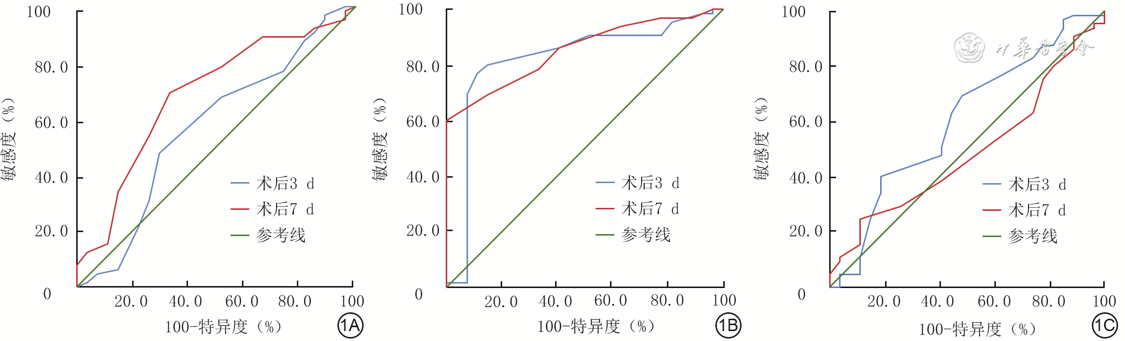
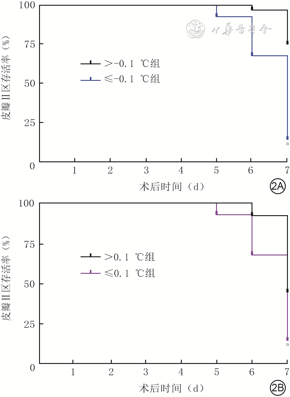
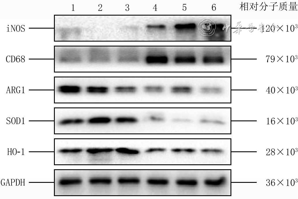
目的 探讨红外热成像(IRT)对小鼠随意型皮瓣坏死风险的早期识别作用及其机制。 方法 该研究为成组设计实验研究。于92只6~8周龄雄性C57BL/6J小鼠背部建立随意型皮瓣模型,将皮瓣从尾部到头部划分为面积相等的Ⅰ、Ⅱ、Ⅲ区,用红外热成像仪测量3个区术后0(即刻)、3、7 d的相对温度(目标区与正常对照区的温度差值)。术后3、7 d,通过受试者操作特征(ROC)曲线分析皮瓣Ⅱ区相对温度对皮瓣坏死的预测效能,并确定最佳截断值。根据术后3 d皮瓣Ⅱ区相对温度,将小鼠分为>-0.1 ℃组、≤-0.1 ℃组;根据术后7 d皮瓣Ⅱ区相对温度,将小鼠分为>0.1 ℃组、≤0.1 ℃组。计算前2组小鼠术后3 d和后2组小鼠术后7 d皮瓣存活率,采用激光多普勒血流成像仪测量前述各组相应时间点皮瓣血流灌注量。术后3 d,取>-0.1 ℃组、≤-0.1 ℃组小鼠皮瓣Ⅱ区组织,行转录组测序筛选差异表达显著的差异表达基因(DEG),行免疫荧光染色后计算诱导型一氧化氮合酶(iNOS)/CD68、精氨酸酶1(ARG1)/CD68比值,行免疫组织化学染色检测CC趋化因子配体2(CCL2)的表达,行二氢乙锭染色检测二氢乙锭的表达,采用实时荧光定量反转录PCR法检测肿瘤坏死因子α(TNF-α)、白细胞介素1β(IL-1β)和IL-10的mRNA表达,采用蛋白质印迹法检测CD68、iNOS、ARG1、血红素加氧酶1(HO-1)及超氧化物歧化酶1(SOD1)的蛋白表达。DEG筛选和蛋白表达检测实验样本数为3,其余实验样本数为4。 结果 术后0~7 d,皮瓣Ⅰ区和Ⅱ区相对温度呈升高趋势,而皮瓣Ⅲ区相对温度呈先升高后降低的趋势。术后3、7 d,皮瓣Ⅱ区相对温度预测皮瓣坏死的ROC曲线下面积分别为0.82、0.85(95%CI分别为0.72~0.93、0.78~0.93),最佳截断值分别为-0.1、0.1 ℃。术后3 d,≤-0.1 ℃组与>-0.1 ℃组小鼠皮瓣存活率比及血流灌注量比较,差异均无统计学意义(P>0.05);术后7 d,≤0.1 ℃组小鼠皮瓣存活率显著低于>0.1 ℃组(t=4.19,P<0.05),血流灌注量显著少于>0.1 ℃组(t=5.58,P<0.05)。术后3 d,相较于>-0.1 ℃组,≤-0.1 ℃组小鼠皮瓣Ⅱ区组织中存在2 849个DEG,其中1 509个上调DEG、1 340个下调DEG。术后3 d,>-0.1 ℃组小鼠皮瓣Ⅱ区组织中iNOS/CD68比值显著低于≤-0.1 ℃组(t=4.97,P<0.05),ARG1/CD68比值显著高于≤-0.1 ℃组(t=3.42,P<0.05),CCL2、二氢乙锭的表达和TNF-α、IL-1β的mRNA表达及CD68、iNOS的蛋白表达均显著低于≤-0.1 ℃组(t值分别为4.82、4.51、4.43、5.47、3.69、6.14,P<0.05),IL-10的mRNA表达及ARG1、SOD1与HO-1的蛋白表达均显著高于≤-0.1 ℃组(t值分别为8.58、8.61、6.24、11.03,P<0.05)。 结论 IRT可通过监测皮瓣温度实现小鼠随意型皮瓣坏死风险的早期客观预测。术后3 d皮瓣Ⅱ区相对温度≤-0.1 ℃时,组织呈显著炎症反应、免疫细胞浸润及氧化应激状态。CCL2可能是诱导巨噬细胞极化的关键趋化因子。 Abstract:Objective To investigate the role and mechanism of infrared thermography (IRT) in early identification of necrosis risk in random-pattern flaps (RPFs) of mice. Methods This study was an experimental study with a grouped design. A RPF model was established on the dorsum of 92 male C57BL/6J mice aged 6-8 weeks. The flap was divided into three equal zones (zones Ⅰ, Ⅱ, and Ⅲ) from the caudal to the cephalic end. The relative temperatures (RTs, the difference between the target zones and the normal control zone) of the three zones were measured using an infrared thermal imager at postoperative day (POD) 0 (immediately), 3, and 7. At POD 3 and 7, receiver operating characteristic (ROC) curves were used to evaluate the predictive efficacy of the RT of flap zone Ⅱ for flap necrosis and to determine the optimal cutoff values. Based on the RT of flap zone Ⅱ at POD 3, mice were divided into >-0.1 ℃ group and ≤-0.1 ℃ group; based on the RT of flap zone Ⅱ at POD 7, mice were divided into >0.1 ℃ group and ≤0.1 ℃ group. The survival rate of flaps was calculated for the first two groups of mice at POD 3 and for the latter two groups of mice at POD 7. Laser Doppler perfusion imaging was used to measure blood perfusion volume of flaps in the above groups at the corresponding time points. At POD 3, the tissue of flap zone Ⅱ from >-0.1 ℃ and ≤-0.1 ℃ groups of mice was collected for transcriptomic sequencing to screen for differentially expressed genes (DEGs) with significantly differential expression, for immunofluorescence staining to calculate the ratios of inducible nitric oxide synthase (iNOS)/CD68 and arginase 1 (ARG1)/CD68, for immunohistochemical staining to detect the expression of C-C motif chemokine ligand 2 (CCL2), for dihydroethidium (DHE) staining to detect the expression of DHE, for real-time fluorescence quantitative reverse transcription polymerase chain reaction to measure the mRNA expressions of tumor necrosis factor α (TNF-α), interleukin-1β (IL-1β), and IL-10, and for Western blotting to detect the protein expressions of CD68, iNOS, ARG1, heme oxygenase-1 (HO-1), and superoxide dismutase 1 (SOD1). The sample size was three for the DEGs screening and protein expression detecting experiments, and 4 for the remaining experiments. Results From POD 0 to 7, RTs of flap zones Ⅰ and Ⅱ showed an increasing trend, while RTs in flap zone Ⅲ rose initially and then decreased. At POD 3 and 7, the areas under the ROC curves of the RT of flap zone Ⅱ for predicting flap necrosis were 0.82 and 0.85, respectively (with 95% CIs of 0.72-0.93 and 0.78-0.93, respectively), with optimal cutoff values of -0.1 and 0.1 ℃, respectively. At POD 3, there were no statistically significant differences in the survival rate or blood perfusion volume of flaps between ≤-0.1 ℃ group and >-0.1 ℃ group of mice (P>0.05). At POD 7, the survival rate (t=4.19, P<0.05) and blood perfusion volume (t=5.58, P<0.05) of flaps were significantly decreased in ≤0.1 ℃ group of mice compared with those in >0.1 ℃ group. At POD 3, compared with that in >-0.1 ℃ group, there were 2 849 DEGs in tissue of flap zone Ⅱ in ≤-0.1 ℃ group of mice, including 1 509 upregulated DEGs and 1 340 downregulated DEGs. At POD 3, there were significantly lower iNOS/CD68 ratio (t=4.97, P<0.05) and significantly higher ARG1/CD68 ratio (t=3.42, P<0.05), significantly lower expressions of CCL2 and DHE, mRNA expressions of TNF-α and IL-1β, as well as protein expressions of CD68 and iNOS (with t values of 4.82, 4.51, 4.43, 5.47, 3.69, and 6.14, respectively, P<0.05), and significantly higher mRNA expression of IL-10 and protein expressions of ARG1, SOD1, and HO-1 (with t values of 8.58, 8.61, 6.24, and 11.03, respectively, P<0.05) in tissue of flap zone Ⅱ in >-0.1 ℃ group of mice compared with those in ≤-0.1 ℃ group. Conclusions IRT enables early and objective prediction of necrosis risk of RPFs in mice through monitoring flap temperature. The RT ≤-0.1 ℃ in flap zone Ⅱ at POD 3 is associated with marked inflammatory response, immune cell infiltration, and oxidative stress in the tissue. CCL2 may serve as a key chemokine inducing macrophage polarization. -
参考文献
(36) [1] BerryCE,LeT,AnN,et al.Pharmacological and cell-based treatments to increase local skin flap viability in animal models[J].J Transl Med,2024,22(1):68.DOI: 10.1186/s12967-024-04882-9. [2] 王晓武,朱仙懂,王永强,等.神经生长因子改善糖尿病大鼠随意皮瓣缺血坏死的机制[J].温州医科大学学报,2024,54(2):87-98.DOI: 10.3969/j.issn.2095-9400.2024.02.001. [3] LouJ,ZhangH,QiJ,et al.Cyclic helix B peptide promotes random-pattern skin flap survival via TFE3-mediated enhancement of autophagy and reduction of ROS levels[J].Br J Pharmacol,2022,179(2):301-321.DOI: 10.1111/bph.15702. [4] YuG,ChenY,YangN,et al.Apoptotic bodies derived from fibroblast-like cells in subcutaneous connective tissue inhibit ferroptosis in ischaemic flaps via the miR-339-5p/KEAP1/Nrf2 axis[J].Adv Sci (Weinh),2024,11(24):e2307238.DOI: 10.1002/advs.202307238. [5] AvilaFR,Torres-GuzmanRA,HuayllaniMT,et al.Human stem cells prevent flap necrosis in preclinical animal models: a systematic review[J].J Clin Transl Res,2022,8(2):110-124. [6] ZhangX,JiangX,DengH,et al.Engineering exosomes from fibroblast growth factor 1 pre-conditioned adipose-derived stem cells promote ischemic skin flaps survival by activating autophagy[J].Mater Today Bio,2024,29:101314.DOI: 10.1016/j.mtbio.2024.101314. [7] 黄亚伟,徐停伟,张浩. 皮瓣缺血再灌注损伤大鼠模型的构建及其运用进展[J]. 中国实验动物学报,2023,31(6):827-832. DOI: 10.3969/j.issn.1005-4847.2023.06.016. [8] WuH,DingJ,WangL,et al.Valproic acid enhances the viability of random pattern skin flaps: involvement of enhancing angiogenesis and inhibiting oxidative stress and apoptosis[J].Drug Des Devel Ther,2018,12:3951-3960.DOI: 10.2147/DDDT.S186222. [9] JohnsonBM,CullomME,EganKG,et al.Comparing tissue oximetry to doppler monitoring in 1367 consecutive breast free flaps[J].Microsurgery,2023,43(1):57-62.DOI: 10.1002/micr.30873. [10] TangeFP,VerduijnPS,Sibinga MulderBG,et al.Near-infrared fluorescence angiography with indocyanine green for perfusion assessment of DIEP and msTRAM flaps: a Dutch multicenter randomized controlled trial[J].Contemp Clin Trials Commun,2023,33:101128.DOI: 10.1016/j.conctc.2023.101128. [11] 符健松,刘英,王辉,等.高频彩色多普勒超声引导下精准设计第1掌骨桡背侧皮瓣修复拇指小面积皮肤软组织缺损的疗效[J].中华烧伤与创面修复杂志,2025,41(10):986-993.DOI: 10.3760/cma.j.cn501225-20241130-00468. [12] XiaoW,ZhangS,LiH,et al.The use of infrared thermography in determining timing for early pedicle division of the preexpanded bipedicled visor flap after ischemic preconditioning[J].Appl Bionics Biomech,2022,2022:8564922.DOI: 10.1155/2022/8564922. [13] CaiF,JiangX,HouX,et al.Application of infrared thermography in the early warning of pressure injury: a prospective observational study[J].J Clin Nurs,2021,30(3/4):559-571.DOI: 10.1111/jocn.15576. [14] 张丕红.浅谈薄型穿支皮瓣切取与穿支血管探测和皮瓣血运评估[J].中华烧伤与创面修复杂志,2023,39(10):911-918.DOI: 10.3760/cma.j.cn501225-20230812-00047. [15] PhillipsCJ,BarronMR,KuckelmanJ,et al.Mobile smartphone thermal imaging characterization and identification of microvascular flow insufficiencies in deep inferior epigastric artery perforator free flaps[J].J Surg Res,2021,261:394-399.DOI: 10.1016/j.jss.2020.12.044. [16] JiangX, HouX, DongN, et al. Skin temperature and vascular attributes as early warning signs of pressure injury[J]. J Tissue Viability, 2020,29(4):258-263. DOI: 10.1016/j.jtv.2020.08.001. [17] LiJ,LouJ,YuG,et al.Targeting TFE3 protects against lysosomal malfunction-induced pyroptosis in random skin flaps via ROS elimination[J].Front Cell Dev Biol,2021,9:643996.DOI: 10.3389/fcell.2021.643996. [18] LouJ,WangX,ZhangH,et al.Inhibition of PLA2G4E/cPLA2 promotes survival of random skin flaps by alleviating lysosomal membrane permeabilization-induced necroptosis[J].Autophagy,2022,18(8):1841-1863.DOI: 10.1080/15548627.2021.2002109. [19] Mota-RojasD,Olmos-HernándezA,Verduzco-MendozaA,et al.Infrared thermal imaging associated with pain in laboratory animals[J].Exp Anim,2021,70(1):1-12.DOI: 10.1538/expanim.20-0052. [20] LiJ,ChenH,LouJ,et al.Exenatide improves random-pattern skin flap survival via TFE3 mediated autophagy augment[J].J Cell Physiol,2021,236(5):3641-3659.DOI: 10.1002/jcp.30102. [21] 楼承浩,卢颖枫,王健.褪黑素通过激活Nrf2抑制铁死亡改善随意皮瓣存活[J].温州医科大学学报,2023,53(7):526-531,539.DOI: 10.3969/j.issn.2095-9400.2023.07.002. [22] 赵毅,闫洪伟,王玉琦,等.腺病毒介导的肝细胞生长因子/血管内皮生长因子165对大鼠超比例随意皮瓣成活的影响及机制研究[J].中国美容医学,2024,33(2):60-63,97. [23] 姚兴璋,姚辉,赵秋玥,等.消肿止痛合剂调控PI3K/AKT信号通路对大鼠皮瓣IRI的影响[J].甘肃医药,2024,43(4):289-291,296. [24] GuM,LiC,DengQ,et al.Celastrol enhances the viability of random-pattern skin flaps by regulating autophagy through the AMPK-mTOR-TFEB axis[J].Phytother Res,2024,38(6):3020-3036.DOI: 10.1002/ptr.8198. [25] JiangX,WangY,WangY,et al.Application of an infrared thermography-based model to detect pressure injuries: a prospective cohort study[J].Br J Dermatol,2022,187(4):571-579.DOI: 10.1111/bjd.21665. [26] KimH,KwakSH,ByeonJY,et al.An experimental and clinical study of flap monitoring with an analysis of the clinical course of the flap using an infrared thermal camera[J].Bioengineering (Basel),2024,11(7):688.DOI: 10.3390/bioengineering11070688. [27] 盛淑文 红外热成像技术在皮瓣外科中的应用 南昌 南昌大学医学部 2020 盛淑文. 红外热成像技术在皮瓣外科中的应用[D]. 南昌:南昌大学医学部,2020.
[28] SinglaP,DixitPK,KalaPC,et al.Free flap monitoring using infrared thermography: an objective adjunct to clinical monitoring[J].Indian J Plast Surg,2024,57(3):179-183.DOI: 10.1055/s-0044-1786742. [29] RahemanFJ,RojoaDM,PatelNG.Performance of infrared thermography and thermal stress test in perforator mapping and flap monitoring: a meta-analysis of diagnostic accuracy[J].J Plast Reconstr Aesthet Surg,2021,74(9):2013-2025.DOI: 10.1016/j.bjps.2021.03.088. [30] Casas-AlvaradoA,Mota-RojasD,Hernández-ÁvalosI,et al.Advances in infrared thermography: surgical aspects, vascular changes, and pain monitoring in veterinary medicine[J].J Therm Biol,2020,92:102664.DOI: 10.1016/j.jtherbio.2020.102664. [31] Mota-RojasD,OgiA,Villanueva-GarcíaD,et al.Thermal imaging as a method to indirectly assess peripheral vascular integrity and tissue viability in veterinary medicine: animal models and clinical applications[J].Animals (Basel),2023,14(1):142.DOI: 10.3390/ani14010142. [32] ShenH,ZhaoZ,LiuJ,et al.The application value of early postoperative pain management (EPPM) combined with skin temperature monitoring (STM) after flap repair of soft tissue defects in the lower limbs: a non-randomized controlled trial[J].Ann Palliat Med,2022,11(3):1068-1076.DOI: 10.21037/apm-22-161. [33] ChangAJBA,de Barros PintoEAF,SilvaDR,et al.Photobiomodulation in promoting increased skin flap viability: a systematic review of animal studies[J].Lasers Med Sci,2024,39(1):109.DOI: 10.1007/s10103-024-04055-6. [34] ZhouK,ChenH,LinJ,et al.FGF21 augments autophagy in random-pattern skin flaps via AMPK signaling pathways and improves tissue survival[J].Cell Death Dis,2019,10(12):872.DOI: 10.1038/s41419-019-2105-0. [35] WuH,DingJ,LiS,et al.Metformin promotes the survival of random-pattern skin flaps by inducing autophagy via the AMPK-mTOR-TFEB signaling pathway[J].Int J Biol Sci,2019,15(2):325-340.DOI: 10.7150/ijbs.29009. [36] BhavaniSV,WolfeKS,HruschCL,et al.Temperature trajectory subphenotypes correlate with immune responses in patients with sepsis[J].Crit Care Med,2020,48(11):1645-1653.DOI: 10.1097/CCM.0000000000004610. -

图 1 92只小鼠术后各时间点随意型皮瓣各区相对温度预测皮瓣坏死的受试者操作特征曲线。1A、1B、1C.分别为Ⅰ、Ⅱ、Ⅲ区
注:相对温度指皮瓣各区温度与正常对照区温度的差值

图 2 各组小鼠术后各时间点随意型皮瓣Ⅱ区存活率。2A.术后3 d,≤-0.1 ℃组(53只)和>-0.1 ℃组(39只);2B.术后7 d,≤0.1 ℃组(60只)和>0.1 ℃组(32只)
注:与>-0.1 ℃组或>0.1 ℃组比较,aP<0.05

图 3 各组小鼠术后不同时间点随意型皮瓣存活情况及血流灌注情况。3A、3B.分别为>-0.1 ℃组和≤-0.1 ℃组术后3 d皮瓣存活情况,图3A和图3B皮瓣存活面积相近;3C、3D.分别为>0.1 ℃组和≤0.1 ℃组术后7 d皮瓣存活情况,图3C皮瓣存活面积大于图3D;3E、3F.分别为>-0.1 ℃组和≤-0.1 ℃组术后3 d皮瓣血流灌注情况,图3E和图3F皮瓣血流灌注量相近;3G、3H.分别为>0.1 ℃组和≤0.1 ℃组术后7 d皮瓣血流灌注情况,图3G皮瓣血流灌注量多于图3H
注:图中虚线框标注皮瓣区域,图3E~3H中绿色、黄色、红色代表血流灌注量从低到高,蓝色代表无血流灌注量

图 4 2组小鼠术后7 d随意型皮瓣Ⅱ区组织中CHP荧光强度及CD34和α-SMA双阳性细胞数。4A、4B.分别为>0.1 ℃组和≤0.1 ℃组CHP荧光强度 CHP-4',6-二脒基-2-苯基吲哚 ×200,图4A中CHP荧光强度低于图4B;4C、4D.分别为>0.1 ℃组和≤0.1 ℃组CD34和α-SMA双阳性细胞数 Alexa Fluor 488-Alexa Fluor 594-4',6-二脒基-2-苯基吲哚 ×200,图4C中CD34和α-SMA双阳性细胞数多于图4D
注:胶原蛋白杂交肽(CHP)阳性染色为绿色,CD34和α-平滑肌肌动蛋白(α-SMA)阳性染色分别为红色、绿色

图 5 2组小鼠术后3 d随意型皮瓣Ⅱ区组织中巨噬细胞极化和炎症趋化及氧化应激情况。5A~5D和5E~5H.分别为>-0.1 ℃组和≤-0.1 ℃组,图5A、5E和5B、5F分别为M1型(iNOS/CD68比值)、M2型(ARG1/CD68比值)巨噬细胞情况 Alexa Fluor 488-Alexa Fluor 594-4',6-二脒基-2-苯基吲哚 ×200,图5C和5G为CCL2表达情况 二氨基联苯胺-苏木精 ×200,图5D和5H为二氢乙锭表达情况 二氢乙锭-4',6-二脒基-2-苯基吲哚 ×200;与>-0.1 ℃组比较,≤-0.1 ℃组皮瓣Ⅱ区组织中iNOS/CD68比值升高、ARG1/CD68比值降低、CCL2和二氢乙锭的表达升高
注:CD68阳性染色为绿色,诱导型一氧化氮合酶(iNOS)、精氨酸酶1(ARG1)阳性染色均为红色,CC趋化因子配体2(CCL2)阳性染色为棕色,二氢乙锭阳性染色为红色

图 6 蛋白质印迹法检测的2组小鼠术后3 d皮瓣Ⅱ区组织中巨噬细胞极化相关蛋白及抗氧化酶的蛋白表达
注:iNOS为诱导型一氧化氮合酶,ARG1为精氨酸酶1,SOD1为超氧化物歧化酶1,HO-1为血红素加氧酶1,GAPDH为3-磷酸甘油醛脱氢酶;条带上方1~3为>-0.1 ℃组的3个样本,4~6为≤-0.1 ℃组的3个样本
Table 1. 小鼠术后各时间点随意型皮瓣各区的相对温度(℃,
区域 鼠数(只) 0 d(即刻) 3 d 7 d Ⅰ区 92 -1.71±1.03 -0.71±0.52 -0.44±0.43 Ⅱ区 92 -0.85±0.63 -0.14±0.37 0.03±0.33 Ⅲ区 92 -0.40±0.46 -0.01±0.32 -0.13±0.30 F值 133.20 162.00 77.17 P值 <0.001 <0.001 <0.001 注:相对温度指皮瓣各区温度与正常对照区温度的差值  下载: 导出CSV
-

 下载:
下载:




计量
- 文章访问数: 197
- HTML全文浏览量: 99
- PDF下载量: 26
- 被引次数: 0
