


Research advances on the application of fibroblasts and their derivatives in the treatment of diabetic foot ulcers
-
摘要: 糖尿病足溃疡是糖尿病的严重并发症之一,具有易感染、难治愈、易复发等特点。目前,糖尿病足溃疡的治疗方法包括标准护理治疗、手术治疗、药物治疗等,但临床疗效欠佳。作为创面愈合关键细胞之一的成纤维细胞(Fb),不仅可以通过分泌纤维连接蛋白、胶原蛋白构建角质形成细胞、上皮细胞等细胞迁移、增殖、定植的支架,还能通过分泌炎症因子、生长因子、细胞因子扭转糖尿病足溃疡慢性炎症局面,促进细胞增殖,改善创面微环境,是目前应用于糖尿病足溃疡创面治疗的细胞之一。该文从Fb角度出发,综述Fb及其衍生物在糖尿病足溃疡治疗中的应用情况,以期为糖尿病足溃疡的治疗提供参考。Abstract: Diabetic foot ulcer (DFU) is one of the serious complications of diabetes mellitus, which is characterized by susceptibility to infection, difficult to cure, and easy to relapse. At present, the treatment methods for DFUs include standard nursing care, surgical treatment, and medication, but the clinical efficacy is suboptimal. Fibroblasts (Fbs), as one of the key cells for wound healing, can not only secrete fibronectin and collagen to construct scaffolds for cell migration, proliferation, and colonization of keratinocytes and epithelial cells, but also secrete inflammatory factors, growth factors, and cytokines to reverse the chronic inflammation of DFUs, promote cell proliferation, and improve the wound microenvironment. It is one of the cell types currently used in DFU wound treatment. This article reviews the application of Fbs and their derivatives in DFU treatment from the perspective of Fbs, in order to provide a reference for DFU treatment.
-
Key words:
- Diabetic foot /
- Foot ulcer /
- Fibroblasts /
- Exosomes /
- Skin, artificial /
- Wound repair
-
参考文献
(52) [1] 魏在荣,杨成兰,黄广涛.糖尿病足外科整合治疗的进展评述[J].中国美容整形外科杂志,2020,31(7): 385-389.DOI: 10.3969/j.issn.1673-7040.2020.07.001. [2] GBD 2021 Diabetes Collaborators.Global, regional, and national burden of diabetes from 1990 to 2021, with projections of prevalence to 2050: a systematic analysis for the Global Burden of Disease Study 2021[J].Lancet,2023,402(10397):203-234.DOI: 10.1016/S0140-6736(23)01301-6. [3] 牛少辉,李贝,方毅娜,等.不同细胞来源的外泌体在糖尿病足创面修复中的作用机制和干预前景[J/OL].中国血管外科杂志(电子版),2023,15(1):83-87[2024-12-04].https://d.wanfangdata.com.cn/periodical/ChVQZXJpb2RpY2FsQ0hJMjAyNTA2MjISEXpneGd3a3p6MjAyMzAxMDIyGghmcWNsaGJ1NQ%3D%3D.DOI: 10.3969/j.issn.1674-7429.2023.01.020. [4] KolarićV,SvirčevićV,BijukR,et al.Chronic complications of diabetes and quality of life[J].Acta Clin Croat,2022,61(3):520-527.DOI: 10.20471/acc.2022.61.03.18. [5] GaoR,ZhouP,LiY,et al.High glucose-induced IL-7/IL-7R upregulation of dermal fibroblasts inhibits angiogenesis in a paracrine way in delayed diabetic wound healing[J].J Cell Commun Signal,2023,17(3):1023-1038.DOI: 10.1007/s12079-023-00754-x. [6] 阮琼芳,章思语,席毛毛,等.糖尿病足患者创缘皮肤组织中成纤维细胞与角质形成细胞的相互作用及其机制[J].中华烧伤与创面修复杂志,2024,40(8):762-771.DOI: 10.3760/cma.j.cn501225-20240221-00067. [7] BurgessJL,WyantWA,Abdo AbujamraB,et al.Diabetic wound-healing science[J].Medicina (Kaunas),2021,57(10):1072.DOI: 10.3390/medicina57101072. [8] LiuY,WangP,LiJ,et al.Single-cell RNA sequencing reveals the impaired epidermal differentiation and pathological microenvironment in diabetic foot ulcer[J/OL].Burns Trauma,2025,13:tkae065[2025-08-04].https://pubmed.ncbi.nlm.nih.gov/40040959/.DOI: 10.1093/burnst/tkae065. [9] TalbottHE,MascharakS,GriffinM,et al.Wound healing, fibroblast heterogeneity, and fibrosis[J].Cell Stem Cell,2022,29(8):1161-1180. DOI: 10.1016/j.stem.2022.07.006. [10] VozaFA,HuertaCT,LeN,et al.Fibroblasts in diabetic foot ulcers[J].Int J Mol Sci,2024,25(4):2172.DOI: 10.3390/ijms25042172. [11] 蒋祉萱,姚敏.特殊的成纤维细胞祖细胞亚群通过免疫调节加速黏膜愈合再生[J].中华烧伤与创面修复杂志,2024,40(4):313.DOI: 10.3760/cma.j.issn.2097-1109.2024.04.101. [12] DuJ,LiuX,WongCWY,et al.Direct cellular reprogramming and transdifferentiation of fibroblasts on wound healing-fantasy or reality?[J].Chronic Dis Transl Med,2023,9(3):191-199.DOI: 10.1002/cdt3.77. [13] 王晓雨,张玉杰,曲春安,等.成纤维细胞在糖尿病患者创伤愈合中作用的研究进展[J].中国细胞生物学学报,2019,41(4):710-714.DOI: 10.11844/cjcb.2019.04.0021. [14] KitaA,YamamotoS,SaitoY,et al.Cellular senescence and wound healing in aged and diabetic skin[J].Front Physiol,2024,15:1344116.DOI: 10.3389/fphys.2024.1344116. [15] AltieriA,VisserGV,BuechlerMB.Enter the matrix: fibroblast-immune cell interactions shape extracellular matrix deposition in health and disease[J].F1000Res,2024,13:119.DOI: 10.12688/f1000research.143506.2. [16] LiuY,LiuY,HeW,et al.Fibroblasts: immunomodulatory factors in refractory diabetic wound healing[J].Front Immunol,2022,13:918223.DOI: 10.3389/fimmu.2022.918223. [17] ParkLK,MaioneAG,SmithA,et al.Genome-wide DNA methylation analysis identifies a metabolic memory profile in patient-derived diabetic foot ulcer fibroblasts[J].Epigenetics,2014,9(10):1339-1349.DOI: 10.4161/15592294.2014.967584. [18] VelanderP,TheopoldC,BleizifferO,et al.Cell suspensions of autologous keratinocytes or autologous fibroblasts accelerate the healing of full thickness skin wounds in a diabetic porcine wound healing model[J].J Surg Res,2009,157(1):14-20.DOI: 10.1016/j.jss.2008.10.001. [19] ZabihiA,MahmoodiM.The effects of subdermal injection of fibroblast cells on collagen synthesis and wound healing in diabetic rat[J].Yafte,2021,23(1):149-160. [20] NilforoushzadehMA,JaffaryF,SiavashM,et al.Autologous fibroblast suspension for the treatment of refractory diabetic foot ulcer[J].Indian J Dermatol Venereol Leprol,2016,82(1):105-106.DOI: 10.4103/0378-6323.172905. [21] NilforoushzadehMA,Heidari-KharajiM,ZareM,et al.Combination therapy of trichloroacetic acid, human autologous fibroblast injection and fibroblast seeded microfibrous collagen scaffold as a novel treatment for osteomyelitis diabetic foot ulcer[J].J Diabetes Investig,2021,12(6):1112-1117.DOI: 10.1111/jdi.13454. [22] NilforoushzadehMA,JaffaryF,SiavashM,et al.Treatment of recalcitrant diabetic ulcers with trichloroacetic acid and fibroblasts[J].J Skin Stem Cell,2014,1(2):e23312. DOI: 10.17795/jssc23312. [23] CavalliniM.Autologous fibroblasts to treat deep and complicated leg ulcers in diabetic patients[J].Wound Repair Regen,2007,15(1):35-38.DOI: 10.1111/j.1524-475X.2006.00182.x. [24] YouHJ,HanSK,RhieJW.Randomised controlled clinical trial for autologous fibroblast-hyaluronic acid complex in treating diabetic foot ulcers[J].J Wound Care,2014,23(11):521-522, 524, 526-530.DOI: 10.12968/jowc.2014.23.11.521. [25] 彭颖,赵阳,解英,等.异体皮肤成纤维细胞在糖尿病小鼠创面愈合中的作用及其机制[J].中华烧伤杂志,2018,34(8):532-541.DOI: 10.3760/cma.j.issn.1009-2587.2018.08.011. [26] Kazemi-DarabadiS,Sarrafzadeh-RezaeiF,FarshidAA,et al.Allogenous skin fibroblast transplantation enhances excisional wound healing following alloxan diabetes in sheep, a randomized controlled trial[J].Int J Surg,2014,12(8):751-756.DOI: 10.1016/j.ijsu.2014.06.007. [27] HanSK,ChoiKJ,KimWK.Clinical application of fresh fibroblast allografts for the treatment of diabetic foot ulcers: a pilot study[J].Plast Reconstr Surg,2004,114(7):1783-1789.DOI: 10.1097/01.prs.0000142415.57470.df. [28] HanSK,KimHS,KimWK.Efficacy and safety of fresh fibroblast allografts in the treatment of diabetic foot ulcers[J].Dermatol Surg,2009,35(9):1342-1348.DOI: 10.1111/j.1524-4725.2009.01239.x. [29] 梁结梅,孙铁辉,向鹏安,等.不同源性外泌体在糖尿病足溃疡修复中作用的研究进展[J].感染、炎症、修复,2021,22(4):244-249.DOI: 10.3969/j.issn.1672-8521.2021.04.016. [30] FengJ,YaoY,WangQ,et al.Exosomes: potential key players towards novel therapeutic options in diabetic wounds[J].Biomed Pharmacother,2023,166:115297.DOI: 10.1016/j.biopha.2023.115297. [31] GuoL,XiaoD,XingH,et al.Engineered exosomes as a prospective therapy for diabetic foot ulcers[J/OL].Burns Trauma,2024,12:tkae023[2024-12-04].https://pubmed.ncbi.nlm.nih.gov/39026930/.DOI: 10.1093/burnst/tkae023. [32] HanX,WuP,LiL,et al.Exosomes derived from autologous dermal fibroblasts promote diabetic cutaneous wound healing through the Akt/β-catenin pathway[J].Cell Cycle,2021,20(5/6):616-629.DOI: 10.1080/15384101.2021.1894813. [33] GeigerA,WalkerA,NissenE.Human fibrocyte-derived exosomes accelerate wound healing in genetically diabetic mice[J].Biochem Biophys Res Commun,2015,467(2):303-309.DOI: 10.1016/j.bbrc.2015.09.166. [34] DongJ,WuB,TianW.How to maximize the therapeutic effect of exosomes on skin wounds in diabetes mellitus: review and discussion[J].Front Endocrinol (Lausanne),2023,14:1146991.DOI: 10.3389/fendo.2023.1146991. [35] SabolinskiML,CapotortoJV.Comparative effectiveness of a human fibroblast-derived dermal substitute and a viable cryopreserved placental membrane for the treatment of diabetic foot ulcers[J].J Comp Eff Res,2019,8(14):1229-1238.DOI: 10.2217/cer-2019-0001. [36] FarzanbakhshS,AminiMR,MadaniH,et al.Safety evaluation of bi-layered allogenic keratinocyte and fibroblast skin substitute for diabetic foot ulcers-SAFESKIN-DFU: a phase 1 clinical trial[J].Diabetes Obes Metab,2024,26(11):5078-5086.DOI: 10.1111/dom.15843. [37] HanftJR,SurprenantMS.Healing of chronic foot ulcers in diabetic patients treated with a human fibroblast-derived dermis[J].J Foot Ankle Surg,2002,41(5):291-299.DOI: 10.1016/s1067-2516(02)80047-3. [38] MarstonWA,HanftJ,NorwoodP,et al.The efficacy and safety of Dermagraft in improving the healing of chronic diabetic foot ulcers: results of a prospective randomized trial[J].Diabetes Care,2003,26(6):1701-1705.DOI: 10.2337/diacare.26.6.1701. [39] LandsmanA,RoukisTS,DefronzoDJ,et al. Living cells or collagen matrix: which is more beneficial in the treatment of diabetic foot ulcers?[J].Wounds,2008,20(5):111-116. [40] WarrinerRA3rd,CardinalM,TIDEInvestigators.Human fibroblast-derived dermal substitute: results from a treatment investigational device exemption (TIDE) study in diabetic foot ulcers[J].Adv Skin Wound Care,2011,24(7):306-311.DOI: 10.1097/01.ASW.0000399647.80210.61. [41] MorimotoN,ItoT,TakemotoS,et al.An exploratory clinical study on the safety and efficacy of an autologous fibroblast-seeded artificial skin cultured with animal product-free medium in patients with diabetic foot ulcers[J].Int Wound J,2014,11(2):183-189.DOI: 10.1111/j.1742-481X.2012.01064.x. [42] CerneckisJ,CaiH,ShiY.Induced pluripotent stem cells (iPSCs): molecular mechanisms of induction and applications[J].Signal Transduct Target Ther,2024,9(1):112.DOI: 10.1038/s41392-024-01809-0. [43] ShiY,InoueH,WuJC,et al.Induced pluripotent stem cell technology: a decade of progress[J].Nat Rev Drug Discov,2017,16(2):115-130.DOI: 10.1038/nrd.2016.245. [44] 高春辰,陈金安,王爱萍.诱导多能干细胞促进糖尿病足溃疡愈合的研究进展[J].中华烧伤与创面修复杂志,2022,38(9):864-869.DOI: 10.3760/cma.j.cn501120-20210630-00230. [45] Gerami-NainiB,SmithA,MaioneAG,et al.Generation of induced pluripotent stem cells from diabetic foot ulcer fibroblasts using a nonintegrative Sendai virus[J].Cell Reprogram,2016,18(4):214-223.DOI: 10.1089/cell.2015.0087. [46] KashpurO,SmithA,Gerami-NainiB,et al.Differentiation of diabetic foot ulcer-derived induced pluripotent stem cells reveals distinct cellular and tissue phenotypes[J].FASEB J,2019,33(1):1262-1277.DOI: 10.1096/fj.201801059. [47] PastarI,MarjanovicJ,LiangL,et al.Cellular reprogramming of diabetic foot ulcer fibroblasts triggers pro-healing miRNA-mediated epigenetic signature[J].Exp Dermatol,2021,30(8):1065-1072.DOI: 10.1111/exd.14405. [48] GoreckaJ,KostiukV,FereydooniA,et al.The potential and limitations of induced pluripotent stem cells to achieve wound healing[J].Stem Cell Res Ther,2019,10(1):87.DOI: 10.1186/s13287-019-1185-1. [49] 刘柳,张雪,张文涛,等.胚胎成纤维细胞对糖尿病溃疡成纤维细胞老化表型和功能的影响[J].中华内分泌外科杂志,2014,8(6):447-451.DOI: 10.3760/cma.j.issn.1674-6090.2014.06.003. [50] 苏浩 利用人表皮干细胞和成纤维细胞3D打印皮肤组织的初步研究 扬州 扬州大学 2020 苏浩.利用人表皮干细胞和成纤维细胞3D打印皮肤组织的初步研究[D].扬州:扬州大学,2020.
[51] LiM,SunL,LiuZ,et al.3D bioprinting of heterogeneous tissue-engineered skin containing human dermal fibroblasts and keratinocytes[J].Biomater Sci,2023,11(7):2461-2477.DOI: 10.1039/d2bm02092k. [52] Sierra-SánchezÁ,MagneB,SavardE,et al.In vitro comparison of human plasma-based and self-assembled tissue-engineered skin substitutes: two different manufacturing processes for the treatment of deep and difficult to heal injuries[J/OL].Burns Trauma,2023,11:tkad043[2024-12-04].https://pubmed.ncbi.nlm.nih.gov/37908563/.DOI: 10.1093/burnst/tkad043. -

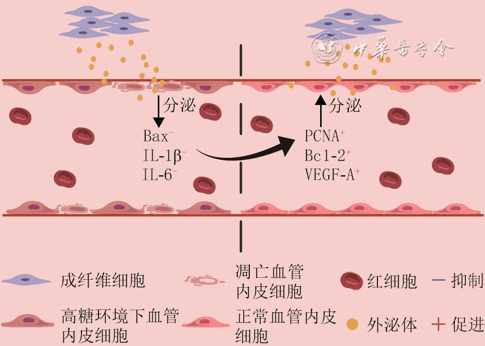
图 1 成纤维细胞来源的外泌体改善高糖环境下血管内皮细胞功能的机制
注:Bcl-2为B淋巴细胞瘤-2,Bax为Bcl-2相关X蛋白,IL为白细胞介素,PCNA为增殖细胞核抗原,VEGF-A为血管内皮生长因子A;图中黑色竖线分隔左侧高糖环境下血管内皮细胞和右侧正常血管内皮细胞,横跨黑色竖线的圆弧形箭头表示左侧细胞向右侧细胞转化
-

 下载:
下载:

图(2)
计量
- 文章访问数: 1091
- HTML全文浏览量: 339
- PDF下载量: 29
- 被引次数: 0
