






Bidirectional two-sample MR analysis of causal relationships between human inflammatory proteins and HS and keloids
-
摘要:
目的 探讨人炎症蛋白与增生性瘢痕(HS)、瘢痕疙瘩之间的因果关系。 方法 该研究为基于双向双样本孟德尔随机化(MR)分析的研究。从全基因组关联分析数据库中获取人炎症蛋白、HS和瘢痕疙瘩的数据,采用逆方差加权(IVW)法评估91种炎症蛋白与HS、瘢痕疙瘩之间的因果关系,即正向MR分析。针对前述关联,采用Cochran Q检验评估异质性,采用MR-Egger回归检验和MR-PRESSO离群值检验评估水平多效性,采用留一法分析结果的稳健性。采用IVW法评估增生性瘢痕、瘢痕疙瘩与前述正向MR分析筛选出的炎症蛋白之间是否存在反向因果关系。 结果 CD6、白血病抑制因子(LIF)、肿瘤坏死因子配体超家族成员12(TNFSF12)、程序性死亡配体1(PD-L1)、白细胞介素-17C(IL-17C)、LIF受体(LIFR)、骨保护素、成纤维细胞生长因子23(FGF23)与HS之间均存在显著因果关系(OR分别为1.365、0.506、1.567、1.683、0.621、1.375、0.623、0.553,95%CI分别为1.100~1.693、0.289~0.887、1.081~2.273、1.090~2.599、0.408~0.947、1.025~1.845、0.402~0.966、0.315~0.971,P<0.05),其中CD6、TNFSF12、PD-L1、LIFR为HS的风险性因素,LIF、IL-17C、骨保护素、FGF23为HS的保护性因素;CD5、IL-10受体α亚基(IL-10RA)、IL-5、LIF、骨保护素与瘢痕疙瘩之间均存在显著因果关系(OR分别为0.744、1.303、0.686、0.603、0.715,95%CI分别为0.573~0.965、1.024~1.660、0.472~0.996、0.431~0.842、0.553~0.924,P<0.05),其中IL-10RA为瘢痕疙瘩的风险性因素,CD5、IL-5、LIF、骨保护素为瘢痕疙瘩的保护性因素。上述关联均不存在显著异质性或显著水平多效性(P>0.05),结果的稳健性未受单个单核苷酸多态性驱动。HS与前述正向MR分析筛选出的与HS之间存在显著因果关系的8种炎症蛋白中的TNFSF12、LIFR之间均存在显著反向因果关系(OR分别为0.972、0.968,95%CI分别为0.949~0.997、0.942~0.994,P<0.05),瘢痕疙瘩与前述正向MR分析筛选出的与瘢痕疙瘩之间存在显著因果关系的5种炎症蛋白之间均不存在反向因果关系(P>0.05)。 结论 CD6、TNFSF12、PD-L1、LIFR可能增加HS患病风险,LIF、IL-17C、骨保护素、FGF23可能降低HS患病风险;IL-10RA可能增加瘢痕疙瘩患病风险,CD5、IL-5、LIF、骨保护素可能降低瘢痕疙瘩患病风险。 Abstract:Objective To explore the causal relationships between human inflammatory proteins and hypertrophic scars (HS) and keloids. Methods This study was conducted based on bidirectional two-sample Mendelian randomization (MR) analysis. Data of human inflammatory proteins, HS, and keloids were acquired from genome-wide association study database. The inverse variance weighted (IVW) method was adopted to evaluate the causal relationships between 91 kinds of human inflammatory proteins and HS and keloids, i.e., a forward MR analysis. For the above associations, Cochran's Q test was used to assess heterogeneity, MR-Egger regression and MR-PRESSO outlier tests were performed to evaluate horizontal pleiotropy, and the leave-one-out method was applied to analyze the robustness of the results. The IVW method was also used to evaluate whether there was a reverse causal relationship between HS, keloids and inflammatory proteins screened out by aforementioned forward MR analysis. Results CD6, leukemia inhibitory factor (LIF), tumor necrosis factor ligand superfamily member 12 (TNFSF12), programmed death-ligand 1 (PD-L1), interleukin-17C (IL-17C), LIF receptor (LIFR), osteoprotegerin (OPG), and fibroblast growth factor 23 (FGF23) had significant causal relationships with HS (with ORs of 1.365, 0.506, 1.567, 1.683, 0.621, 1.375, 0.623, and 0.553, respectively, 95% CIs of 1.100-1.693, 0.289-0.887, 1.081-2.273, 1.090-2.599, 0.408-0.947, 1.025-1.845, 0.402-0.966, and 0.315-0.971, respectively, P<0.05). Among them, CD6, TNFSF12, PD-L1, and LIFR were risk factors for HS, while LIF, IL-17C, OPG, and FGF23 were protective factors for HS. CD5, IL-10 receptor subunit alpha (IL-10RA), IL-5, LIF, and OPG had significant causal relationships with keloids (with ORs of 0.744, 1.303, 0.686, 0.603, and 0.715, respectively, 95% CIs of 0.573-0.965, 1.024-1.660, 0.472-0.996, 0.431-0.842, and 0.553-0.924, respectively, P<0.05). Among them, IL-10RA was a risk factor for keloids, whereas CD5, IL-5, LIF, and OPG were protective factors for keloids. No significant heterogeneity or horizontal pleiotropy was observed in the above associations (P>0.05), and the robustness of the results was not driven by any single nucleotide polymorphism. Significant reverse causal relationships existed between HS and TNFSF12 and LIFR of the 8 inflammatory proteins which had significant causal relationships with HS screened out by aforementioned forward MR analysis (with ORs of 0.972 and 0.968, respectively, 95% CIs of 0.949-0.997 and 0.942-0.994, respectively, P<0.05). No reverse causal relationship was found between keloids and the 5 inflammatory proteins which had significant causal relationships with keloids screened out by aforementioned forward MR analysis (P>0.05). Conclusions CD6, TNFSF12, PD-L1, and LIFR may increase the risk of HS, while LIF, IL-17C, OPG, and FGF23 may decrease the risk of HS. IL-10RA may increase the risk of keloids, while CD5, IL-5, LIF, and OPG may decrease the risk of keloids. -
参考文献
(40) [1] LiT,ZhangM,LiY,et al.Twist-related protein 1 promotes transforming growth factor β receptor 1 in keloid fibroblasts via regulating the stability of myocyte enhancer factor 2A[J/OL].Burns Trauma,2024,12:tkae024[2025-01-17].https://pubmed.ncbi.nlm.nih.gov/39429644/.DOI: 10.1093/burnst/tkae024. [2] 臧梦青.增生性瘢痕和瘢痕疙瘩的诊治现状[J].中华医学杂志,2023,103(7):469-472.DOI: 10.3760/cma.j.cn112137-20220627-01415. [3] 王洪涛,韩军涛,胡大海.炎症反应在增生性瘢痕和瘢痕疙瘩形成中的作用及其机制研究进展[J].中华烧伤杂志,2021,37(5):490-494.DOI: 10.3760/cma.j.cn501120-20200310-00143. [4] ZhaoJH,StaceyD,ErikssonN,et al.Genetics of circulating inflammatory proteins identifies drivers of immune-mediated disease risk and therapeutic targets[J].Nat Immunol,2023,24(9):1540-1551.DOI: 10.1038/s41590-023-01588-w. [5] WangZC,ZhaoWY,CaoY,et al.The roles of inflammation in keloid and hypertrophic scars[J].Front Immunol,2020,11:603187.DOI: 10.3389/fimmu.2020.603187. [6] ZhaoY,WeiQ,ZengR,et al.Natural killer cell dysfunction drives keloid pathogenesis[J].Cell Rep,2026,45(4):117129.DOI: 10.1016/j.celrep.2026.117129. [7] AbdouAG,MaraeeAH,SaifHF.Immunohistochemical evaluation of COX-1 and COX-2 expression in keloid and hypertrophic scar[J].Am J Dermatopathol,2014,36(4):311-317.DOI: 10.1097/DAD.0b013e3182a27b83. [8] TanakaR,UmeyamaY,HagiwaraH,et al.Keloid patients have higher peripheral blood endothelial progenitor cell counts and CD34+ cells with normal vasculogenic and angiogenic function that overexpress vascular endothelial growth factor and interleukin-8[J].Int J Dermatol,2019,58(12):1398-1405.DOI: 10.1111/ijd.14575. [9] NishiguchiMA,SpencerCA,LeungDH,et al.Aging suppresses skin-derived circulating SDF1 to promote full-thickness tissue regeneration[J].Cell Rep,2018,24(13):3383-3392.e5.DOI: 10.1016/j.celrep.2018.08.054. [10] ZhangJ,QiaoQ,LiuM,et al.IL-17 promotes scar formation by inducing macrophage infiltration[J].Am J Pathol,2018,188(7):1693-1702.DOI: 10.1016/j.ajpath.2018.04.005. [11] XuM,ShaoQ,ZhouY,et al.Potential effects of specific gut microbiota on periodontal disease: a two-sample bidirectional Mendelian randomization study[J].Front Microbiol,2024,15:1322947.DOI: 10.3389/fmicb.2024.1322947. [12] LarssonSC,ButterworthAS,BurgessS.Mendelian randomization for cardiovascular diseases: principles and applications[J].Eur Heart J,2023,44(47):4913-4924.DOI: 10.1093/eurheartj/ehad736. [13] EmdinCA,KheraAV,KathiresanS.Mendelian randomization[J].JAMA,2017,318(19):1925-1926.DOI: 10.1001/jama.2017.17219. [14] SakaueS,KanaiM,TanigawaY,et al.A cross-population atlas of genetic associations for 220 human phenotypes[J].Nat Genet,2021,53(10):1415-1424.DOI: 10.1038/s41588-021-00931-x. [15] LongY,TangL,ZhouY,et al.Causal relationship between gut microbiota and cancers: a two-sample Mendelian randomisation study[J].BMC Med,2023,21(1):66.DOI: 10.1186/s12916-023-02761-6. [16] LiW,XuJW,ChaiJL,et al.Complex causal association between genetically predicted 731 immunocyte phenotype and osteonecrosis: a bidirectional two-sample Mendelian randomization analysis[J].Int J Surg,2024,110(6):3285-3293.DOI: 10.1097/JS9.0000000000001327. [17] WangQ,SunY,ZhouT,et al.Gut microbiota-dependent trimethylamine N-oxide pathway contributes to the bidirectional relationship between intestinal inflammation and periodontitis[J].Front Cell Infect Microbiol,2023,12:1125463.DOI: 10.3389/fcimb.2022.1125463. [18] 娄家祺,李吉良,崔胜勇,等.人肠道菌群特征和免疫细胞表型与HS之间因果关系的两步双样本中介MR分析[J].中华烧伤与创面修复杂志,2026,42(4):383-392.DOI: 10.3760/cma.j.cn501225-20241226-00509. [19] 甘文军,王婧薷,何佳,等.人免疫细胞表型与瘢痕疙瘩之间因果关系的双样本孟德尔随机化分析[J].中华烧伤与创面修复杂志,2025,41(1):84-93.DOI: 10.3760/cma.j.cn501225-20231130-00219. [20] HongYK,ChangYH,LinYC,et al.Inflammation in wound healing and pathological scarring[J].Adv Wound Care (New Rochelle),2023,12(5):288-300.DOI: 10.1089/wound.2021.0161. [21] HassanshahiA,MoradzadM,GhalamkariS,et al.Macrophage-mediated inflammation in skin wound healing[J].Cells,2022,11(19):2953.DOI: 10.3390/cells11192953. [22] Moreno-ManuelA,Jantus-LewintreE,SimõesI,et al.CD5 and CD6 as immunoregulatory biomarkers in non-small cell lung cancer[J].Transl Lung Cancer Res,2020,9(4):1074-1083.DOI: 10.21037/tlcr-19-445. [23] Gurrea-RubioM,FoxDA,CastresanaJS.CD6 in human disease[J].Cells,2025,14(4):272.DOI: 10.3390/cells14040272. [24] DalloulA.CD5: a safeguard against autoimmunity and a shield for cancer cells[J].Autoimmun Rev,2009,8(4):349-353.DOI: 10.1016/j.autrev.2008.11.007. [25] DongJ,ZhangK,HongJ,et al.The multiple functions of CD5 in diseases related to immune disorders[J].Ann Med,2025,57(1):2519682.DOI: 10.1080/07853890.2025.2519682. [26] ChenJ,SheY,FengC,et al.TNFSF12 is associated with breast cancer prognosis and immune cell infiltration[J].Am J Transl Res,2024,16(8):4120-4133.DOI: 10.62347/IDTK3218. [27] SonA,OshioT,KawamuraYI,et al.TWEAK/Fn14 pathway promotes a T helper 2-type chronic colitis with fibrosis in mice[J].Mucosal Immunol,2013,6(6):1131-1142.DOI: 10.1038/mi.2013.10. [28] MatellanC,KennedyC,Santiago-VelaMI,et al.The TNFSF12/TWEAK modulates colonic inflammatory fibroblast differentiation and promotes fibroblast-monocyte interactions[J].J Immunol,2024,212(12):1958-1970.DOI: 10.4049/jimmunol.2300762. [29] CaiY,XiaoM,LiX,et al.BMS-202, a PD-1/PD-L1 inhibitor, decelerates the pro-fibrotic effects of fibroblasts derived from scar tissues via ERK and TGFβ1/Smad signaling pathways[J].Immun Inflamm Dis,2022,10(10):e693.DOI: 10.1002/iid3.693. [30] ShenC,WuN,ChenX,et al.Interleukin-5 alleviates cardiac remodelling via the STAT3 pathway in angiotensin II-infused mice[J].J Cell Mol Med,2024,28(13):e18493.DOI: 10.1111/jcmm.18493. [31] SubudhiI,KoniecznyP,PrystupaA,et al.Metabolic coordination between skin epithelium and type 17 immunity sustains chronic skin inflammation[J].Immunity,2024,57(7):1665-1680.e7.DOI: 10.1016/j.immuni.2024.04.022. [32] ShiJ,ShiS,XieW,et al.IL-10 alleviates lipopolysaccharide-induced skin scarring via IL-10R/STAT3 axis regulating TLR4/NF-κB pathway in dermal fibroblasts[J].J Cell Mol Med,2021,25(3):1554-1567.DOI: 10.1111/jcmm.16250. [33] YuY,WangY,NiuY,et al.Leukemia inhibitory factor attenuates renal fibrosis through Stat3-miR-29c[J].Am J Physiol Renal Physiol,2015,309(7):F595-F603.DOI: 10.1152/ajprenal.00634.2014. [34] HooshiarSH,TobeihaM,JafarnejadS.Soy isoflavones and bone health: focus on the RANKL/RANK/OPG pathway[J].Biomed Res Int,2022,2022:8862278.DOI: 10.1155/2022/8862278. [35] MohamadHE,AskerME,ShaheenMA,et al.Secukinumab and black garlic downregulate OPG/RANK/RANKL axis and devitalize myocardial interstitial fibrosis induced by sunitinib in experimental rats[J].Life (Basel),2023,13(2):308.DOI: 10.3390/life13020308. [36] KohlhauserM,MayrhoferM,KamolzLP,et al.An update on molecular mechanisms of scarring-a narrative review[J].Int J Mol Sci,2024,25(21):11579.DOI: 10.3390/ijms252111579. [37] ChenG,ChenL,LiX,et al.FGF-based drug discovery: advances and challenges[J].Nat Rev Drug Discov,2025,24(5):335-357.DOI: 10.1038/s41573-024-01125-w. [38] DongQ,LiS,WangW,et al.FGF23 regulates atrial fibrosis in atrial fibrillation by mediating the STAT3 and SMAD3 pathways[J].J Cell Physiol,2019,234(11):19502-19510.DOI: 10.1002/jcp.28548. [39] SmithER,TanSJ,HoltSG,et al.FGF23 is synthesised locally by renal tubules and activates injury-primed fibroblasts[J].Sci Rep,2017,7(1):3345.DOI: 10.1038/s41598-017-02709-w. [40] 李涛,朱晨晨,陈今源,等.人炎症蛋白与瘢痕疙瘩之间因果关系的孟德尔随机化分析[J].中华烧伤与创面修复杂志,2025,41(2):180-187.DOI: 10.3760/cma.j.cn501225-20240526-00198. -

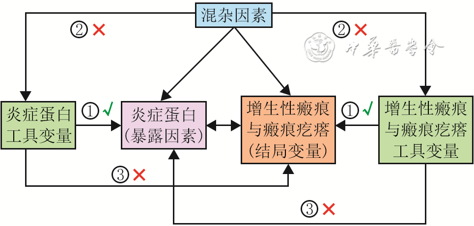
图 1 人炎症蛋白与增生性瘢痕及瘢痕疙瘩之间因果关系的双向双样本孟德尔随机化分析的核心假设和分析流程图
注:“√”表示存在关联或有效路径,“×”表示该路径不存在或已被排除;①表示关联性假设,即假设工具变量与暴露因素之间存在稳固的强相关;②表示独立性假设,即假设工具变量独立于混杂因素;③表示排他性假设,即假设工具变量只能通过暴露因素对结局产生影响

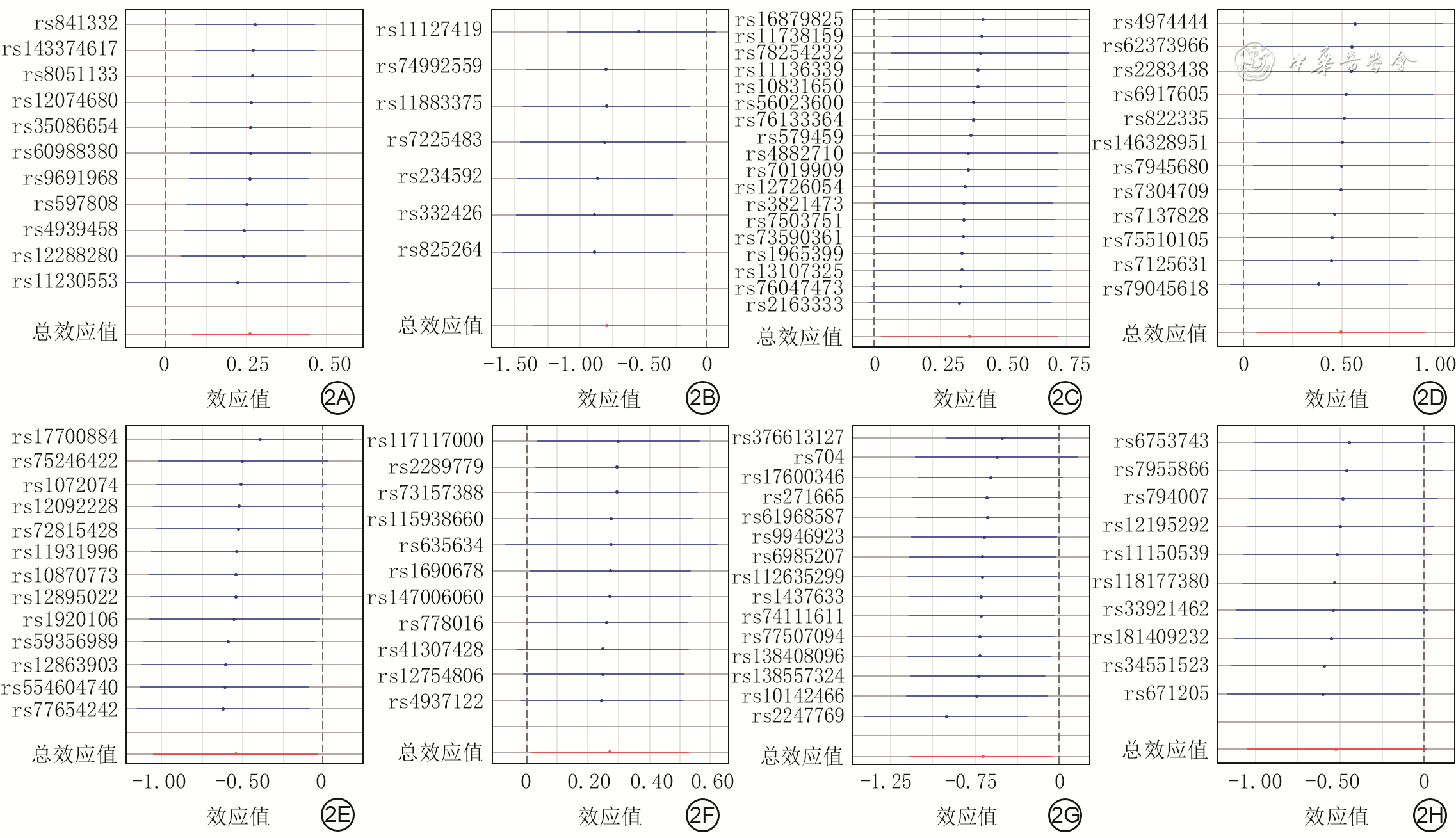
图 2 留一法分析的与8种人炎症蛋白存在显著因果关系的增生性瘢痕SNP的可靠性。2A.SNP与CD6的因果关系;2B.SNP与白血病抑制因子的因果关系;2C.SNP与肿瘤坏死因子配体超家族成员12的因果关系;2D.SNP与程序性死亡配体1的因果关系;2E.SNP与白细胞介素-17C的因果关系;2F.SNP与白血病抑制因子受体的因果关系;2G.SNP与骨保护素的因果关系;2H.SNP与成纤维细胞生长因子23的因果关系
注:SNP为单核苷酸多态性;每个SNP对应的线段为误差线,线段中的圆点代表β值;β值均在0点同侧说明结果可靠,误差线均在0点同侧说明结果非常可靠

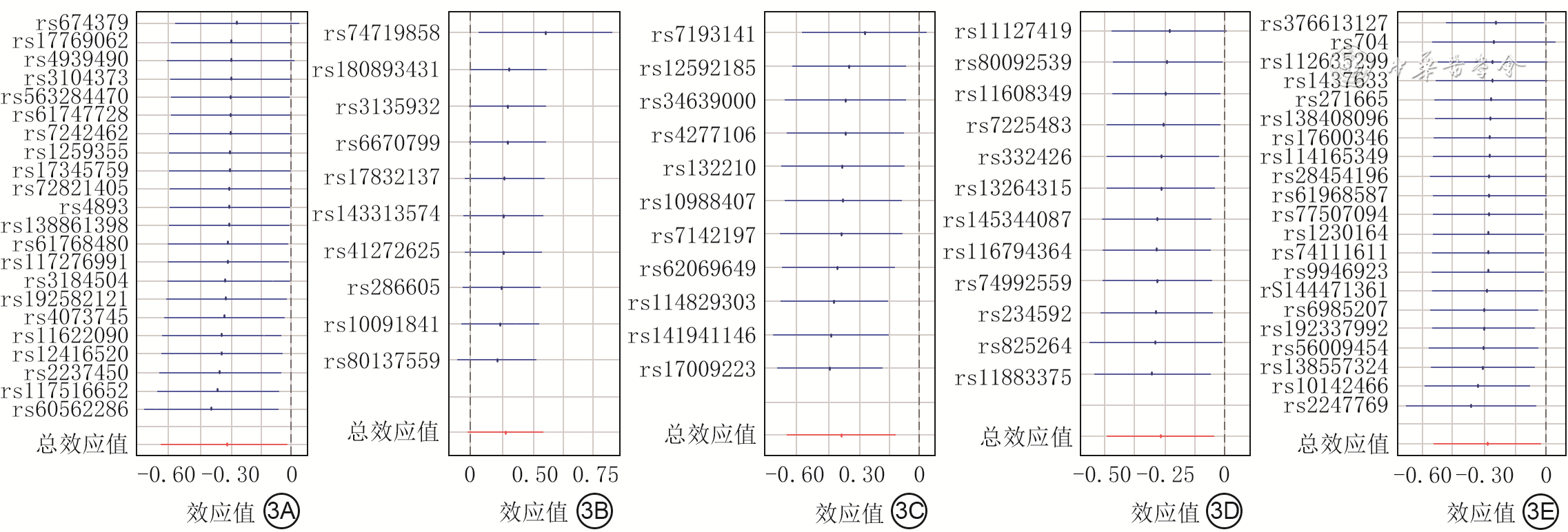
图 3 留一法分析的与5种人炎症蛋白存在显著因果关系的瘢痕疙瘩SNP的可靠性。3A.SNP与CD5的因果关系;3B.SNP与白细胞介素-10受体α亚基的因果关系;3C.SNP与白细胞介素-5的因果关系;3D.SNP与白血病抑制因子的因果关系;3E.SNP与骨保护素的因果关系
注:SNP为单核苷酸多态性;每个SNP对应的线段为误差线,线段中的圆点代表β值;β值均在0点同侧说明结果可靠,误差线均在0点同侧说明结果非常可靠
Table 1. 逆方差加权法分析显示8种人炎症蛋白与增生性瘢痕之间存在显著因果关系
炎症蛋白 SNP数(个) OR 95%CI P值 CD6 11 1.365 1.100~1.693 0.005 成纤维细胞生长因子23 10 0.553 0.315~0.971 0.039 白细胞介素-17C 13 0.621 0.408~0.947 0.027 白血病抑制因子 7 0.506 0.289~0.887 0.017 白血病抑制因子受体 11 1.375 1.025~1.845 0.034 骨保护素 15 0.623 0.402~0.966 0.034 程序性死亡配体1 12 1.683 1.090~2.599 0.019 肿瘤坏死因子配体超家族成员12 18 1.567 1.081~2.273 0.018 注:SNP为单核苷酸多态性  下载: 导出CSV
Table 2. 与增生性瘢痕之间存在显著因果关系的8种人炎症蛋白SNP的异质性和水平多效性分析结果
炎症蛋白 SNP数(个) Cochran Q检验 MR-Egger回归检验 MR-PRESSO离群值检验 Q值 P值 截距 P值 RSSobs P值 CD6 11 6.990 0.726 -0.011 0.746 7.479 0.810 成纤维细胞生长因子23 10 2.451 0.982 -0.026 0.699 3.072 0.987 白细胞介素-17C 13 5.230 0.950 -0.042 0.541 6.451 0.947 白血病抑制因子 7 5.573 0.473 -0.043 0.544 7.422 0.502 白血病抑制因子受体 11 3.849 0.954 0.015 0.728 4.246 0.978 骨保护素 15 22.231 0.074 0.030 0.645 26.590 0.092 程序性死亡配体1 12 7.124 0.789 -0.027 0.560 8.285 0.798 肿瘤坏死因子配体超家族成员12 18 11.064 0.853 -0.025 0.592 12.030 0.868 注:SNP为单核苷酸多态性,MR为孟德尔随机化;Cochran Q检验评估异质性,另2种检验评估水平多效性
下载: 导出CSV
Table 3. 增生性瘢痕与8种人炎症蛋白之间反向因果关系的逆方差加权法分析结果
炎症蛋白 SNP数(个) OR 95%CI P值 CD6 13 0.989 0.965~1.014 0.391 成纤维细胞生长因子23 13 0.995 0.970~1.021 0.713 白细胞介素-17C 13 0.998 0.971~1.026 0.892 白血病抑制因子 13 0.983 0.949~1.017 0.327 白血病抑制因子受体 13 0.968 0.942~0.994 0.016 骨保护素 13 0.998 0.974~1.021 0.842 程序性死亡配体1 13 1.013 0.982~1.044 0.419 肿瘤坏死因子配体超家族成员12 13 0.972 0.949~0.997 0.026 注:SNP为单核苷酸多态性
下载: 导出CSV
Table 4. 逆方差加权法分析得出5种人炎症蛋白与瘢痕疙瘩之间存在显著因果关系
炎症蛋白 SNP数(个) OR 95%CI P值 CD5 22 0.744 0.573~0.965 0.026 白细胞介素-10受体α亚基 10 1.303 1.024~1.660 0.032 白细胞介素-5 11 0.686 0.472~0.996 0.048 白血病抑制因子 12 0.603 0.431~0.842 0.003 骨保护素 21 0.715 0.553~0.924 0.010 注:SNP为单核苷酸多态性
下载: 导出CSV
Table 5. 补充双样本MR分析方法得出3种人炎症蛋白与瘢痕疙瘩之间存在因果关系
分析方法及炎症蛋白 SNP数(个) OR 95%CI P值 MR-Egger法 骨保护素 21 0.537 0.311~0.927 0.038 加权中位数法 CD5 22 0.679 0.467~0.986 0.042 白细胞介素-5 11 0.604 0.372~0.980 0.041 骨保护素 21 0.688 0.502~0.944 0.020 简单模式法 CD5 22 0.502 0.273~0.924 0.038 骨保护素 21 0.546 0.326~0.917 0.033 注:MR为孟德尔随机化,SNP为单核苷酸多态性
下载: 导出CSV
Table 6. 与瘢痕疙瘩之间存在显著因果关系的5种人炎症蛋白SNP的异质性和水平多效性分析结果
炎症蛋白 SNP数(个) Cochran Q检验 MR-Egger回归检验 MR-PRESSO离群值检验 Q值 P值 截距 P值 RSSobs P值 CD5 22 15.487 0.798 0.018 0.626 16.842 0.826 白细胞介素-10受体α亚基 10 8.551 0.480 0.044 0.185 14.737 0.469 白细胞介素-5 11 11.178 0.344 -0.017 0.713 13.570 0.363 白血病抑制因子 12 6.504 0.838 -0.020 0.608 7.321 0.864 骨保护素 21 24.469 0.223 0.038 0.260 26.167 0.305 注:SNP为单核苷酸多态性,MR为孟德尔随机化;Cochran Q检验评估异质性,另2种检验评估水平多效性
下载: 导出CSV
Table 7. 瘢痕疙瘩与5种人炎症蛋白之间反向因果关系的逆方差加权法分析结果
炎症蛋白 SNP数(个) OR 95%CI P值 CD5 23 0.988 0.969~1.008 0.245 白细胞介素-10受体α亚基 23 0.994 0.972~1.017 0.593 白细胞介素-5 23 1.016 0.993~1.039 0.171 白血病抑制因子 23 1.006 0.979~1.033 0.664 骨保护素 23 0.995 0.975~1.015 0.638 注:SNP为单核苷酸多态性
下载: 导出CSV
-
 王思思.mp4

-

 下载:
下载:

计量
- 文章访问数: 449
- HTML全文浏览量: 302
- PDF下载量: 14
- 被引次数: 0
