


Effects and mechanism of tannic acid/magnesium nanocomplex on wound healing in rats with full-thickness scald
-
摘要:
目的 探讨单宁酸/镁纳米复合物(MgTA NC)对Ⅲ度烫伤大鼠创面愈合的作用及其机制。 方法 该研究为实验研究。通过水热法制备了生物相容性良好的MgTA NC。取小鼠RAW 264.7细胞,分为单纯内毒素/脂多糖(LPS)组、低MgTA NC组、中MgTA NC组、高MgTA NC组,均先用终质量浓度1 μg/mL的LPS处理,再分别采用终质量浓度0(不含)、2.5、5.0、7.5 μg/mL的MgTA NC培养24 h后,采用蛋白质印迹法检测细胞中M1型巨噬细胞标志物诱导型一氧化氮合酶(iNOS)、M2型巨噬细胞标志物CD163和糖酵解代谢相关的蛋白M2型丙酮酸激酶(PKM2)、己糖激酶的蛋白表达,采用酶联免疫吸附测定法检测细胞中琥珀酸脱氢酶和异柠檬酸脱氢酶的表达水平。细胞实验样本数均为3。取12只6周龄雄性SD大鼠,使用控温电烫伤仪在其背部造成Ⅲ度烫伤,采用随机数字表法将其分为对照组、单纯水凝胶组和复合水凝胶组(每组4只),分别采用磷酸盐缓冲液、甲基丙烯酸酐化明胶水凝胶、负载MgTA NC的甲基丙烯酸酐化明胶水凝胶处理创面,计算伤后3、7、14 d创面愈合率(样本数为4)。伤后14 d,行免疫组织化学染色观察创面组织中炎症因子肿瘤坏死因子α(TNF-α)的表达。 结果 培养24 h后,单纯LPS组、低MgTA NC组、中MgTA NC组、高MgTA NC组RAW 264.7细胞中M1型巨噬细胞标志物iNOS的蛋白表达依次减少,M2型巨噬细胞标志物CD163的蛋白表达依次增加,PKM2和己糖激酶的蛋白表达均依次减少。培养24 h后,与单纯LPS组比较,低MgTA NC组RAW 264.7细胞中琥珀酸脱氢酶和异柠檬酸脱氢酶的表达水平均明显升高(P<0.05);与低MgTA NC组比较,中MgTA NC组RAW 264.7细胞中琥珀酸脱氢酶和异柠檬酸脱氢酶的表达水平均明显升高(P<0.05);与中MgTA NC组比较,高MgTA NC组RAW 264.7细胞中琥珀酸脱氢酶和异柠檬酸脱氢酶的表达水平均明显升高(P<0.05)。伤后3 d,3组大鼠创面愈合率比较,差异无统计学意义(P>0.05)。伤后7、14 d,单纯水凝胶组[(52.28±5.11)%、(81.11±2.09)%]、复合水凝胶组[(76.81±2.68)%、(98.93±0.29)%]大鼠创面愈合率均明显高于对照组[(32.75±6.86)%、(60.10±2.10)%,P<0.05],复合水凝胶组大鼠创面愈合率均明显高于单纯水凝胶组(P<0.05)。伤后14 d,单纯水凝胶组和复合水凝胶组大鼠创面组织中TNF-α的表达均明显少于对照组,复合水凝胶组大鼠创面组织中TNF-α的表达明显少于单纯水凝胶组。 结论 MgTA NC具有良好的生物相容性,能调控巨噬细胞向M2型极化,有效降低巨噬细胞糖酵解水平并提高其氧化磷酸化水平,抑制组织过度炎症反应并增强组织的再生及修复能力,从而加速Ⅲ度烫伤大鼠创面愈合。 Abstract:Objective To investigate the effects and mechanism of tannic acid/magnesium nanocomplex (MgTA NC) on wound healing in rats with full-thickness scald. Methods This study was an experimental study. The MgTA NC with good biocompatibility was synthesized using the hydrothermal method. Mouse RAW 264.7 cells were divided into endotoxins/lipopolysaccharides (LPS) alone group, low MgTA NC group, medium MgTA NC group, and high MgTA NC group, which were all treated with LPS at final mass concentration of 1 μg/mL, and then cultured respectively with MgTA NC at the final mass concentration of 0 (without), 2.5, 5.0, or 7.5 μg/mL for 24 hours. The protein expressions of M1 type macrophage marker inducible nitric oxide synthase (iNOS), M2 type macrophage marker CD163, as well as glycolysis metabolism-related proteins pyruvate kinase type M2 (PKM2) and hexokinase in cells were detected by Western blotting; the expression levels of succinate dehydrogenase and isocitrate dehydrogenase in cells were detected by enzyme-linked immunosorbent assay method. The sample size in cell experiment was 3. Twelve six-week-old male Sprague-Dawley rats were selected and subjected to full-thickness scald on their backs using a temperature-controlled electrothermal burn device. The rats were assigned to control group, simple hydrogel group, and composite hydrogel group according to the random number table method, with 4 rats in each group. The wounds were treated with phosphate buffered saline, methacrylated gelatin (GelMA) hydrogel, or GelMA hydrogel loaded with MgTA NC, respectively. The wound healing rates were calculated at post-injury day 3, 7, and 14 (with the sample size of 4), and the expression level of inflammatory factor tumor necrosis factor α (TNF-α) in the wound tissue at post-injury day 14 was detected by immunohistochemical staining. Results After 24 hours of culture, the protein expressions of iNOS, the M1 type macrophage marker in RAW 264.7 cells decreased successively in LPS alone group, low MgTA NC group, medium MgTA NC group, and high MgTA NC group, while the protein expressions of CD163, the M2 type macrophage marker increased successively, and the protein expressions of PKM2 and hexokinase decreased successively. After 24 hours of culture, compared with those in LPS alone group, the expression levels of succinate dehydrogenase and isocitrate dehydrogenase in RAW 264.7 cells in low MgTA NC group were significantly increased (P<0.05); compared with those in low MgTA NC group, the expression levels of succinate dehydrogenase and isocitrate dehydrogenase in RAW 264.7 cells in medium MgTA NC group were significantly increased (P<0.05); compared with those in medium MgTA NC group, the expression levels of succinate dehydrogenase and isocitrate dehydrogenase in RAW 264.7 cells in high MgTA NC group were significantly increased (P<0.05). At post-injury day 3, there was no statistically significant difference in the wound healing rate among the three groups of rats (P>0.05). At post-injury day 7 and 14, the wound healing rates of rats in simple hydrogel group were (52.28±5.11)% and (81.11±2.09)%, and those in composite hydrogel group were (76.81±2.68)% and (98.93±0.29)%, which were significantly higher than (32.75±6.86)% and (60.10±2.10)% in control group, respectively (P<0.05); the wound healing rates of rats in composite hydrogel group were significantly higher than those in simple hydrogel group (P<0.05). At post-injury day 14, the expression of TNF-α in the wound tissue of rats in simple hydrogel group and composite hydrogel group was significantly reduced compared with that in control group, and the expression of TNF-α in the wound tissue of rats in composite hydrogel group was significantly reduced compared with that in simple hydrogel group. Conclusions MgTA NC exhibits excellent biocompatibility, and it is capable of modulating macrophage polarization toward the M2 type, effectively reducing glycolysis level and enhancing oxidative phosphorylation level of macrophages, suppressing excessive inflammatory responses and enhancing the ability of tissue regeneration and repair, therefore significantly accelerating wound healing in rats with full-thickness scald. -
参考文献
(40) [1] LiS,PanW,ZhangM,et al.Chitosan-based dressing materials for burn wound healing[J].Polymers (Basel),2025,17(12):1647.DOI: 10.3390/polym17121647. [2] MaJ, LiS, ZhangL, et al. Oxidativestress-scavenging thermo-activated MXene hydrogel for rapid repair of MRSA impaired wounds and burn wounds[J]. Materials Today, 2024, 80: 139-155. DOI: 10.1016/j.mattod.2024.08.010. [3] KangX, LiY, DuanZ, et al. A Mxene@TA/Fe dual-nanozyme composited antifouling hydrogel for burn wound repair[J]. Chemical Engineering Journal, 2023, 476: 146420. DOI: 10.1016/j.cej.2023.146420. [4] LanJ,ShiL,XiaoW,et al.A rapid self-pumping organohydrogel dressing with hydrophilic fractal microchannels to promote burn wound healing[J].Adv Mater,2023,35(38):e2301765.DOI: 10.1002/adma.202301765. [5] ZhangJ,PanT,LeeJ,et al.Enabling tumor-specific drug delivery by targeting the Warburg effect of cancer[J].Cell Rep Med,2025,6(1):101920.DOI: 10.1016/j.xcrm.2024.101920. [6] ShahaA,WangY,WangX,et al.CMTM6 mediates the Warburg effect and promotes the liver metastasis of colorectal cancer[J].Exp Mol Med,2024,56(9):2002-2015.DOI: 10.1038/s12276-024-01303-1. [7] RyanDG,O'NeillL.Krebs cycle reborn in macrophage immunometabolism[J].Annu Rev Immunol,2020,38:289-313.DOI: 10.1146/annurev-immunol-081619-104850. [8] WillenborgS,SaninDE,JaisA,et al.Mitochondrial metabolism coordinates stage-specific repair processes in macrophages during wound healing[J].Cell Metab,2021,33(12):2398-2414.e9.DOI: 10.1016/j.cmet.2021.10.004. [9] PilchovaI,KlacanovaK,TatarkovaZ,et al.The involvement of Mg2+ in regulation of cellular and mitochondrial functions[J].Oxid Med Cell Longev,2017,2017:6797460.DOI: 10.1155/2017/6797460. [10] WangJ,MaXY,FengYF,et al.Magnesium ions promote the biological behaviour of rat calvarial osteoblasts by activating the PI3K/Akt signalling pathway[J].Biol Trace Elem Res,2017,179(2):284-293.DOI: 10.1007/s12011-017-0948-8. [11] YamanakaR,TabataS,ShindoY,et al.Mitochondrial Mg2+ homeostasis decides cellular energy metabolism and vulnerability to stress[J].Sci Rep,2016,6:30027.DOI: 10.1038/srep30027. [12] TatarkovaZ,de BaaijJHF,GrendarM,et al.Dietary Mg2+ intake and the Na+/Mg2+ exchanger SLC41A1 influence components of mitochondrial energetics in murine cardiomyocytes[J].Int J Mol Sci,2020,21(21):8221.DOI: 10.3390/ijms21218221. [13] HuX, HeJ, QiaoL, et al.Multifunctional dual network hydrogel loaded with novel tea polyphenol magnesium nanoparticles accelerates wound repair of MRSA infected diabetes[J].Adv Funct Mater, 2024, 34(22): 2312140.DOI: 10.1002/adfm.202312140. [14] BravoM,SimónJ,González-RecioI,et al.Magnesium and liver metabolism through the lifespan[J].Adv Nutr,2023,14(4):739-751.DOI: 10.1016/j.advnut.2023.05.009. [15] MudrykK,LeeC,TomaníkL,et al.How does Mg2+(aq) interact with ATP(aq)? Biomolecular structure through the lens of liquid-jet photoemission spectroscopy[J].J Am Chem Soc,2024,146(23):16062-16075.DOI: 10.1021/jacs.4c03174. [16] ShenJ, YongL, ChenB, et al. Effect of biocomposite mediated magnesium ionic micro-homeostasis on cell fate regulation and bone tissue regeneration[J]. Composites Part B: Engineering, 2023, 265: 110961. DOI: 10.1016/j.compositesb.2023.110961. [17] ChenJ, GuanY, YangY, et al. Tannic acid-modified magnesium oxychloride bone cement with high water resistance and osteogenic properties[J]. Ceramics International, 2024, 50(24): 53407-53420. DOI: 10.1016/j.ceramint.2024.10.191. [18] JeongH,ByunH,LeeJ,et al.Enhancement of bone tissue regeneration with multi-functional nanoparticles by coordination of immune, osteogenic, and angiogenic responses[J].Adv Healthc Mater,2025,14(5):e2400232.DOI: 10.1002/adhm.202400232. [19] LiD,LiJ,WangS,et al.Dually crosslinked copper-poly(tannic acid) nanoparticles with microenvironment-responsiveness for infected wound treatment[J].Adv Healthc Mater,2023,12(17):e2203063.DOI: 10.1002/adhm.202203063. [20] SuR,LiP,ZhangY,et al.Polydopamine/tannic acid/chitosan/poloxamer 407/188 thermosensitive hydrogel for antibacterial and wound healing[J].Carbohydr Polym,2023,302:120349.DOI: 10.1016/j.carbpol.2022.120349. [21] 罗高兴,詹日兴,袁志强,等. 建议应用自来水冲洗代替生理盐水清洁皮肤创面[J]. 中华烧伤与创面修复杂志,2025,41(3):201-205. DOI: 10.3760/cma.j.cn501225-20250108-00015. [22] 张婷,刘佳琦,杨云舒,等. Meek皮片移植联合富血小板血浆修复大面积深度烧伤创面的临床效果[J]. 中华烧伤与创面修复杂志,2024,40(12):1150-1157. DOI: 10.3760/cma.j.cn501225-20231124-00206. [23] TannahillGM,CurtisAM,AdamikJ,et al.Succinate is an inflammatory signal that induces IL-1β through HIF-1α[J].Nature,2013,496(7444):238-242.DOI: 10.1038/nature11986. [24] O'NeillLAJ,ArtyomovMN.Itaconate: the poster child of metabolic reprogramming in macrophage function[J].Nat Rev Immunol,2019,19(5):273-281.DOI: 10.1038/s41577-019-0128-5. [25] LiuS,WangH,LiJ,et al.Loss of Bcl-3 regulates macrophage polarization by promoting macrophage glycolysis[J].Immunol Cell Biol,2024,102(7):605-617.DOI: 10.1111/imcb.12785. [26] ShaoY,ZhouX,ZhouS,et al.Injectable DMM/GelMA hydrogel for diabetic wound healing via regulating mitochondrial metabolism and macrophage repolarization[J].Colloids Surf B Biointerfaces,2025,248:114488.DOI: 10.1016/j.colsurfb.2024.114488. [27] LiQ,SongH,LiS,et al.Macrophage metabolism reprogramming EGCG-Cu coordination capsules delivered in polyzwitterionic hydrogel for burn wound healing and regeneration[J].Bioact Mater,2023,29:251-264.DOI: 10.1016/j.bioactmat.2023.07.011. [28] LuL,LiaoJ,XuC,et al.Kinsenoside-loaded microneedle accelerates diabetic wound healing by reprogramming macrophage metabolism via inhibiting IRE1α/XBP1 signaling axis[J].Adv Sci (Weinh),2025,12(26):e2502293.DOI: 10.1002/advs.202502293. [29] AndreicaBI,AnisieiA,RoscaI,et al.Quaternized chitosan/chitosan nanofibrous mats: an approach toward bioactive materials for tissue engineering and regenerative medicine[J].Carbohydr Polym,2023,302:120431.DOI: 10.1016/j.carbpol.2022.120431. [30] ZhaoH, WangL, XiK, et al. In-situ pore forming bone adhesives targeting disordered osteoimmune microenvironment and bone metabolic balance to promote osteoporotic fracture healing[J]. Chemical Engineering Journal, 2025, 510: 161315. DOI: 10.1016/j.cej.2025.161315. [31] YuanW,LuG,ZhaoY,et al.Intranuclear TCA and mitochondrial overload: the nascent sprout of tumors metabolism[J].Cancer Lett,2025,613:217527.DOI: 10.1016/j.canlet.2025.217527. [32] 苟伟茗,杨鹏,卢毅飞,等. 大鲵皮肤黏液多糖对糖尿病小鼠全层皮肤缺损创面愈合的作用及其机制[J]. 中华烧伤与创面修复杂志,2025,41(2):127-136. DOI: 10.3760/cma.j.cn501225-20240725-00280. [33] XuN, GaoY, LiZ, et al. Immunoregulatory hydrogel decorated with tannic acid/ferric ion accelerates diabetic wound healing via regulating macrophage polarization[J]. Chemical Engineering Journal, 2023, 466:143173. DOI: 10.1016/j.cej.2023.143173. [34] IshidaK,NagatakeT,SaikaA,et al.Induction of unique macrophage subset by simultaneous stimulation with LPS and IL-4[J].Front Immunol,2023,14:1111729.DOI: 10.3389/fimmu.2023.1111729. [35] GillenJ,OndeeT,GurusamyD,et al.LPS tolerance inhibits cellular respiration and induces global changes in the macrophage secretome[J].Biomolecules,2021,11(2):164.DOI: 10.3390/biom11020164. [36] WuQ,LuZ,WangL,et al.Konjac glucomannan/xanthan gum hydrogels loaded with metal-phenolic networks encapsulated probiotic to promote infected wound healing[J].Carbohydr Polym,2025,353:123243.DOI: 10.1016/j.carbpol.2025.123243. [37] UllahS,HussainZ,MehmoodS,et al.Metal-phenolic network (MPN) modified Janus fibrous hydrogel scaffold for infected diabetic wound healing[J].ACS Appl Mater Interfaces,2025,17(7):10470-10484.DOI: 10.1021/acsami.4c20592. [38] WengZ,YeJ,CaiC,et al.Inflammatory microenvironment regulation and osteogenesis promotion by bone-targeting calcium and magnesium repletion nanoplatform for osteoporosis therapy[J].J Nanobiotechnology,2024,22(1):314.DOI: 10.1186/s12951-024-02581-7. [39] HuangL,CaiP,BianM,et al.Injectable and high-strength PLGA/CPC loaded ALN/MgO bone cement for bone regeneration by facilitating osteogenesis and inhibiting osteoclastogenesis in osteoporotic bone defects[J].Mater Today Bio,2024,26:101092.DOI: 10.1016/j.mtbio.2024.101092. [40] LiX,LiX,YangJ,et al.Living and injectable porous hydrogel microsphere with paracrine activity for cartilage regeneration[J].Small,2023,19(17):e2207211.DOI: 10.1002/smll.202207211. -

图 2 蛋白质印迹法检测的4组小鼠RAW 264.7细胞培养24 h后极化和代谢相关蛋白的表达。2A.细胞极化相关蛋白的表达;2B.细胞代谢相关蛋白的表达
注:iNOS为诱导型一氧化氮合酶,PKM2为M2型丙酮酸激酶;图2A和2B条带上方1、2、3、4分别代表均用终质量浓度为1 μg/mL的内毒素/脂多糖(LPS)处理后,再分别用终质量浓度为0(不含)、2.5、5.0、7.5 μg/mL的单宁酸/镁纳米复合物(MgTA NC)培养的单纯LPS组、低MgTA NC组、中MgTA NC组、高MgTA NC组

图 3 3组Ⅲ度烫伤大鼠伤后各时间点创面愈合情况。3A、3B、3C、3D、3E.分别为对照组伤后1、3、7、14 d的创面情况及创面愈合过程模拟图;3F、3G、3H、3I、3J.分别为单纯水凝胶组伤后1、3、7、14 d的创面情况及创面愈合过程模拟图,图3H、3I创面面积分别明显小于图3C、3D;3K、3L、3M、3N、3O.分别为复合水凝胶组伤后1、3、7、14 d的创面情况及创面愈合过程模拟图,图3M创面面积明显小于图3C、3H,图3N创面面积明显小于图3D、3I
注:对照组、单纯水凝胶组和复合水凝胶组大鼠创面分别采用磷酸盐缓冲液、甲基丙烯酸酐化明胶水凝胶、负载单宁酸/镁纳米复合物的甲基丙烯酸酐化明胶水凝胶处理;图3E、3J、3O中蓝色、黄色、橙色、绿色分别表示伤后1、3、7、14 d的创面

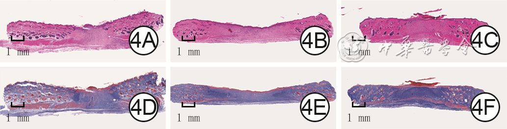
图 4 3组Ⅲ度烫伤大鼠伤后14 d创面新生肉芽组织、上皮化程度和胶原沉积情况。4A、4B、4C.分别为对照组、单纯水凝胶组和复合水凝胶组新生肉芽组织和上皮化情况,图4C新生肉芽组织和上皮化情况优于图4A、4B 苏木精-伊红×40;4D、4E、4F.分别为对照组、单纯水凝胶组和复合水凝胶组胶原沉积情况,图4F较图4D、4E胶原分布更加有序和紧实 Masson×40
注:对照组、单纯水凝胶组和复合水凝胶组大鼠创面分别采用磷酸盐缓冲液、甲基丙烯酸酐化明胶水凝胶、负载单宁酸/镁纳米复合物的甲基丙烯酰化明胶水凝胶处理

图 5 3组Ⅲ度烫伤大鼠伤后14 d创面组织中炎症因子TNF-α的表达情况 二氨基联苯胺-苏木精×200。5A、5B、5C.分别为对照组、单纯水凝胶组和复合水凝胶组,图5C中TNF-α的表达明显少于图5A、5B
注:对照组、单纯水凝胶组和复合水凝胶组大鼠创面分别采用磷酸盐缓冲液、甲基丙烯酸酐化明胶水凝胶、负载单宁酸/镁纳米复合物的甲基丙烯酸酐化明胶水凝胶处理;肿瘤坏死因子α(TNF-α)阳性染色为棕褐色
Table 1. 4组小鼠RAW 264.7细胞培养24 h后琥珀酸脱氢酶和异柠檬酸脱氢酶的表达水平比较(%,
组别 样本数 琥珀酸脱氢酶 异柠檬酸脱氢酶 单纯LPS组 3 30.5±2.1 26.7±2.0 低MgTA NC组 3 35.5±1.7 33.6±1.2 中MgTA NC组 3 41.8±1.8 39.5±1.7 高MgTA NC组 3 48.3±2.7 46.2±0.8 F值 168.90 53.54 P值 <0.001 <0.001 P1值 <0.001 <0.001 P2值 0.005 0.001 P3值 0.004 <0.001 注:表中数据为以未经任何处理细胞结果为1,计算的各组细胞相对表达百分比;单纯内毒素/脂多糖(LPS)组、低单宁酸/镁纳米复合物(MgTA NC)组、中MgTA NC组、高MgTA NC组细胞均用终质量浓度为1 μg/mL的LPS处理后,再分别用终质量浓度为0(不含)、2.5、5.0、7.5 μg/mL的MgTA NC培养;F值和P值为4组间各指标总体比较所得;P1值为低MgTA NC组和单纯LPS组各指标比较所得,P2值为中MgTA NC组和低MgTA NC组各指标比较所得,P3值为高MgTA NC组和中MgTA NC组各指标比较所得  下载: 导出CSV
Table 2. 3组Ⅲ度烫伤大鼠伤后各时间点创面愈合率比较(%,
组别 鼠数(只) 3 d 7 d 14 d 对照组 4 13.81±0.93 32.75±6.86 60.10±2.10 单纯水凝胶组 4 12.97±1.05 52.28±5.11 81.11±2.09 复合水凝胶组 4 11.70±1.11 76.81±2.68 98.93±0.29 F值 4.23 72.81 511.90 P值 0.051 <0.001 <0.001 P1值 — 0.001 <0.001 P2值 — <0.001 <0.001 P3值 — <0.001 <0.001 注:对照组、单纯水凝胶组和复合水凝胶组大鼠创面分别采用磷酸盐缓冲液、甲基丙烯酸酐化明胶水凝胶、负载单宁酸/镁纳米复合物的甲基丙烯酸酐化明胶水凝胶处理;时间因素主效应,F=59.44,P<0.001;处理因素主效应,F=568.60,P<0.001;两者交互作用,F=15.56,P<0.001;F值和P值为3组间各时间点总体比较所得;P1值为单纯水凝胶组与对照组各时间点比较所得,P2值为复合水凝胶组与单纯水凝胶组各时间点比较所得,P3值为复合水凝胶组与对照组各时间点比较所得;“—”表示无此项
下载: 导出CSV
-

 下载:
下载:


计量
- 文章访问数: 2969
- HTML全文浏览量: 352
- PDF下载量: 27
- 被引次数: 0
