


Clinical effects of extracorporeal shock wave combined with complex decongestive therapy in the treatment of lower limb lymphedema after cervical cancer surgery
-
摘要:
目的 探讨体外冲击波疗法联合综合消肿治疗对宫颈癌术后下肢淋巴水肿的临床疗效。 方法 该研究为前瞻性随机对照试验。2023年4月—2024年12月,深圳市大鹏新区南澳人民医院肿瘤康复科收治64例符合入选标准的宫颈癌术后下肢淋巴水肿患者,患者均为女性,年龄33~75岁。将患者按随机数字表法分为仅进行综合消肿治疗的对照组及进行体外冲击波疗法联合综合消肿治疗的联合治疗组,每组32例。研究过程中4例患者退出研究,最终每组纳入30例。于治疗前及4周治疗结束时(下称治疗后),采用便携式MyotonPRO仪测量股外侧肌、股内侧肌、股中间肌、腓肠肌内侧头、腓肠肌外侧头体表投射位置的皮肤硬度,以反映下肢皮肤纤维化程度;测量膝中、髌骨上缘上10 cm、髌骨上缘上20 cm、踝中、髌骨下缘下20 cm、髌骨下缘下10 cm处的周径,以反映下肢水肿程度;采用简体中文版下肢淋巴水肿功能、残疾与健康问卷(Lymph-ICF-LL)进行生活质量评分,采用视觉模拟评分法(VAS)进行下肢疼痛程度评分。计算上述指标治疗前后差值。 结果 联合治疗组患者治疗后股外侧肌、股中间肌、股内侧肌、腓肠肌内侧头、腓肠肌外侧头体表投射位置的皮肤硬度均明显小于对照组(t值分别为2.78、2.04、3.12、2.01、2.35,P < 0.05);联合治疗组患者治疗前后以上肌肉体表投射位置的皮肤硬度差值分别为(65±23)、(24±8)、(25±8)、(65±27)、(69±34)N/m,均明显大于对照组的(49±23)、(16±19)、(8±9)、(45±39)、(43±42)N/m(t值分别为-2.75、-2.35、-7.47、-2.33、-2.64,P < 0.05),组间均数差值(95%置信区间)分别为16(5~28)、9(1~17)、17(12~21)、20(3~36)、26(4~49)N/m。联合治疗组患者治疗后髌骨下缘下20 cm、髌骨下缘下10 cm、膝中、髌骨上缘上10 cm、髌骨上缘上20 cm处的周径均明显小于对照组(t值分别为-2.41、-2.49、-2.44、-2.21、-2.36,P < 0.05);联合治疗组患者治疗前后以上位置周径差值均明显大于对照组(t值分别为2.21、3.62、3.35、4.14、3.89,P < 0.05),组间均数差值(95%置信区间)分别为2.3(0.1~4.6)、2.4(1.0~3.8)、2.1(0.8~3.4)、3.5(1.6~5.4)、3.4(1.5~5.2)cm。联合治疗组患者治疗后下肢VAS评分和Lymph-ICF-LL总得分均明显低于对照组(t值分别为-2.46、-2.63,P < 0.05);联合治疗组患者治疗前后下肢VAS评分和Lymph-ICF-LL总得分差值均明显高于对照组(t值分别为2.34、3.32,P < 0.05),组间均数差值(95%置信区间)分别为0.5(0~0.9)、6(2~9)分。 结论 体外冲击波疗法联合综合消肿治疗在改善宫颈癌术后下肢淋巴水肿和皮肤纤维化方面具有显著疗效,且能缓解疼痛,提高患者生活质量。 Abstract:Objective To explore the clinical effects of extracorporeal shock wave therapy (ESWT) combined with complex decongestive therapy (CDT) in the treatment of lower limb lymphedema after cervical cancer surgery. Methods This study was a prospective randomized controlled trial. From April 2023 to December 2024, 64 patients were admitted to the Department of Oncology Rehabilitation of Nan'ao People's Hospital of Dapeng New District of Shenzhen. All patients were female, aged 33-75 years. The patients were divided into control group treated with CDT alone and combined treatment group treated with ESWT and CDT according to the random number table method, with 32 patients in each group. Four patients withdrew in the research process, and 30 patients were included in each group finally. Before treatment and at the end of 4 weeks of treatment (hereinafter referred to as after treatment), the skin stiffness was measured using a portable MyotonPRO device at the superficial projection position of vastus lateralis, vastus medialis, vastus intermedius, medial gastrocnemius, and lateral gastrocnemius to reflect the degree of skin fibrosis of lower limb, the circumferences were measured at the mid-knee, 10 cm and 20 cm above the superior patellar border, and the mid-ankle, 10 cm and 20 cm below the inferior patellar border to reflect the severity degree of edema of lower limb, the life quality was scored using the simplified Chinese version of the lower limb lymphedema functioning, disability, and health questionnaire (Lymph-ICF-LL), and the pain intensity was scored using the visual analogue scale (VAS). The differences of the above indexes were calculated between before and after treatment. Results The skin stiffness at the superficial projection position of vastus lateralis, vastus medialis, vastus intermedius, medial gastrocnemius, and lateral gastrocnemius of patients in combined treatment group after treatment was significantly smaller than that in control group (with t values of 2.78, 2.04, 3.12, 2.01, and 2.35, respectively, P < 0.05). The differences in skin stiffness between before and after treatment at the superficial projection position of the above-mentioned muscles of patients in combined treatment group was (65±23), (24±8), (25±8), (65±27), and (69±34) N/m, respectively, which were significantly larger than (49±23), (16±19), (8±9), (45±39), and (43±42) N/m in control group (with t values of -2.75 -2.35, -7.47, -2.33, and -2.64, respectively, P < 0.05), and the mean differences between groups (95% confidence intervals) were 16 (5 to 28), 9 (1 to 17), 17 (12 to 21), 20 (3 to 36), and 26 (4 to 49) N/m, respectively. The circumferences at 10 cm and 20 cm below the inferior patellar border, mid-knee, and 10 cm and 20 cm above the superior patellar border of patients in combined treatment group after treatment were significantly smaller than those in control group (with t values of -2.41, -2.49, -2.44, -2.21, and -2.36, respectively, P < 0.05). The circumference differences between before and after treatment at the above-mentioned locations of patients in combined treatment group were significantly larger than those in control group (with t values of 2.21, 3.62, 3.35, 4.14, and 3.89, respectively, P < 0.05), and the mean differences between groups (95% confidence intervals) were 2.3 (0.1 to 4.6), 2.4 (1.0 to 3.8), 2.1 (0.8 to 3.4), 3.5 (1.6 to 5.4), and 3.4 (1.5 to 5.2) cm, respectively. The VAS scores of lower limb and the total Lymph-ICF-LL scores of patients in combined treatment group after treatment were significantly lower than those in control group (with t values of -2.46 and -2.63, respectively, P < 0.05); the differences of VAS scores of lower limb and the total Lymph-ICF-LL scores between before and after treatment of patients in combined treatment group were significantly higher than those in control group (with t values of 2.34 and 3.32, respectively, P < 0.05), and the mean differences between groups (95% confidence intervals) were 0.5 (0 to 0.9) and 6 (2 to 9), respectively. Conclusions Combined application of ESWT and CDT shows superior efficacy in alleviating lower limb lymphedema and skin fibrosis after cervical cancer surgery, and can relieve pain and improve patients' quality of life. 本文亮点采用前瞻性随机对照试验证实体外冲击波疗法联合综合消肿治疗能更显著减轻宫颈癌术后下肢淋巴水肿患者的水肿和皮肤纤维化程度,提高患者生活质量。体外冲击波疗法作为一种无创、创新且高效的治疗手段,为这种慢性、渐进性、难治性的疾病提供了极具前景的治疗选择。 -
参考文献
(40) [1] 刘宗超, 李哲轩, 张阳, 等. 2020全球癌症统计报告解读[J/OL]. 肿瘤综合治疗电子杂志, 2021, 7(2): 1-13[2025-02-05]. http://www.jmcm2018.com/CN/10.12151/JMCM.2021.02-01 . DOI:10.12151/JMCM.2021.02-01 .[2] Dayan JH, Ly CL, Kataru RP, et al. Lymphedema: pathogenesis and novel therapies[J]. Annu Rev Med, 2018, 69(1): 263-276. DOI: 10.1146/annurev-med-060116-022900. [3] Lee KW, Kim SB, Lee JH, et al. Effects of extracorporeal shockwave therapy on improvements in lymphedema, quality of life, and fibrous tissue in breast cancer-related lymphedema[J]. Ann Rehabil Med, 2020, 44(5): 386-392. DOI: 10.5535/arm.19213. [4] Auersperg V, Trieb K. Extracorporeal shock wave therapy: an update[J]. EFORT Open Rev, 2020, 5(10): 584-592. DOI: 10.1302/2058-5241.5.190067. [5] 廖冬发, 权毅, 潘显明, 等. 体外冲击波治疗部队伤病员末端病疗效观察[J]. 解放军医学杂志, 2009, 34(6): 806. DOI: 10.3321/j.issn:0577-7402.2009.06.054. [6] 傅聪颖, 黎宁, 李茂君, 等. 非药物干预治疗患者慢性创面疼痛效果的系统评价与贝叶斯网状荟萃分析[J]. 中华烧伤与创面修复杂志, 2025, 41(5): 491-500. DOI: 10.3760/cma.j.cn501225-20241213-00486. [7] Miccinilli S, Bravi M, Maselli M, et al. The effectiveness of extracorporeal shock wave therapy on breast cancer-related lymphedema: a literature review[J]. Lymphology, 2020, 53(3): 118-135. [8] Tsai YL, I TJ, Chuang YC, et al. Extracorporeal shock wave therapy combined with complex decongestive therapy in patients with breast cancer-related lymphedema: a systemic review and meta-analysis[J]. J Clin Med, 2021, 10(24): 5970. DOI: 10.3390/jcm10245970. [9] Lee JH, Kim SB, Lee KW, et al. Long-term effects of extracorporeal shock wave therapy on breast cancer-related lymphedema[J]. J Clin Med, 2022, 11(22): 6747. DOI: 10.3390/jcm11226747. [10] 谢幸, 孔北华, 段涛. 妇产科学[M]. 9版. 北京: 人民卫生出版社, 2018. [11] Executive Committee of the International Society of Lymphology. The diagnosis and treatment of peripheral lymphedema: 2023 Consensus Document of The International Society of Lymphology[J]. Lymphology, 2023, 56(4): 133-151. [12] 周煦川, 马戈甲, 王文飞, 等. 简体中文版下肢淋巴水肿功能、残疾与健康问卷的信度和效度分析[J]. 中华整形外科杂志, 2023, 39(6): 634-641. DOI: 10.3760/cma.j.cn114453-20220309-00060. [13] Bona AF, Ferreira KR, Carvalho RBM, et al. Incidence, prevalence, and factors associated with lymphedema after treatment for cervical cancer: a systematic review[J]. Int J Gynecol Cancer, 2020, 30(11): 1697-1704. DOI: 10.1136/ijgc-2020-001682. [14] Finnane A, Janda M, Hayes SC. Review of the evidence of lymphedema treatment effect[J]. Am J Phys Med Rehabil, 2015, 94(6): 483-498. DOI: 10.1097/PHM.0000000000000246. [15] 黄成, 胡学庆. 淋巴水肿治疗的研究进展[J]. 组织工程与重建外科, 2022, 18 (1): 38-41. DOI: 10.3969/j.issn.1673-0364.2022.01.009. [16] Carroll BJ, Singhal D. Advances in lymphedema: an under-recognized disease with a hopeful future for patients [J]. Vasc Med, 2024, 29(1): 70-84. DOI: 10.1177/1358863X231215329. [17] Zhang HZ, Zhong QL, Zhang HT, et al. Effectiveness of six-step complex decongestive therapy for treating upper limb lymphedema after breast cancer surgery[J]. World J Clin Cases, 2022, 10(25): 8827-8836. DOI: 10.12998/wjcc.v10.i25.8827. [18] Zeng Y, Liu G, Peng Z, et al. Application of complete decongestive therapy in patients with secondary bilateral lower limb lymphedema after comprehensive treatment of gynecological malignant tumor[J]. Lymphat Res Biol, 2024, 22(1): 60-65. DOI: 10.1089/lrb.2023.0029. [19] Thompson B, Gaitatzis K, Janse de Jonge X, et al. Manual lymphatic drainage treatment for lymphedema: a systematic review of the literature[J]. J Cancer Surviv, 2021, 15(2): 244-258. DOI: 10.1007/s11764-020-00928-1. [20] 陈佳佳, 高敏哲, 汪立, 等. 妇科相关肿瘤术后下肢淋巴水肿合并会阴部水肿的综合治疗初探[J]. 组织工程与重建外科杂志, 2022, 18(3): 242-246. DOI: 10.3969/j.issn.1673-0364.2022.03.008. [21] Chen F, Dellalana LE, Gandelman JS, et al. Non-invasive measurement of sclerosis in cutaneous cGVHD patients with the handheld device Myoton: a cross-sectional study [J]. Bone Marrow Transplant, 2019, 54(4): 616-619. DOI: 10.1038/s41409-018-0346-7. [22] John AJUK, Galdo FD, Gush R, et al. An evaluation of mechanical and biophysical skin parameters at different body locations[J]. Skin Res Technol, 2023, 29(2): e13292. DOI: 10.1111/srt.13292. [23] Junker HJ, Thumm B, Halvachizadeh S, et al. A quantitative comparison of devices for in vivo biomechanical characterization of human skin[J]. Mech Soft Mater, 2023, 5(1): 5. DOI: 10.1007/s42558-023-00053-w. [24] Glassman GE, Dellalana L, Tkaczyk ER, et al. Measuring biomechanical properties using a noninvasive Myoton device for lymphedema detection and tracking: a pilot study[J]. Eplasty, 2022, 22: e54. [25] Dellalana LE, Chen F, Vain A, et al. Reproducibility of the durometer and myoton devices for skin stiffness measurement in healthy subjects[J]. Skin Res Technol, 2019, 25(3): 289-293. DOI: 10.1111/srt.12646. [26] Lettner J, Królikowska A, Ramadanov N, et al. Evaluating the reliability of MyotonPro in assessing muscle properties: a systematic review of diagnostic test accuracy[J]. Medicina (Kaunas), 2024, 60(6): 851. DOI: 10.3390/medicina60060851. [27] Lin YP, Huang HY, Wu MH, et al. Effects of extracorporeal shock wave therapy on motor recovery, arm circumference, subcutaneous tissue thickness and skin thickness in a case with breast cancer related lymphedema portrayed with sonographic technique[J]. Rehabil Pract Sci, 2023, 1: 49-56. [28] Grushina TI, Orlov II. Pilot study of oncological safety of extracorporeal shock wave therapy for post-mastectomy lymphedema in patients with breast cancer[J]. Vopr Kurortol Fizioter Lech Fiz Kult, 2022, 99(6): 30-33. DOI: 10.17116/kurort20229906130. [29] Kojima M, Yamauchi C, Oyamada S, et al. Assessment of upper limb physiological features in patients with lymphedema after breast surgery using multiple instruments[J]. Lymphat Res Biol, 2020, 18(3): 239-246. DOI: 10.1089/lrb.2019.0039. [30] Cui HS, Joo SY, Cho YS, et al. Effect of extracorporeal shock wave therapy on keratinocytes derived from human hypertrophic scars[J]. Sci Rep, 2021, 11(1): 17296. DOI: 10.1038/s41598-021-96537-8. [31] Simplicio CL, Purita J, Murrell W, et al. Extracorporeal shock wave therapy mechanisms in musculoskeletal regenerative medicine[J]. J Clin Orthop Trauma, 2020, 11(Suppl 3): S309-318. DOI: 10.1016/j.jcot.2020.02.004. [32] 陈伟, 徐广超, 黄仲路, 等. 神经再生相关蛋白在皮肤纤维化中的作用机制研究进展[J]. 中华烧伤与创面修复杂志, 2023, 39(5): 491-495. DOI: 10.3760/cma.j.cn501225-20220701-00278. [33] Cebicci MA, Sutbeyaz ST, Goksu SS, et al. Extracorporeal shock wave therapy for breast cancer-related lymphedema: a pilot study[J]. Arch Phys Med Rehabil, 2016, 97(9): 1520-1525. DOI: 10.1016/j.apmr.2016.02.019. [34] Planas-Paz L, Lammert E. Mechanical forces in lymphatic vascular development and disease[J]. Cell Mol Life Sci, 2013, 70(22): 4341-4354. DOI: 10.1007/s00018-013-1358-5. [35] Wang M, Yang D, Hu Z, et al. Extracorporeal cardiac shock waves therapy improves the function of endothelial progenitor cells after hypoxia injury via activating PI3K/ Akt/eNOS signal pathway[J]. Front Cardiovasc Med, 2021, 8: 747497. DOI: 10.3389/fcvm.2021.747497. [36] Cho HK, Sung WJ, Lee YJ, et al. Two methods of extracorporeal shock-wave therapy in a rat model of secondary lymphedema: a pilot study[J]. J Int Med Res, 2021, 49(6): 3000605211024473. DOI: 10.1177/03000605211024473. [37] Lee SO, Kim IK. Molecular pathophysiology of secondary lymphedema[J]. Front Cell Dev Biol, 2024, 12: 1363811. DOI: 10.3389/fcell.2024.1363811. [38] Serizawa F, Ito K, Matsubara M, et al. Extracorporeal shock wave therapy induces therapeutic lymphangiogenesis in a rat model of secondary lymphoedema[J]. Eur J Vasc Endovasc Surg, 2011, 42(2): 254-260. DOI: 10.1016/j.ejvs.2011.02.029. [39] Kubo M, Li TS, Kamota T, et al. Extracorporeal shock wave therapy ameliorates secondary lymphedema by promoting lymphangiogenesis[J]. J Vasc Surg, 2010, 52(2): 429-434. DOI: 10.1016/j.jvs.2010.03.017. [40] Bae H, Kim HJ. Clinical outcomes of extracorporeal shock wave therapy in patients with secondary lymphedema: a pilot study[J]. Ann Rehabil Med, 2013, 37(2): 229-234. DOI: 10.5535/arm.2013.37.2.229. -

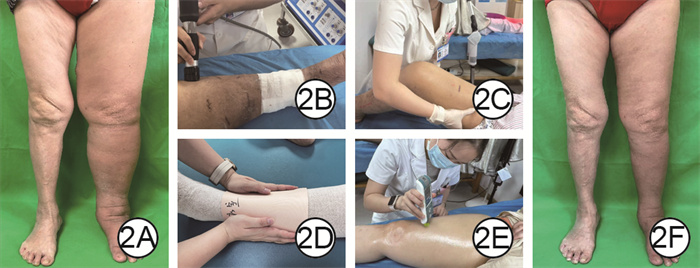
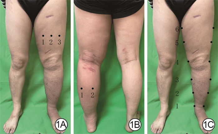
图 1 下肢皮肤纤维化程度与水肿程度的测量点。1A.大腿皮肤纤维化程度测量点,1表示股内侧肌测量点,2表示股中间肌测量点,3表示股外侧肌测量点;1B.小腿皮肤纤维化程度测量点,1表示腓肠肌外侧头测量点,2表示腓肠肌内侧头测量点;1C.下肢水肿程度的测量点,1表示踝中,2表示髌骨下缘下20 cm,3表示髌骨下缘下10 cm,4表示膝中,5表示髌骨上缘上10 cm,6表示髌骨上缘上20 cm
Figure 1. Measurement points for the degree of skin fibrosis and degree of edema of lower limb

图 2 体外冲击波疗法联合综合消肿治疗对联合治疗组宫颈癌术后患者左下肢淋巴水肿的效果。2A.治疗前左下肢水肿明显;2B.体外冲击波治疗中;2C.徒手淋巴引流技术手法治疗中;2D.绷带包扎治疗中;2E.用MyotonPRO仪评估皮肤纤维化程度中;2F.4周治疗结束时左下肢水肿情况明显改善
Figure 2. Effects of extracorporeal shock wave therapy combined with complex decongestive therapy on left lower limb lymphedema in patients after cervical cancer surgery in combined treatment group
表 1 2组宫颈癌术后下肢淋巴水肿患者一般资料比较
Table 1. Comparison of general data of two groups of patients with lower limb lymphedema after cervical cancer surgery
组别 例数 年龄(岁,x±s) 身高(cm,x±s) 体重(kg,x±s) 体重指数(kg/m2,x±s) 淋巴水肿病程(d,x±s) 淋巴水肿分期(例) Ⅱ期 Ⅲ期 对照组 30 54±8 159±4 61±8 24±3 22±11 23 7 联合治疗组 30 54±12 159±5 60±9 24±4 21±7 26 4 统计量值 t=0.02 t=0.49 t=0.67 t=0.38 t=0.26 χ²=1.00 P值 0.981 0.623 0.507 0.705 0.796 0.316 注:对照组患者仅行综合消肿治疗,联合治疗组患者行体外冲击波疗法联合综合消肿治疗  下载: 导出CSV
表 2 2组宫颈癌术后下肢淋巴水肿患者治疗前后下肢不同肌肉体表投射位置的皮肤硬度比较(N/m,x±s)
Table 2. Comparison of the skin stiffness at the superficial projection location of different muscles of the lower limbs before and after treatment in 2 groups of patients with lower limb lymphedema after cervical cancer surgery
组别与时间点 例数 股外侧肌 股中间肌 股内侧肌 腓肠肌内侧头 腓肠肌外侧头 对照组 30 治疗前 325±41 272±37 244±28 325±53 379±74 治疗后 276±29 256±36 236±25 279±47 337±54 治疗前后差值 49±23 16±19 8±9 45±39 43±42 联合治疗组 30 治疗前 327±47 270±34 241±25 332±57 380±69 治疗后 262±38 245±36 216±26 267±46 311±50 治疗前后差值 65±23 24±8 25±8 65±27 69±34 t1值 -0.21 0.21 0.51 -0.51 -0.06 P1值 0.834 0.836 0.610 0.610 0.954 t2值 2.78 2.04 3.12 2.01 2.35 P2值 0.007 0.046 0.003 0.049 0.022 t3值 -2.75 -2.35 -7.47 -2.33 -2.64 P3值 0.008 0.024 < 0.001 0.023 0.011 均数差值(95%置信区间) 16(5~28) 9(1~17) 17(12~21) 20(3~36) 26(4~49) 注:对照组患者仅行综合消肿治疗,联合治疗组患者行体外冲击波疗法联合综合消肿治疗;治疗后指4周治疗结束时;t1值、P1值和t2值、P2值分别为2组间治疗前、后各指标比较所得;t3值、P3值、均数差值(95%置信区间)为2组间治疗前后差值比较所得
下载: 导出CSV
表 3 2组宫颈癌术后下肢淋巴水肿患者治疗前后下肢不同位置周径比较(cm,x±s)
Table 3. Comparison of the circumferences at different locations of the lower limbs before and after treatment in 2 groups of patients with lower limb lymphedema after cervical cancer surgery
组别与时间点 例数 踝中 髌骨下缘下20 cm 髌骨下缘下10 cm 膝中 髌骨上缘上10 cm 髌骨上缘上20 cm 对照组 30 治疗前 26.6±4.1 30.6±5.0 37.5±4.4 38.6±4.1 48.1±5.4 55.2±5.2 治疗后 25.4±3.4 29.2±4.3 35.7±4.1 37.4±3.9 46.0±5.6 53.0±5.7 治疗前后差值 1.2±1.4 1.4±1.5 1.7±1.0 1.2±1.3 2.1±1.8 2.1±1.9 联合治疗组 30 治疗前 26.3±3.3 30.6±6.2 37.4±4.9 38.6±3.7 48.6±5.4 55.2±4.5 治疗后 25.4±3.0 26.9±2.8 33.2±3.7 35.3±2.7 43.0±5.0 49.7±5.1 治疗前后差值 1.0±1.0 3.8±5.6 4.1±3.5 3.3±3.2 5.6±4.3 5.5±4.4 t1值 -0.28 0.05 -0.08 -0.02 0.31 0.05 P1值 0.781 0.960 0.938 0.987 0.759 0.958 t2值 0.15 -2.41 -2.49 -2.44 -2.21 -2.36 P2值 0.883 0.019 0.016 0.018 0.031 0.021 t3值 -0.85 2.21 3.62 3.35 4.14 3.89 P3值 0.401 0.031 0.001 0.001 < 0.001 < 0.001 均数差值(95%置信区间) -0.3(-0.9~0.4) 2.3(0.1~4.6) 2.4(1.0~3.8) 2.1(0.8~3.4) 3.5(1.6~5.4) 3.4(1.5~5.2) 注:对照组患者仅行综合消肿治疗,联合治疗组患者行体外冲击波疗法联合综合消肿治疗;治疗后指4周治疗结束时;t1值、P1值和t2值、P2值分别为2组间治疗前、后各指标比较所得;t3值、P3值、均数差值(95%置信区间)为2组间各指标治疗前后差值比较所得
下载: 导出CSV
表 4 2组宫颈癌术后下肢淋巴水肿患者治疗前后下肢VAS评分和Lymph-ICF-LL总得分比较(分,x±s)
Table 4. Comparison of the VAS scores of lower limb and total Lymph-ICF-LL questionnaire scores before and after treatment in 2 groups of patients with lower limb lymphedema after cervical cancer surgery
组别与时间点 例数 VAS评分 Lymph-ICF-LL总得分 对照组 30 治疗前 3.5±1.4 54±12 治疗后 1.8±1.0 39±9 治疗前后差值 1.7±0.7 15±5 联合治疗组 30 治疗前 3.4±1.2 56±9 治疗后 1.2±0.7 35±7 治疗前后差值 2.2±0.8 21±8 t1值 -0.29 0.55 P1值 0.770 0.586 t2值 -2.46 -2.63 P2值 0.017 0.011 t3值 2.34 3.32 P3值 0.023 0.002 均数差值(95%置信区间) 0.5(0~0.9) 6(2~9) 注:对照组患者仅行综合消肿治疗,联合治疗组患者行体外冲击波疗法联合综合消肿治疗;治疗后指4周治疗结束时;VAS为视觉模拟评分法,Lymph-ICF-LL为下肢淋巴水肿功能、残疾与健康问卷;t1值、P1值和t2值、P2值分别为2组间治疗前、后各指标比较所得;t3值、P3值、均数差值(95%置信区间)为2组间各指标治疗前后差值比较所得
下载: 导出CSV
-

 下载:
下载:

计量
- 文章访问数: 2198
- HTML全文浏览量: 628
- PDF下载量: 24
- 被引次数: 0
