


Performance of hyaluronic acid biogel and its effect on healing of infected burn wounds in mice
-
摘要:
目的 探讨透明质酸生物胶的性能及其对小鼠烧伤感染性创面愈合的影响。 方法 该研究为实验研究。将用磷酸盐缓冲液(PBS)配制的透明质酸生物胶浸泡在PBS中,计算浸泡1、2、3、4、5 min时透明质酸生物胶的吸液率。取粉末状态的透明质酸生物胶与PBS混合成胶后,使用旋转流变仪对透明质酸生物胶的机械模量进行测试。将终质量浓度分别为5、10、20 g/L的透明质酸生物胶溶液和PBS分别加入金黄色葡萄球菌和大肠埃希菌菌液中,孵育12 h后稀释涂布于LB固体培养基上,培养24 h后用菌落计数仪观察培养基上的菌落生长情况。取24只8~10周龄雄性BALB/c小鼠,在背部制造烧伤区域并切除坏死组织后,局部滴注金黄色葡萄球菌菌液构建烧伤感染性创面模型。饲养24 h确认造模成功后,将小鼠按随机数字表法分为4组(每组6只),包括对创面滴加生理盐水的生理盐水组,以及分别用京万红软膏、水凝胶敷料、透明质酸生物胶治疗创面的京万红软膏组、水胶体敷料组和透明质酸生物胶组,持续治疗14 d。于治疗7、10、14 d,测量剩余创面面积并计算剩余创面面积百分比。于治疗7 d,用菌落计数仪统计创面中金黄色葡萄球菌菌落数。以上实验样本数均为3。 结果 透明质酸生物胶在PBS中浸泡2 min内就可达到最大387.9%的吸液率,浸泡5 min内其吸液率稳定在300.0%以上。机械模量的频率扫描显示,透明质酸生物胶的弹性模量稳定在250 Pa以上并随着角频率增加而增加;机械模量的幅度扫描显示,在10 rad/s的角频率下,透明酸质生物胶的线性黏弹区接近1 000%。培养24 h后,加入PBS的培养基上几乎长满了金黄色葡萄球菌和大肠埃希菌菌落,加入终质量浓度分别为5、10、20 g/L的透明质酸生物胶溶液的培养基上均未见明显的金黄色葡萄球菌或大肠埃希菌菌落。治疗7、10、14 d,透明质酸生物胶组小鼠剩余创面面积百分比明显低于生理盐水组、京万红软膏组和水胶体敷料组(P值均<0.05);治疗14 d,水胶体敷料组小鼠剩余创面面积百分比明显低于生理盐水组和京万红软膏组(P值均<0.05)。治疗7 d,透明质酸生物胶组小鼠创面中金黄色葡萄球菌的菌落数[(4.3±0.6)个]明显少于生理盐水组、京万红软膏组、水胶体敷料组[分别为(2 400.7±225.4)、(899.0±57.0)、(11.7±5.7)个,P值均<0.05]。 结论 透明质酸生物胶具有良好的吸收液体并形成水凝胶的能力,可通过消除金黄色葡萄球菌促进小鼠烧伤感染性创面愈合。 Abstract:Objective To explore the performance of hyaluronic acid biogel and its effect on healing of infected burn wounds in mice. Methods This study was an experimental study. The hyaluronic acid biogel prepared in phosphate buffered saline (PBS) was soaked in PBS. Then the liquid absorption rate of hyaluronic acid biogel at 1, 2, 3, 4, and 5 min of soaking was calculated. After mixing hyaluronic acid biogel in powder form with PBS to form a gel, the mechanical modulus of the hyaluronic acid biogel was tested using a rotational rheometer. The hyaluronic acid biogel solution at final mass concentrations of 5, 10, and 20 g/L along with PBS were added to Staphylococcus aureus and Escherichia coli bacteria culture solution, respectively, then after incubation for 12 h, dilution plating was performed on Lysogeny Broth agar medium. The colony growth on the medium was observed by a colony counter after culture for 24 h. Twenty-four male BALB/c mice aged 8-10 weeks underwent dorsal burn wound creation followed by necrotic tissue excision. A infected burn wound model was established by locally instilling Staphylococcus aureus bacteria culture solution. With the successful modeling being confirmed at 24 h after incubation, the mice were divided into 4 groups (n=6) according to the random number table method. The wounds in normal saline group received normal saline, while the wounds in Jingwanhong ointment group, hydrocolloid dressing group, and hyaluronic acid biogel group received Jingwanhong ointment, hydrocolloid dressing, and hyaluronic acid biogel, respectively, for 14 d. At treatment day 7, 10, and 14, the remaining wound area was measured and the percentage of residual wound area was calculated. At treatment day 7, the number of Staphylococcus aureus colonies on wounds were counted by a colony counter. All the experiments used a sample size of 3. Results The hyaluronic acid biogel achieved the highest liquid absorption rate of 387.9% within 2 min of soaking in PBS, and stabilized at over 300.0% within 5 min of soaking. Frequency scanning of the mechanical modulus revealed that the hyaluronic acid biogel's elastic modulus remained stable above 250 Pa and increased with rising angular frequency. Amplitude scanning of the mechanical modulus indicated that at an angular frequency of 10 rad/s, the linear viscoelastic range of the hyaluronic acid biogel was close to 1 000%. After culture for 24 h, the medium added with PBS was almost completely covered with Staphylococcus aureus and Escherichia coli. The medium added with hyaluronic acid biogel solution at final mass concentrations of 5, 10, and 20 g/L showed no significant growth of Staphylococcus aureus or Escherichia coli. At treatment day 7, 10, and 14, the percentage of residual wound area of mice in hyaluronic acid biogel group was significantly lower than that in normal saline, Jingwanhong ointment, and hydrocolloid dressing groups (with P values all <0.05). At treatment day 14, the percentage of residual wound area of mice in hydrocolloid dressing group was significantly lower than that in normal saline and Jingwanhong ointment groups (with P values both <0.05). At treatment day 7, the number of Staphylococcus aureus colonies on wounds of mice in hyaluronic acid biogel group ((4.3±0.6) colonies) was significantly lower than that in normal saline group ((2 400.7±225.4) colonies), Jingwanhong ointment group ((899.0±57.0) colonies), and hydrocolloid dressing group ((11.7±5.7) colonies), with P values all <0.05. Conclusions Hyaluronic acid biogel exhibits an excellent capacity to absorb liquid and form hydrogel, and promotes healing of infected burn wounds in mice by eliminating Staphylococcus aureus. -
Key words:
- Burns /
- Bacterial infections /
- Hyaluronic acid /
- Biocompatible materials /
- Wound healing /
- Bacterial resistance
-
参考文献
(42) [1] WangY, BeekmanJ, HewJ, et al. Burn injury: challenges and advances in burn wound healing, infection, pain and scarring[J]. Adv Drug Deliv Rev, 2018, 123: 3-17. DOI: 10.1016/j.addr.2017.09.018. [2] CuiW, GongC, LiuY, et al. Composite antibacterial hydrogels based on two natural products pullulan and ε-poly-l-lysine for burn wound healing[J]. Int J Biol Macromol, 2024,277(Pt 2):134208. DOI: 10.1016/j.ijbiomac.2024.134208. [3] SinghH, HassanS, NabiSU, et al. Multicomponent decellularized extracellular matrix of caprine small intestine submucosa based bioactive hydrogel promoting full-thickness burn wound healing in rabbits[J]. Int J Biol Macromol, 2024,255:127810. DOI: 10.1016/j.ijbiomac.2023.127810. [4] PereiraRF, BarriasCC, GranjaPL, et al. Advanced biofabrication strategies for skin regeneration and repair[J]. Nanomedicine (Lond), 2013,8(4):603-621. DOI: 10.2217/nnm.13.50. [5] YaoY, ZhangA, YuanC, et al. Recent trends on burn wound care: hydrogel dressings and scaffolds[J]. Biomater Sci, 2021,9(13):4523-4540. DOI: 10.1039/d1bm00411e. [6] PalackicA, JayJW, DugganRP, et al. Therapeutic strategies to reduce burn wound conversion[J]. Medicina (Kaunas), 2022, 58(7):922. DOI: 10.3390/medicina58070922. [7] NuhijiE. Trends and innovation in negative pressure wound therapy: a review of burn wound management[J]. Adv Wound Care (New Rochelle), 2024,13(8):391-399. DOI: 10.1089/wound.2023.0114. [8] RoseLF, ChanRK. The burn wound microenvironment[J]. Adv Wound Care (New Rochelle), 2016,5(3):106-118. DOI: 10.1089/wound.2014.0536. [9] SongJH, GuJT, DangGP, et al. DNA-collagen dressing for promoting scarless healing in early burn wound management[J]. Adv Compos Hybrid Mater, 2025, 8: 212. DOI: 10.1007/s42114-025-01295-0. [10] LiuW, HuangH, ShuF, et al. AntagomiR-192-5p-engineered exosomes encapsulated in MXene-modified GelMA hydrogel facilitated epithelization of burn wounds by targeting OLFM4[J]. Bioact Mater, 2025,52:318-337. DOI: 10.1016/j.bioactmat.2025.06.013. [11] LiX, WangW, GaoQ, et al. Intelligent bacteria-targeting ZIF-8 composite for fluorescence imaging-guided photodynamic therapy of drug-resistant superbug infections and burn wound healing[J]. Exploration (Beijing), 2024,4(6):20230113. DOI: 10.1002/EXP.20230113. [12] MaJ, LiS, ZhangL, et al. Oxidative stress-scavenging thermo-activated MXene hydrogel for rapid repair of MRSA impaired wounds and burn wounds[J]. Mater Today, 2024, 80: 139-155. DOI: 10.1016/j.mattod.2024.08.010. [13] HataY, MiyazakiH, OkamotoS, et al. Nanospiked cellulose gauze that attracts bacteria with biomolecules for reducing bacterial load in burn wounds[J]. Nano Letters, 2025, 25(3): 1177-1184. DOI: 10.1021/acs.nanolett.4c05773. [14] MaslovaE, EisaiankhongiL, SjöbergF, et al. Burns and biofilms: priority pathogens and in vivo models[J]. NPJ Biofilms Microbiomes, 2021,7(1):73. DOI: 10.1038/s41522-021-00243-2. [15] 董云青,李琳琳,朱宣儒,等. 含银黏性水凝胶的制备及其在小鼠细菌定植全层皮肤缺损创面愈合中的作用[J]. 中华烧伤杂志, 2021, 37(11): 1036-1047. DOI: 10.3760/cma.j.cn501120-20210906-00304. [16] RibeiroMM, SimõesM, VitorinoC, et al. Physical crosslinking of hydrogels: the potential of dynamic and reversible bonds in burn care[J]. Coord Chem Rev, 2025, 542: 216868. DOI: 10.1016/j.ccr.2025.216868. [17] LiM, NieJ, LiX, et al. Exudate management, facile detachment, and immunometabolism regulation for wound healing using breathable dressings[J]. ACS Appl Mater Interfaces, 2025, 17(15): 22394-22409. DOI: 10.1021/acsami.5c01729. [18] 顾雅男, 徐翔昊, 王彦平, 等. 氧化铈纳米酶-甲基丙烯酸酐化明胶水凝胶在小鼠全层皮肤缺损感染创面修复中的作用[J].中华烧伤与创面修复杂志,2024,40(2):131-140. DOI: 10.3760/cma.j.cn501225-20231120-00201. [19] ZhangZ, LiW, LiuY, et al. Design of a biofluid-absorbing bioactive sandwich-structured Zn-Si bioceramic composite wound dressing for hair follicle regeneration and skin burn wound healing[J]. Bioact Mater, 2021,6(7):1910-1920. DOI: 10.1016/j.bioactmat.2020.12.006. [20] WangX, SunX, ZengY, et al. Membrane fusion strategy boosts immune homeostasis, mobilizing macrophages to eliminate bacteria and acccelerate skin regeneration in infected burn wound[J]. Adv Funct Mater, 2025, 35(10): 2416791. DOI: 10.1002/adfm.202416791. [21] GallaherJR, BandaW, LachiewiczAM, et al. Predictors of multi-drug resistance in burn wound colonization following burn injury in a resource-limited setting[J]. Burns, 2021,47(6):1308-1313. DOI: 10.1016/j.burns.2020.12.007. [22] NoorA, AfzalA, MasoodR, et al. Dressings for burn wound: a review[J]. J Mater Sci, 2022, 57: 6536-6572. DOI: 10.1007/s10853-022-07056-4. [23] YeongEK, ShengWH, HsuehPR, et al. The wound microbiology and the outcomes of the systemic antibiotic prophylaxis in a mass burn casualty incident[J]. J Burn Care Res, 2020, 41(1): 95-103. DOI: 10.1093/jbcr/irz077. [24] HuangY, MuL, ZhaoX, et al. Bacterial growth-induced tobramycin smart release self-healing hydrogel for Pseudomonas aeruginosa-infected burn wound healing[J]. ACS Nano, 2022,16(8):13022-13036. DOI: 10.1021/acsnano.2c05557. [25] KantakNA, MistryR, VaronDE, et al. Negative pressure wound therapy for burns[J]. Clin Plast Surg, 2017, 44(3): 671-677. DOI: 10.1016/j.cps.2017.02.023. [26] SalwowskaNM, BebenekKA, ŻądłoDA, et al. Physiochemical properties and application of hyaluronic acid: a systematic review[J]. J Cosmetic Dermatol, 2016, 15(4): 520-526. DOI: 10.1111/jocd.12237. [27] SionkowskaA, GadomskaM, MusiałK, et al. Hyaluronic acid as a component of natural polymer blends for biomedical applications: a review[J]. Molecules, 2020, 25(18):4035.DOI: 10.3390/molecules25184035. [28] 黄日中,王裔惟,黄何艳,等. 雄激素及其拮抗剂双重释放系统在小鼠Ⅲ度烧伤创面修复中的应用效果[J]. 中华烧伤与创面修复杂志,2024,40(2): 180-189. DOI: 10.3760/cma.j.cn501225-20230802-00033. [29] HeC, BiS, ZhangR, et al. A hyaluronic acid hydrogel as a mild photothermal antibacterial, antioxidant, and nitric oxide release platform for diabetic wound healing[J]. J Control Release, 2024,370:543-555. DOI: 10.1016/j.jconrel.2024.05.011. [30] LiuS, LiuX, RenY, et al. Mussel-inspired dual-cross-linking hyaluronic acid/ε-polylysine hydrogel with self-healing and antibacterial properties for wound healing[J]. ACS Appl Mater Interfaces, 2020, 12(25): 27876-27888. DOI: 10.1021/acsami.0c00782. [31] Ter HorstB, ChouhanG, MoiemenNS, et al. Advances in keratinocyte delivery in burn wound care[J]. Adv Drug Deliv Rev, 2018,123:18-32. DOI: 10.1016/j.addr.2017.06.012. [32] ShanY, CaoF, ZhaoX, et al. Procoagulant, antibacterial and antioxidant high-strength porous hydrogel adhesives in situ formed via self-gelling hemostatic microsheets for emergency hemostasis and wound repair[J]. Biomaterials, 2025,315:122936. DOI: 10.1016/j.biomaterials.2024.122936. [33] MurakamiK, AokiH, NakamuraS, et al. Hydrogel blends of chitin/chitosan, fucoidan and alginate as healing-impaired wound dressings[J]. Biomaterials, 2010,31(1):83-90. DOI: 10.1016/j.biomaterials.2009.09.031. [34] ChaiL, HuangJ, WangM, et al. Injectable deferoxamine-loaded microsphere hydrogels for inhibition of ferroptosis and promotion of third-degree burn wound healing[J]. Mater Today Bio, 2025,32:101806. DOI: 10.1016/j.mtbio.2025.101806. [35] LeeJ, ShinD, RohJL. Use of a pre-vascularised oral mucosal cell sheet for promoting cutaneous burn wound healing[J]. Theranostics, 2018,8(20):5703-5712. DOI: 10.7150/thno.28754. [36] LanJ, ShiL, XiaoW, et al. A rapid self-pumping organohydrogel dressing with hydrophilic fractal microchannels to promote burn wound healing[J]. Adv Mater, 2023, 35(38): e2301765. DOI: 10.1002/adma.202301765. [37] LiW, YuQ, YaoH, et al. Superhydrophobic hierarchical fiber/bead composite membranes for efficient treatment of burns[J]. Acta Biomater, 2019,92:60-70. DOI: 10.1016/j.actbio.2019.05.025. [38] 刘颖, 程凤, 王泽薇, 等. 负载小鼠脂肪干细胞的甲壳素/透明质酸/胶原水凝胶的制备及其对大鼠全层皮肤缺损创面愈合的作用[J].中华烧伤与创面修复杂志,2024,40(1):50-56. DOI: 10.3760/cma.j.cn501225-20230928-00101. [39] KonieczynskaMD, Villa-CamachoJC, GhobrilC, et al. On-demand dissolution of a dendritic hydrogel-based dressing for second-degree burn wounds through thiol-thioester exchange reaction[J]. Angew Chem Int Ed Engl, 2016, 55(34): 9984-9987. DOI: 10.1002/anie.201604827. [40] DehariD, KumarDN, ChaudhuriA, et al. Bacteriophage entrapped chitosan microgel for the treatment of biofilm-mediated polybacterial infection in burn wounds[J]. Int J Biol Macromol, 2023,253(Pt 5):127247. DOI: 10.1016/j.ijbiomac.2023.127247. [41] HeX, LiuL, GuF, et al. Exploration of the anti-inflammatory, analgesic, and wound healing activities of Bletilla Striata polysaccharide[J]. Int J Biol Macromol, 2024,261(Pt 2):129874. DOI: 10.1016/j.ijbiomac.2024.129874. [42] GongY, WangP, CaoR, et al. Exudate absorbing and antimicrobial hydrogel integrated with multifunctional curcumin-loaded magnesium polyphenol network for facilitating burn wound healing[J]. ACS Nano, 2023, 17(22): 22355-22370. DOI: 10.1021/acsnano.3c04556. -

图 2 透明质酸生物胶的吸液能力。2A.加入磷酸盐缓冲液(PBS)前,粉末状态的透明质酸生物胶呈离散的颗粒状态 偏光显微镜×20;2B、2C、2D.分别为粉末状态的透明质酸生物胶加入PBS后15、30、60 s时的状态,逐渐溶解交联形成水凝胶 偏光显微镜×20;2E.透明质酸生物胶在浸泡于PBS 5 min以内可以达到最大387.9%的吸液率,并稳定在300.0%以上(样本数为3,


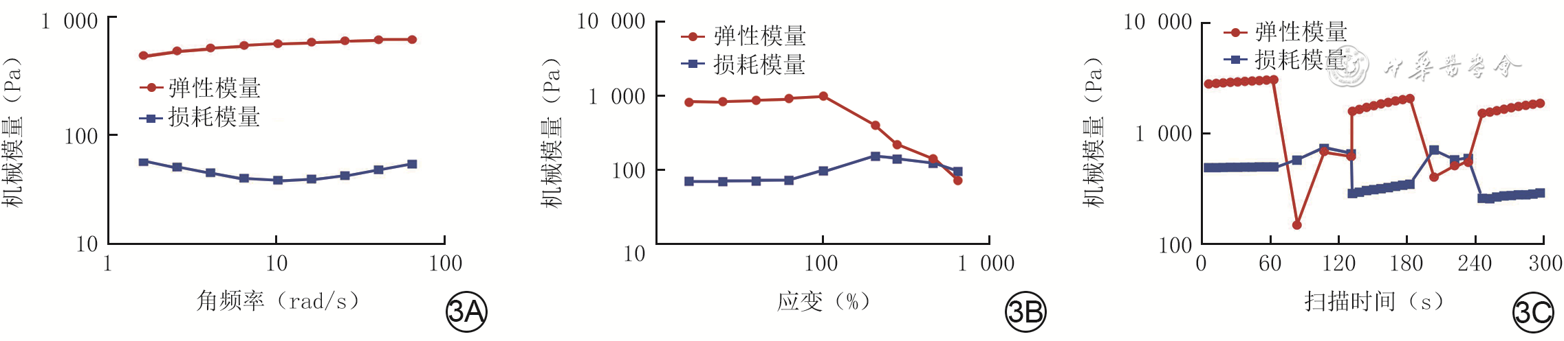
图 3 透明质酸生物胶的机械模量和自愈性。3A.透明质酸生物胶行频率扫描测定的机械模量结果显示,弹性模量在250 Pa以上;3B.透明质酸生物胶行幅度扫描测定的机械模量结果显示,线性黏弹区接近1 000%;3C.透明质酸生物胶在1%与600%交替应变后,机械模量仍然能够恢复
注:该图为经过lg处理的数据形成的描记图,坐标轴数据为未经lg处理的原始数据

图 4 2种细菌在加入4种溶液培养24 h后的生长情况。4A、4B、4C、4D.分别为加入磷酸盐缓冲液和质量浓度分别为20、10、5 g/L的透明质酸生物胶溶液后,LB固体培养基上的金黄色葡萄球菌菌落(黄色)情况,其中图4B、4C中无明显菌落,图4B~4D中菌落明显少于图4A;4E、4F、4G、4H.分别为加入磷酸盐缓冲液和质量浓度分别为20、10、5 g/L的透明质酸生物胶溶液后,LB固体培养基上的大肠埃希菌菌落(黄色)情况,其中图4F、4G、4H中均无明显菌落,显著少于图4E

图 5 4组烧伤感染性创面小鼠治疗各时间点创面愈合情况。图5A、5B、5C、5D.分别为生理盐水组、京万红软膏组、水胶体敷料组和透明质酸生物胶组治疗7 d的创面,其中图5D剩余创面面积明显小于图5A、5B、5C;图5E、5F、5G、5H.分别为生理盐水组、京万红软膏组、水胶体敷料组和透明质酸生物胶组治疗10 d的创面,其中图5H剩余创面面积明显小于图5E、5F、5G;图5I、5J、5K、5L.分别为生理盐水组、京万红软膏组、水胶体敷料组和透明质酸生物胶组治疗14 d的创面,其中图5L剩余创面面积明显小于图5I、5J、5K
注:生理盐水组、京万红软膏组、水胶体敷料组、透明质酸生物胶组小鼠创面分别采用组名中药物治疗;图中圆环直径1.2 cm

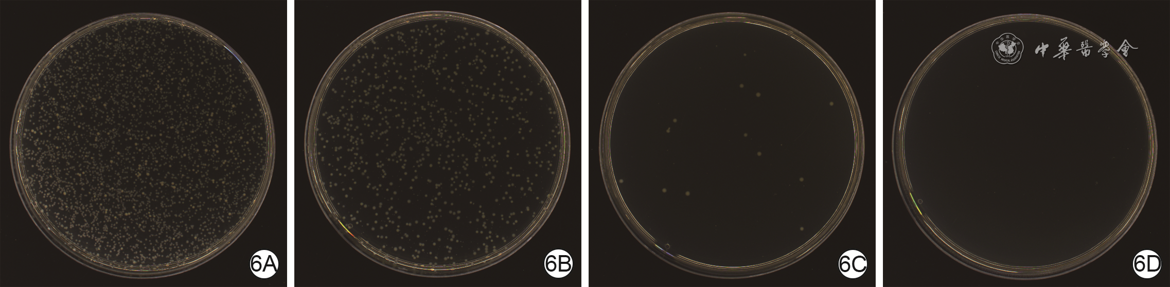
图 6 4组烧伤感染性创面小鼠治疗7 d创面中金黄色葡萄球菌分布情况。6A.生理盐水组菌落已经长满;6B.京万红软膏组菌落较多;6C.水胶体敷料组有少量菌落;6D.透明质酸生物胶组几乎无菌落
注:生理盐水组、京万红软膏组、水胶体敷料组和透明质酸生物胶组小鼠创面分别采用组名中药物治疗;金黄色葡萄球菌菌落为黄色
Table 1. 4组烧伤感染性创面小鼠治疗各时间点剩余创面面积百分比比较(%,
组别 样本数 7 d 10 d 14 d 生理盐水组 3 119.8±8.9 78.7±17.9 31.9±5.4 京万红软膏组 3 118.4±13.9 78.9±5.0 25.8±5.1 水胶体敷料组 3 104.0±10.7 64.6±10.4 10.9±2.0 透明质酸生物胶组 3 76.3±7.1 32.1±10.4 2.4±0.6 P1值 0.998 >0.999 0.292 P2值 0.319 0.500 <0.001 P3值 0.004 0.005 <0.001 P4值 0.391 0.490 0.006 P5值 0.005 0.005 <0.001 P6值 0.048 0.040 0.043 注:生理盐水组、京万红软膏组、水胶体敷料组和透明质酸生物胶组小鼠创面分别采用组名中药物治疗;处理因素主效应,F=28.73,P<0.001;时间因素主效应,F=1 131.05,P<0.001;两者交互作用,F=1.36,P=0.030;P1值、P2值、P3值分别为生理盐水组与京万红软膏组、水胶体敷料组、透明质酸生物胶组比较所得,P4值、P5值分别为京万红软膏组与水胶体敷料组、透明质酸生物胶组比较所得,P6值为水胶体敷料组与透明质酸生物胶组比较所得  下载: 导出CSV
-
 石明生.mp4

-

 下载:
下载:


计量
- 文章访问数: 1758
- HTML全文浏览量: 433
- PDF下载量: 40
- 被引次数: 0
