


-
摘要:
目的 探讨谷胱甘肽对小鼠皮肤急性光损伤的作用及其机制。 方法 该研究为成组设计实验研究。取15只8周龄雄性C57BL/6小鼠,剃去背部毛发后按随机数字表法分为空白对照组、模型组、低剂量干预组、中剂量干预组、高剂量干预组,每组3只。模型组小鼠背部皮肤每日接受中波紫外线联合长波紫外线照射造成急性光损伤,然后经腹腔注射磷酸盐缓冲液(PBS);空白对照组小鼠不照射紫外线做假伤处理,仅每日经腹腔注射PBS;低剂量干预组、中剂量干预组、高剂量干预组小鼠每日同模型组接受紫外线照射后分别经腹腔注射50、100、200 mg/kg的谷胱甘肽。伤后第7天末次注射后2 h时(下称伤后7 d),大体观察各组小鼠背部皮肤色泽、形态,然后切取小鼠背部皮肤组织进行下述检测。采用苏木精-伊红染色检测皮肤组织的角质层、表皮层和真皮层结构,毛囊、汗腺、皮脂腺等附属器官的形态,有无出血现象及炎症细胞浸润等情况,并统计表皮厚度;采用Masson染色检测皮肤组织中胶原纤维沉积情况;采用蛋白质印迹法检测皮肤组织中的炎症相关蛋白白细胞介素-1β(IL-1β)、IL-6、肿瘤坏死因子-α(TNF-α)、基质金属蛋白酶1(MMP-1)的蛋白表达情况。 结果 伤后7 d,与空白对照组相比,模型组小鼠皮肤出现大范围皮屑、红肿、痂壳;与模型组相比,低剂量干预组、中剂量干预组、高剂量干预组小鼠皮肤的光损伤程度依次减轻。伤后7 d,与空白对照组相比,模型组小鼠皮肤组织结构紊乱,角质层增厚、剥脱,表皮层细胞层数增多、排列紊乱,真皮层水肿,毛囊、汗腺、皮脂腺等附属器官形态异常,可见散在出血灶及大量炎症细胞浸润。与模型组相比,低剂量干预组、中剂量干预组、高剂量干预组小鼠皮肤组织结构紊乱程度依次减轻。伤后7 d,模型组小鼠皮肤的表皮厚度为(116.4±6.4)μm,明显厚于空白对照组的(20.9±1.6)μm,P<0.05;与模型组相比,中剂量干预组、高剂量干预组小鼠皮肤的表皮厚度[分别为(77.7±5.6)、(56.9±0.8)μm]明显变薄(P值均<0.05)。伤后7 d,与空白对照组相比,模型组小鼠皮肤组织中胶原纤维含量显著增加,且纤维排列紊乱,呈现出一定程度的胶原纤维增生现象;与模型组相比,低剂量干预组、中剂量干预组、高剂量干预组小鼠皮肤组织中胶原纤维的排列依次趋于规则,整体组织结构逐渐恢复至接近正常皮肤。伤后7 d,与空白对照组相比,模型组小鼠皮肤组织中IL-1β、IL-6、TNF-α和MMP-1的蛋白表达水平显著升高(P值均<0.05);与模型组相比,低剂量干预组小鼠皮肤组织中IL-1β、IL-6的蛋白表达水平明显降低(P值均<0.05),中剂量干预组、高剂量干预组小鼠皮肤组织中IL-1β、IL-6、TNF-α和MMP-1的蛋白表达水平显著降低(P值均<0.05)。 结论 谷胱甘肽可显著减轻中波紫外线与长波紫外线联合诱导的小鼠皮肤急性光损伤,且具有明显的剂量依赖性保护作用,其机制可能与抑制炎症因子表达、减少MMP-1介导的胶原降解及改善真皮胶原结构有关。 Abstract:Objective To investigate the effects of glutathione on acute cutaneous photodamage in mice and its mechanism. Methods This study was a group-designed experimental study. Fifteen 8-week-old male C57BL/6 mice were shaved on the dorsal skin and divided into blank control group, model group, low-dose intervention group, medium-dose intervention group, and high-dose intervention group using a random number table, with 3 mice in each group. Mice in model group received daily combined irradiation of ultraviolet B and ultraviolet A on the dorsal skin to cause acute photodamage, followed by intraperitoneal injection of phosphate-buffered saline (PBS). Mice in blank control group were subjected to sham injury without ultraviolet irradiation and received daily intraperitoneal injection of PBS only. Mice in low-dose intervention group, medium-dose intervention group, and high-dose intervention group received daily ultraviolet irradiation as the model group, followed by intraperitoneal injection of 50, 100, and 200 mg/kg of glutathione, respectively. Two hours after the last injection on day 7 post injury (hereinafter referred to as day 7 post injury), the color and morphology of the dorsal skin of mice in each group were observed grossly. Then, dorsal skin tissue was excised for the following assays. Hematoxylin-eosin staining was used to examine the structure of the stratum corneum, epidermis, and dermis, the morphology of appendages (hair follicles, sweat glands, and sebaceous glands), the presence of hemorrhage, inflammatory cell infiltration, and the thickness of epidermis were also measured. Masson staining was used to detect collagen fiber deposition in the skin tissue. Western blotting was used to detect the protein expressions of inflammation-related proteins (interleukin-1β (IL-1β), IL-6, tumor necrosis factor-α (TNF-α), and matrix metalloproteinase 1 (MMP-1)) in the skin tissue. Results On day 7 post injury, compared with that in blank control group, the skin of mice in model group showed extensive scaling, redness, swelling, and crusting. Compared with that in model group, the severity of photodamage of the skin of mice in low-dose intervention group, medium-dose intervention group, and high-dose intervention group was alleviated successively. On day 7 post injury, compared with that in blank control group, the skin tissue structure of mice in model group was disorganized, characterized by thickened and detached stratum corneum, increased number and disordered arrangement of epidermal cell layers, dermal edema, abnormal morphology of appendages (hair follicles, sweat glands, sebaceous glands), scattered hemorrhagic foci, and extensive inflammatory cell infiltration. Compared with that in model group, the degree of tissue disorganization in the skin of mice in low-dose intervention group, medium-dose intervention group, and high-dose intervention group was alleviated successively. On day 7 post injury, the epidermal thickness of the skin of mice in model group was (116.4±6.4) μm, which was significantly greater than (20.9±1.6) μm in blank control group (P<0.05). Compared with that in model group, the epidermal thickness of the skin of mice in medium-dose intervention group and high-dose intervention group ((77.7±5.6) μm and (56.9±0.8) μm, respectively) was significantly decreased (with P values both <0.05). On day 7 post injury, compared with that in blank control group, the skin tissue of mice in model group showed a significant increase in collagen fiber content and disorganized fiber arrangement, indicating a certain degree of collagen fiber proliferation. Compared with that in model group, the arrangement of collagen fibers in the skin tissue of mice in low-dose intervention group, medium-dose intervention group, and high-dose intervention group became progressively more regular, and the overall tissue structure gradually recovered to normal. On day 7 post injury, compared with those in blank control group, the protein expression levels of IL-1β, IL-6, TNF-α, and MMP-1 in the skin tissue of mice in model group were significantly increased (with P values all <0.05). Compared with those in model group, the protein expression levels of IL-1β and IL-6 in the skin tissue of mice in low-dose intervention group were significantly decreased (with P values both <0.05), while the protein expression levels of IL-1β, IL-6, TNF-α, and MMP-1 in the skin tissue of mice in medium-dose intervention group and high-dose intervention group were significantly decreased (with P values all <0.05). Conclusions Glutathione can significantly alleviate acute cutaneous photodamage induced by combined irradiation of ultraviolet B and ultraviolet A in mice, with a clear dose-dependent protective effect, and its mechanism may be related to inhibiting the expression of inflammatory factors, reducing MMP-1-mediated collagen degradation, and improving dermal collagen structure. -
Key words:
- Ultraviolet rays /
- Glutathione /
- Skin /
- Inflammation /
- Oxidative stress /
- Photodamage /
- Wound repair
-
参考文献
(40) [1] YuanX, LiH, LeeJS, et al. Role of mitochondrial dysfunction in UV-induced photoaging and skin cancers[J]. Exp Dermatol, 2025,34(5):e70114. DOI: 10.1111/exd.70114. [2] GolloglyJM, NguyenJK, LauD, et al. Updates on the molecular basis of photoaging in all skin types[J]. J Drugs Dermatol, 2024,23(7):504-509. DOI: 10.36849/JDD.7438. [3] PellacaniG, LimHW, StockflethE, et al. Photoprotection: current developments and controversies[J]. J Eur Acad Dermatol Venereol, 2024,38Suppl 5:12-20. DOI: 10.1111/jdv.19677. [4] SongS, LiF, ZhaoB, et al. Ultraviolet light causes skin cell senescence: from mechanism to prevention principle[J]. Adv Biol (Weinh), 2025,9(2):e2400090. DOI: 10.1002/adbi.202400090. [5] MullendersLHF. Solar UV damage to cellular DNA: from mechanisms to biological effects[J]. Photochem Photobiol Sci, 2018,17(12):1842-1852. DOI: 10.1039/c8pp00182k. [6] LuS, LiuJ, ZhangX, et al. Protective effect of γ-glutamylcysteine against UVB radiation in NIH-3T3 cells[J]. Photodermatol Photoimmunol Photomed, 2022,38(6):522-530. DOI: 10.1111/phpp.12782. [7] Quintana-CabreraR, BolañosJP. Glutathione and γ-glutamylcysteine in the antioxidant and survival functions of mitochondria[J]. Biochem Soc Trans, 2013,41(1):106-110. DOI: 10.1042/BST20120252. [8] CuiX, MiT, XiaoX, et al. Topical glutathione amino acid precursors protect skin against environmental and oxidative stress[J]. J Eur Acad Dermatol Venereol, 2024,38Suppl 3:3-11. DOI: 10.1111/jdv.19717. [9] CuiX, MiT, XiaoX, et al. Metabolomic reprogramming induced by benzo[a]pyene in skin keratinocytes and protective effects of glutathione amino acid precursors[J]. J Cosmet Dermatol, 2025,24(4):e70168. DOI: 10.1111/jocd.70168. [10] LeeAY. Skin pigmentation abnormalities and their possible relationship with skin aging[J]. Int J Mol Sci, 2021,22(7):3727. DOI: 10.3390/ijms22073727. [11] NahhasAF, Abdel-MalekZA, KohliI, et al. The potential role of antioxidants in mitigating skin hyperpigmentation resulting from ultraviolet and visible light-induced oxidative stress[J]. Photodermatol Photoimmunol Photomed, 2019,35(6):420-428. DOI: 10.1111/phpp.12423. [12] KuoYH, WuPY, ChenCW, et al. N-(4-bromophenethyl) caffeamide protects skin from UVB-induced inflammation through MAPK/IL-6/NF-κB-dependent signaling in human skin fibroblasts and hairless mouse skin[J]. Molecules, 2017,22(10):1639. DOI: 10.3390/molecules22101639. [13] GaoP, XiaoX, CuiX, et al. Lysine carboxymethyl cysteinate, as a topical glutathione precursor, protects against oxidative stress and UVB radiation-induced skin damage[J]. Antioxidants (Basel), 2025,14(5):606. DOI: 10.3390/antiox14050606. [14] GaoP, XiaoX, ZhouZ, et al. A triple-precursor blend as a topical solution to protect the skin against environmental damage[J]. Biology (Basel), 2025,14(3):266. DOI: 10.3390/biology14030266. [15] YoshidaM, ShinKO, MuraokaS, et al. The epidermal environment's influence on the dermal environment in response to external stress[J]. Skin Pharmacol Physiol, 2023,36(3):149-159. DOI: 10.1159/000529743. [16] TangZ, TongX, HuangJ, et al. Research progress of keratinocyte-programmed cell death in UV-induced Skin photodamage[J]. Photodermatol Photoimmunol Photomed, 2021,37(5):442-448. DOI: 10.1111/phpp.12679. [17] Yardman-FrankJM, FisherDE. Skin pigmentation and its control: from ultraviolet radiation to stem cells[J]. Exp Dermatol, 2021,30(4):560-571. DOI: 10.1111/exd.14260. [18] BrarG, DhaliwalA, BrarAS, et al. A comprehensive review of the role of UV radiation in photoaging processes between different types of skin[J]. Cureus, 2025,17(3):e81109. DOI: 10.7759/cureus.81109. [19] SalminenA,KaarnirantaK,KauppinenA. Photoaging: UV radiation-induced inflammation and immunosuppression accelerate the aging process in the skin[J]. Inflamm Res, 2022,71(7/8):817-831. DOI: 10.1007/s00011-022-01598-8. [20] GentileP, GarcovichS. Adipose-derived mesenchymal stem cells (AD-MSCs) against ultraviolet (UV) radiation effects and the skin photoaging[J]. Biomedicines, 2021,9(5):532. DOI: 10.3390/biomedicines9050532. [21] ZargaranD, ZollerF, ZargaranA, et al. Facial skin ageing: key concepts and overview of processes[J]. Int J Cosmet Sci, 2022,44(4):414-420. DOI: 10.1111/ics.12779. [22] DoyleAD, NazariSS, YamadaKM. Cell-extracellular matrix dynamics[J]. Phys Biol, 2022,19(2): 10.1088/1478-3975/ac4390. DOI: 10.1088/1478-3975/ac4390. [23] ZhangM, ZhangT, TangY, et al. Concentrated growth factor inhibits UVA-induced photoaging in human dermal fibroblasts via the MAPK/AP-1 pathway[J]. Biosci Rep, 2020,40(7):BSR20193566. DOI: 10.1042/BSR20193566. [24] ForresterSJ, KikuchiDS, HernandesMS, et al. Reactive oxygen species in metabolic and inflammatory signaling[J]. Circ Res, 2018,122(6):877-902. DOI: 10.1161/CIRCRESAHA.117.311401. [25] OyamaM, MurataK, OgataM, et al. Saireito improves lymphatic function and prevents UVB-induced acute inflammation and photodamage in HR-1 hairless mice[J]. Evid Based Complement Alternat Med, 2021,2021:3707058. DOI: 10.1155/2021/3707058. [26] XiongW, SunKY, ZhuY, et al. Metformin alleviates inflammation through suppressing FASN-dependent palmitoylation of Akt[J]. Cell Death Dis, 2021,12(10):934. DOI: 10.1038/s41419-021-04235-0. [27] HungSJ, TangSC, LiaoPY, et al. Photoprotective potential of glycolic acid by reducing NLRC4 and AIM2 inflammasome complex proteins in UVB radiation-induced normal human epidermal keratinocytes and mice[J]. DNA Cell Biol, 2017,36(2):177-187. DOI: 10.1089/dna.2016.3471. [28] LiX, ZhangY, WangJ, et al. Complement C3aR signaling drives mitochondrial dysfunction in light-stressed retinal neurons[J]. Cell Rep, 2024,43(2):110702.DOI: 10.1016/j.celrep.2024.110702. [29] DoeJ, SmithA, JohnsonB, et al. UVB-induced skin inflammation is driven by NLRP3/IL-1β and sustained by IL-17A[J]. Nat Immunol, 2023,24(5):567-578. DOI: 10.1038/s41590-023-01478-9. [30] ZhangL, WangY, ChenX, et al. ROS-dependent TLR4/NF-κB/NLRP3 axis drives UV-induced photoaging[J]. Redox Biol, 2023,62:102798. DOI: 10.1016/j.redox.2023.102798. [31] ChenR, LiX, WangH, et al. Single-cell RNA-seq reveals TLR4+ fibroblast subset drives UV-induced skin fibrosis via NF-κB/CCL2[J]. bioRxiv, 2024,15: 2024.03.15.585203. DOI: 2024.03.15.585203. [32] WangJ, LiuY, ZhangQ, et al. Non-canonical NF-κB signaling amplifies UV-induced skin inflammation through RELB/IL-1β feedback[J]. Nat Commun, 2023,14:567. DOI: 10.1038/s41467-023-36213-5. [33] 宋金婉,任献青,邢琼琼,等.基于ROS介导NLRP3炎症小体活化探讨凉血退紫方治疗过敏性紫癜大鼠作用机制研究[J].中国比较医学杂志,2025,35(4):21-30.DOI: 10.3969/j.issn.1671-7856.2025.04.003. [34] ZengY, LuoM, YaoZ, et al. Adiponectin inhibits ROS/NLRP3 inflammatory pathway through FOXO3A to ameliorate oral submucosal fibrosis[J]. Odontology, 2024,112(3):811-825. DOI: 10.1007/s10266-023-00891-0. [35] ZhaoY, WuJ, LiuX, et al. Decoding nature: multi-target anti-inflammatory mechanisms of natural products in the TLR4/NF-κB pathway[J]. Front Pharmacol, 2024,15:1467193. DOI: 10.3389/fphar.2024.1467193. [36] 朱雅文.基于TLR4/NF-κB通路探讨芪黄健脾滋肾颗粒对系统性红斑狼疮肾足细胞自噬的机制研究[D/OL]. 合肥:安徽中医药大学,2025[2025-06-28].https://xueshu.baidu.com/ndscholar/browse/detail?paperid=16690td0uu2k0j90695x0rv0a2550655&site=xueshu_se.DOI: 10.26922/d.cnki.ganzc.2025.000434. [37] YangY, LiL, HangQ, et al. γ-glutamylcysteine exhibits anti-inflammatory effects by increasing cellular glutathione level[J]. Redox Biol, 2019,20:157-166. DOI: 10.1016/j.redox.2018.09.019. [38] 李玉骞, 张婷婷, 邹桂连, 等. 线粒体移植对糖尿病大鼠全层皮肤缺损的影响[J].中华烧伤与创面修复杂志,2025,41(10):937-948. DOI: 10.3760/cma.j.cn501225-20250721-00315. [39] 刘潇, 蒙健林, 王明刚, 等. 解毒化瘀升散方抑制NLRP3信号通路缓解慢加急性肝衰竭大鼠炎症损伤的实验研究[J].中华肝脏病杂志,2024,32(4):354-362. DOI: 10.3760/cma.j.cn501113-20230816-00060. [40] 肖红艳, 苏珊, 安嘉伟, 等. 氨基胍对急性肝损伤小鼠的作用及其机制[J].中华烧伤与创面修复杂志,2026,42(2):163-172. DOI: 10.3760/cma.j.cn501225-20251016-00431. -

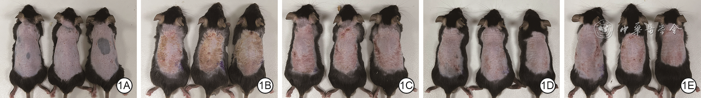
图 1 5组小鼠伤后7 d皮肤外观。1A、1B、1C、1D、1E.分别为空白对照组、模型组、低剂量干预组、中剂量干预组、高剂量干预组,图1B中小鼠皮肤组织的光损伤程度最严重,图1C、1D、1E光损伤程度依次减轻,其中图1E的外观最接近图1A
注:模型组小鼠背部皮肤每日接受中波紫外线联合长波紫外线照射,然后经腹腔注射磷酸盐缓冲液(PBS);空白对照组小鼠不照射紫外线致假伤,仅每日经腹腔注射PBS;低剂量干预组、中剂量干预组、高剂量干预组小鼠每日同模型组接受紫外线照射后分别经腹腔注射50、100、200 mg/kg的谷胱甘肽

图 2 5组小鼠伤后7 d皮肤组织病理情况 苏木精-伊红 ×50。2A、2B、2C、2D、2E.分别为空白对照组、模型组、低剂量干预组、中剂量干预组、高剂量干预组,图2B中的小鼠皮肤组织结构紊乱,角质层增厚、剥脱,表皮层细胞层数增多、排列紊乱,真皮层(箭头所示)水肿,毛囊、汗腺、皮脂腺等附属器官形态异常,可见散在出血灶及大量炎症细胞浸润,图2C、2D、2E中的小鼠皮肤组织结构紊乱程度依次减轻,表皮层依次趋于规整,真皮层水肿及炎症细胞浸润情况依次减轻
注:模型组小鼠背部皮肤每日接受中波紫外线联合长波紫外线照射,然后经腹腔注射磷酸盐缓冲液(PBS);空白对照组小鼠不照射紫外线致假伤,仅每日经腹腔注射PBS;低剂量干预组、中剂量干预组、高剂量干预组小鼠每日同模型组接受紫外线照射后分别经腹腔注射50、100、200 mg/kg的谷胱甘肽

图 3 5组小鼠伤后7 d皮肤组织中胶原纤维沉积情况 Masson ×50。3A、3B、3C、3D、3E.分别为空白对照组、模型组、低剂量干预组、中剂量干预组、高剂量干预组,图3B中的小鼠皮肤组织中胶原纤维大量增生、排列疏松紊乱,胶原沉积明显增多,图3C、3D、3E中小鼠皮肤组织中胶原纤维沉积与排列紊乱程度依次减轻
注:模型组小鼠背部皮肤每日接受中波紫外线联合长波紫外线照射,然后经腹腔注射磷酸盐缓冲液(PBS);空白对照组小鼠不照射紫外线致假伤,仅每日经腹腔注射PBS;低剂量干预组、中剂量干预组、高剂量干预组小鼠每日同模型组接受紫外线照射后分别经腹腔注射50、100、200 mg/kg的谷胱甘肽;箭头指示胶原纤维

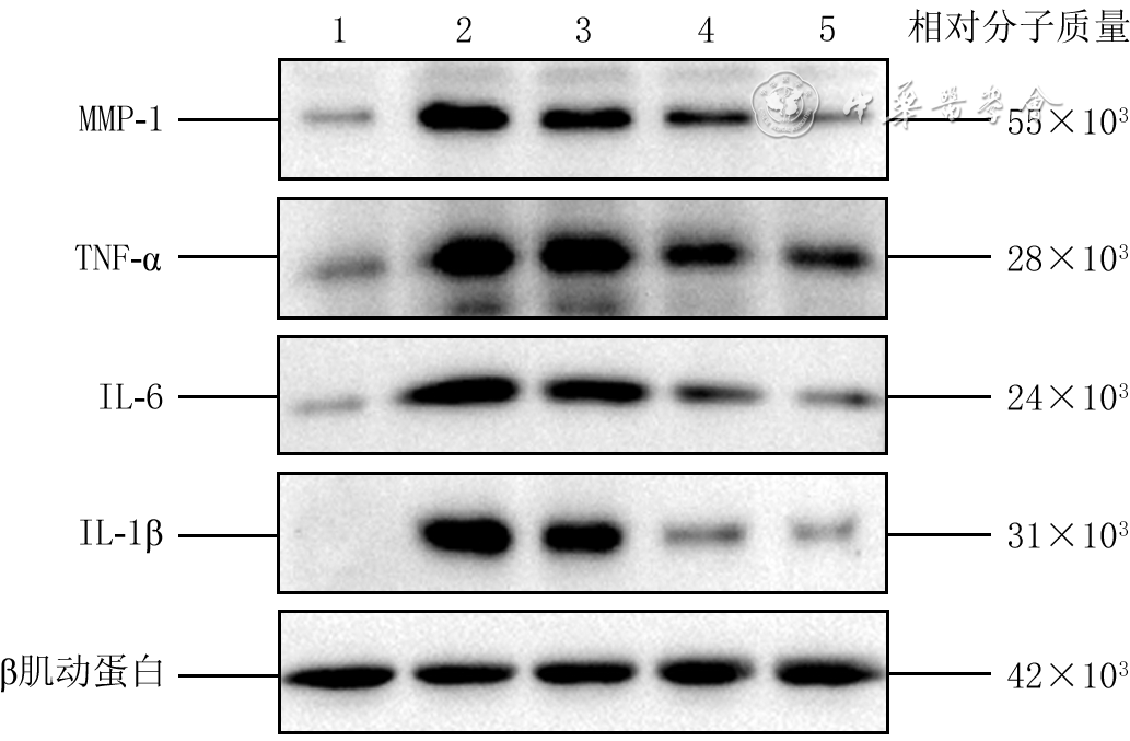
图 4 蛋白质印迹法检测的5组小鼠伤后7 d皮肤组织中炎症相关蛋白的蛋白表达水平
注:条带图上方的1、2、3、4、5分别指空白对照组、模型组、低剂量干预组、中剂量干预组、高剂量干预组;模型组小鼠接受中波紫外线联合长波紫外线照射后经腹腔注射磷酸盐缓冲液(PBS);空白对照组不进行紫外线照射致假伤,仅经腹腔注射PBS;低剂量干预组、中剂量干预组、高剂量干预组小鼠每日同模型组接受紫外线照射后分别经腹腔注射50、100、200 mg/kg的谷胱甘肽
Table 1. 5组小鼠伤后7 d皮肤组织中炎症相关蛋白的蛋白表达水平比较
组别 样本数 白细胞介素-1β 白细胞介素-6 肿瘤坏死因子-α 基质金属蛋白酶1 空白对照组 5 1.00±0.00 1.00±0.00 1.00±0.00 1.00±0.00 模型组 5 5.97±0.17a 11.17±1.56a 3.62±0.18a 5.74±1.20a 低剂量干预组 5 4.17±0.44b 5.86±0.64b 3.69±0.40 5.56±1.35 中剂量干预组 5 2.88±1.02b 5.03±2.37b 2.60±0.27b 2.61±0.08b 高剂量干预组 5 2.31±0.38b 1.80±0.24b 1.80±0.47b 1.76±0.63b F值 37.983 28.727 41.566 19.669 P值 <0.001 <0.001 <0.001 <0.001 注:模型组小鼠背部皮肤每日接受中波紫外线联合长波紫外线照射,然后经腹腔注射磷酸盐缓冲液(PBS);空白对照组小鼠不照射紫外线致假伤,仅每日经腹腔注射PBS;低剂量干预组、中剂量干预组、高剂量干预组小鼠每日同模型组接受紫外线照射后分别经腹腔注射50、100、200 mg/kg的谷胱甘肽;与空白对照组相比,aP<0.05;与模型组相比,bP<0.05  下载: 导出CSV
-

 下载:
下载:


计量
- 文章访问数: 329
- HTML全文浏览量: 172
- PDF下载量: 18
- 被引次数: 0
