


Mechanisms of surgical incision scar and technological innovations for scar tension reduction based the mechano-chemo-biological theory
-
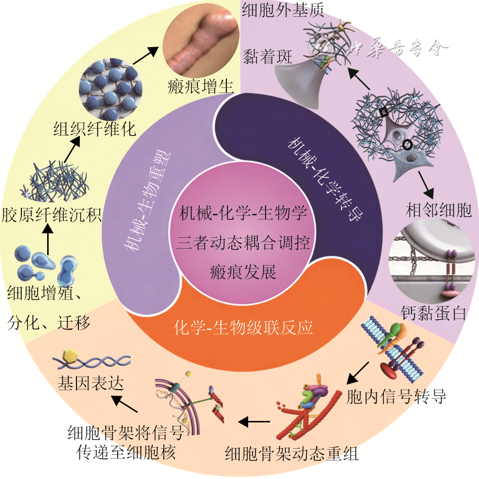
摘要: 外科切口愈合后易形成线性瘢痕,且可能发展为增生性瘢痕或瘢痕疙瘩。研究表明,机械力与基质刚度等生物力学因素通过影响细胞行为和胞外基质重塑来调控瘢痕形成。笔者团队提出机械-化学-生物学三者的动态耦合与协同作用共同调控瘢痕发展的观点,并强调切口闭合后持续控制张力对防止瘢痕变宽及增生至关重要。目前常用减张方法存在局限:术中缝合效果短暂;外用减张装置(如减张胶带、拉链)易脱落、刺激皮肤,且患者依从性低;点阵激光在高张力部位单独使用时仍可能导致瘢痕变宽。为此,笔者团队提出基于慢吸收线原位回针的皮内缝合方案,旨在实现长期有效的张力管理。初步临床应用结果显示,该方案可实现外科切口的持续减张,与点阵激光联用有望协同改善瘢痕宽度,并克服传统方法的不足。未来笔者团队将通过进一步研究优化该方案,推动其标准化与普及,逐步建立以持续减张为核心的瘢痕防治体系,最终促进外科切口的理想愈合。Abstract: Surgical incisions often heal with linear scars, which may further develop into hypertrophic scars or keloids. Research indicates that biomechanical factors, particularly mechanical forces and matrix stiffness, regulate scar formation by influencing cellular behavior and extracellular matrix remodeling. Our team proposes that the dynamic coupling and synergistic interactions among mechanical, chemical, and biological factors jointly regulate scar development. We emphasize that sustained tension control following wound closure is crucial to prevent scar widening and hyperplasia. Currently used tension-reducing methods have limitations: the effect of intraoperative sutures is short-lived; external tension-reducing devices (e.g., tension-reducing tape or zipper) are prone to falling off and causing skin irritation, and patient compliance is low; fractional laser may still cause the scar to widen when used alone in high-tension areas. Therefore, our team proposes a scheme utilizing an intradermal suturing technique based on slow-absorbing sutures with in-situ backstitch, which aims to achieve long-term and effective tension management. Preliminary results from clinical applications showed that this scheme achieved continuous tension reduction in surgical incisions, and its combination with fractional laser therapy is expected to synergistically improve scar width, overcoming the shortcomings of conventional methods. Moving forward, further research will optimize this scheme and promote its standardization and widespread adoption, and gradually establish a scar prevention system centered on continuous tension reduction, ultimately promoting the ideal healing of surgical incisions.
-
参考文献
(74) [1] LiY, LiuA, WangJ, et al. Suture-anchored cutaneous tension induces persistent hypertrophic scarring in a novel murine model[J/OL]. Burns Trauma, 2024,12:tkae051[2025-10-13]. https://pubmed.ncbi.nlm.nih.gov/39429643/. DOI: 10.1093/burnst/tkae051. [2] HosseiniM, BrownJ, KhosrotehraniK, et al. Skin biomechanics: a potential therapeutic intervention target to reduce scarring[J/OL]. Burns Trauma, 2022,10:tkac036[2025-10-13].https://pubmed.ncbi.nlm.nih.gov/36017082/. DOI: 10.1093/burnst/tkac036. [3] SongvasinS, WinaikosolK, KarunasumettaC. A randomized controlled trial comparison of subcuticular suture and adhesive strip for cosmetic outcome in median sternotomy closure in patient undergoing cardiac surgery[J/OL]. Aesthetic Plast Surg, 2025(2025-07-05)[2025-10-13].https://pubmed.ncbi.nlm.nih.gov/40473789/. DOI: 10.1007/s00266-025-04885-x.[published online ahead of print]. [4] ChenZ, JinY, ZouY, et al. Scar prevention with prolonged use of tissue adhesive zipper immediately after facial surgery: a randomized controlled trial[J]. Aesthet Surg J, 2022,42(5):NP265-NP272. DOI: 10.1093/asj/sjab407. [5] JiQ, LuoL, NiJ, et al. Fractional CO2 laser to treat surgical scars: a system review and meta-analysis on optimal timing[J]. J Cosmet Dermatol, 2025,24(1):e16708. DOI: 10.1111/jocd.16708. [6] RothmanSS. Physiology and biochemistry of the skin[M]. London:Cambridge University Press,1954. [7] KangM, KoUH, OhEJ, et al. Tension-sensitive HOX gene expression in fibroblasts for differential scar formation[J]. J Transl Med, 2025,23(1):168. DOI: 10.1186/s12967-025-06191-1. [8] Medina-LombarderoS, BainC, CharltonL, et al. The biomechanics of wounds at physiologically relevant levels: understanding skin's stress-shielding effect for the quantitative assessment of healing[J]. Mater Today Bio, 2024, 25:100963.DOI: 10.1016/j.mtbio.2024.100963. [9] RognoniE, PiscoAO, HiratsukaT, et al. Fibroblast state switching orchestrates dermal maturation and wound healing[J]. Mol Syst Biol, 2018,14(8):e8174. DOI: 10.15252/msb.20178174. [10] AarabiS, BhattKA, ShiY, et al. Mechanical load initiates hypertrophic scar formation through decreased cellular apoptosis[J]. FASEB J, 2007,21(12):3250-3261. DOI: 10.1096/fj.07-8218com. [11] RossR, GuoY, WalkerRN, et al. Biomechanical activation of keloid fibroblasts promotes lysosomal remodeling and exocytosis[J]. J Invest Dermatol, 2024,144(12):2730-2741. DOI: 10.1016/j.jid.2024.04.015. [12] CaoG, YeM, WangH, et al. The role of biomechanical forces in the formation and treatment of pathological scars[J]. Clin Cosmet Investig Dermatol, 2024,17:2565-2571. DOI: 10.2147/CCID.S496253. [13] MascharakS, GuoJL, GriffinM, et al. Modelling and targeting mechanical forces in organ fibrosis[J]. Nat Rev Bioeng, 2024,2(4):305-323. DOI: 10.1038/s44222-023-00144-3. [14] MartinoF, PerestreloAR, VinarskýV, et al. Cellular mechanotransduction: from tension to function[J]. Front Physiol, 2018,9:824. DOI: 10.3389/fphys.2018.00824. [15] KatohK. Integrin and its associated proteins as a mediator for mechano-signal transduction[J]. Biomolecules, 2025, 15(2):166.DOI: 10.3390/biom15020166. [16] WangR, ChenB, WeiH, et al. Collecting and deactivating TGF-β1 hydrogel for anti-scarring therapy in post-glaucoma filtration surgery[J]. Mater Today Bio, 2022,14:100260. DOI: 10.1016/j.mtbio.2022.100260. [17] ZhangQ, ShiL, HeH, et al. Down-regulating scar formation by microneedles directly via a mechanical communication pathway[J]. ACS Nano, 2022,16(7):10163-10178. DOI: 10.1021/acsnano.1c11016. [18] LiYY, JiSF, FuXB, et al. Biomaterial-based mechanical regulation facilitates scarless wound healing with functional skin appendage regeneration[J]. Mil Med Res, 2024, 11(1):13.DOI: 10.1186/s40779-024-00519-6. [19] WenD, GaoY, LiuY, et al. Matrix stiffness-induced α-tubulin acetylation is required for skin fibrosis formation through activation of Yes-associated protein[J]. MedComm (2020), 2023, 4(4):e319.DOI: 10.1002/mco2.319. [20] RagazziniS, ScocozzaF, BernavaG, et al. Mechanosensor YAP cooperates with TGF-β1 signaling to promote myofibroblast activation and matrix stiffening in a 3D model of human cardiac fibrosis[J]. Acta Biomater, 2022,152:300-312. DOI: 10.1016/j.actbio.2022.08.063. [21] MascharakS, desJardins-ParkHE, DavittMF, et al. Preventing Engrailed-1 activation in fibroblasts yields wound regeneration without scarring[J]. Science, 2021,372(6540):eaba2374.DOI: 10.1126/science.aba2374. [22] PorteJ, JenkinsG, TatlerAL. Myofibroblast TGF-β activation measurement in vitro[J]. Methods Mol Biol, 2021, 2299:99-108.DOI: 10.1007/978-1-0716-1382-5_6. [23] ChavulaT, ToS, AgarwalSK. Cadherin-11 and its role in tissue fibrosis[J]. Cells Tissues Organs, 2023, 212(4):293-303.DOI: 10.1159/000525359. [24] Viji BabuPK, MirastschijskiU, BelgeG, et al. Homophilic and heterophilic cadherin bond rupture forces in homo- or hetero-cellular systems measured by AFM-based single-cell force spectroscopy[J]. Eur Biophys J, 2021, 50(3/4):543-559.DOI: 10.1007/s00249-021-01536-2. [25] BagheriL, JavanbakhtM, MalekianS, et al. Antifibrotic therapeutic strategies in systemic sclerosis: critical role of the Wnt/β-catenin and TGF-β signal transduction pathways as potential targets[J]. Eur J Pharmacol, 2025, 999:177607.DOI: 10.1016/j.ejphar.2025.177607. [26] YinJ, ZhangS, YangC, et al. Mechanotransduction in skin wound healing and scar formation: potential therapeutic targets for controlling hypertrophic scarring[J]. Front Immunol, 2022, 13:1028410.DOI: 10.3389/fimmu.2022.1028410. [27] ZhouDW, LeeTT, WengS, et al. Effects of substrate stiffness and actomyosin contractility on coupling between force transmission and vinculin-paxillin recruitment at single focal adhesions[J]. Mol Biol Cell, 2017,28(14):1901-1911. DOI: 10.1091/mbc.E17-02-0116. [28] LiH, RaghunathanV, StamerWD, et al. Extracellular matrix stiffness and TGFβ2 regulate YAP/TAZ activity in human trabecular meshwork cells[J]. Front Cell Dev Biol, 2022, 10:844342.DOI: 10.3389/fcell.2022.844342. [29] TuS, LiY, LiJ, et al. Mechanical stretch-mediated fibroblast activation: the pivotal role of Piezo1 channels[J]. Biochim Biophys Acta Mol Cell Res, 2025,1872(7):120008. DOI: 10.1016/j.bbamcr.2025.120008. [30] Elosegui-ArtolaA, AndreuI, BeedleAEM, et al. Force triggers YAP nuclear entry by regulating transport across nuclear pores[J]. Cell, 2017,171(6):1397-1410.e14. DOI: 10.1016/j.cell.2017.10.008. [31] HoffmanLM, SmithMA, JensenCC, et al. Mechanical stress triggers nuclear remodeling and the formation of transmembrane actin nuclear lines with associated nuclear pore complexes[J]. Mol Biol Cell, 2020,31(16):1774-1787. DOI: 10.1091/mbc.E19-01-0027. [32] LangevinHM, BouffardNA, BadgerGJ, et al. Dynamic fibroblast cytoskeletal response to subcutaneous tissue stretch ex vivo and in vivo[J]. Am J Physiol Cell Physiol, 2005,288(3):C747-756. DOI: 10.1152/ajpcell.00420.2004. [33] StewardRL, ChengCM, WangDL, et al. Probing cell structure responses through a shear and stretching mechanical stimulation technique[J]. Cell Biochem Biophys, 2010,56(2/3):115-124. DOI: 10.1007/s12013-009-9075-2. [34] BecerraN, SalisB, TedescoM, et al. AFM and fluorescence microscopy of single cells with simultaneous mechanical stimulation via electrically stretchable substrates[J]. Materials (Basel), 2021, 14(15):4131.DOI: 10.3390/ma14154131. [35] NagayamaK, FukueiT. Cyclic stretch-induced mechanical stress to the cell nucleus inhibits ultraviolet radiation-induced DNA damage[J]. Biomech Model Mechanobiol, 2020,19(2):493-504. DOI: 10.1007/s10237-019-01224-3. [36] GurusaranM, ErlandsenBS, DaviesOR. The crystal structure of SUN1-KASH6 reveals an asymmetric LINC complex architecture compatible with nuclear membrane insertion[J]. Commun Biol, 2024, 7(1):138.DOI: 10.1038/s42003-024-05794-6. [37] SoboJM, AlagnaNS, SunSX, et al. Lamins: the backbone of the nucleocytoskeleton interface[J]. Curr Opin Cell Biol, 2024, 86:102313.DOI: 10.1016/j.ceb.2023.102313. [38] DonnalojaF, CarnevaliF, JacchettiE, et al. Lamin A/C mechanotransduction in laminopathies[J]. Cells, 2020, 9(5):1306.DOI: 10.3390/cells9051306. [39] FengXQ, LiB, LinSZ, et al. Mechano-chemo-biological theory of cells and tissues: review and perspectives[J]. Acta Mechanica Sinica, 2025, 41(7):625315.DOI: 10.1007/s10409-025-25315-x. [40] GeM, ZhengW, YaoP, et al. Progress in tension-relieving suturing surgery: revolutionary surgical techniques and patient prognosis evaluation methods[J]. Front Surg, 2025,12:1587582. DOI: 10.3389/fsurg.2025.1587582. [41] ZitelliJA, MoyRL. Buried vertical mattress suture[J]. J Dermatol Surg Oncol, 1989,15(1):17-19. DOI: 10.1111/j.1524-4725.1989.tb03107.x. [42] XieY, ChenY, HongY, et al. Effect of trapezoidal excision combined with modified embedded vertical mattress suture technique on postoperative scar formation after cesarean section[J]. Am J Transl Res, 2024, 16(8):3812-3821.DOI: 10.62347/MGKQ5295. [43] ZhangX, DiaoJS, GuoSZ, et al. Wedge-shaped excision and modified vertical mattress suture fully buried in a multilayered and tensioned wound closure[J]. Aesthetic Plast Surg, 2009,33(3):457-460. DOI: 10.1007/s00266-009-9311-6. [44] SuX, ZhouX, TangY, et al. Modified buried vertical mattress suture combined with tension-reducing tape in forearm tattoo resection-a retrospective study[J]. J Cosmet Dermatol, 2025, 24(6):e70266.DOI: 10.1111/jocd.70266. [45] YangD, YaoL, ZhanY, et al. Application of remote buried dermal super-tension-reducing sutures for incisional scar prevention[J]. Clin Cosmet Investig Dermatol, 2025, 18:2749-2756.DOI: 10.2147/CCID.S549932. [46] SeeA, SmithHR. Partially buried horizontal mattress suture: modification of the Haneke-Marini suture[J]. Dermatol Surg, 2004,30(12Pt 1):1491-1492. DOI: 10.1111/j.1524-4725.2004.30508.x. [47] AlamM, GoldbergLH. Utility of fully buried horizontal mattress sutures[J]. J Am Acad Dermatol, 2004,50(1):73-76. DOI: 10.1016/s0190-9622(03)02097-8. [48] MengF, AndreaS, ChengS, et al. Modified subcutaneous buried horizontal mattress suture compared with vertical buried mattress suture[J]. Ann Plast Surg, 2017,79(2):197-202. DOI: 10.1097/SAP.0000000000001043. [49] MinP, ZhangS, SinakiDG, et al. Using Zhang's supertension-relieving suture technique with slowly-absorbable barbed sutures in the management of pathological scars: a multicenter retrospective study[J/OL]. Burns Trauma, 2023,11:tkad026[2025-10-13]. https://pubmed.ncbi.nlm.nih.gov/37334139/. DOI: 10.1093/burnst/tkad026. [50] ChenJ, MoY, ChenY, et al. Application and effect of tension-reducing suture in surgical treatment of hypertrophic scar[J]. BMC Surg, 2024,24(1):119. DOI: 10.1186/s12893-024-02390-7. [51] 刘航, 胡铭, 饶明军, 等. 阶梯状递进式超减张缝合法闭合胸背部及四肢高张力创面[J].中国修复重建外科杂志,2024, 38(12):1505-1509. D0I:10.7507/1002-1892.202409048. [52] ChenW, JiangT, ZhongZ, et al. The effect of double W tension-reduced suture technique on the abdominal scars following the da Vinci robot-assisted gastrectomy for severely obese patients[J]. BMC Surg, 2023,23(1):115. DOI: 10.1186/s12893-023-01979-8. [53] WuF, TianY, WangF, et al. The suture effect of butterfly suture combined with the looped, broad, and deep buried suture in patients with pigmented naevus receiving surgery excision[J]. Arch Dermatol Res, 2025,317(1):433. DOI: 10.1007/s00403-025-03957-x. [54] ZhangY, LeiZ, LinB, et al. Split-level folding, step-type tension-relieving suture technique, and the evaluation on scar minimization[J]. J Cosmet Dermatol, 2024,23(6):2199-2208. DOI: 10.1111/jocd.16236. [55] HuangC, LiuOG. Using a zipper device to minimize scarring after excision of facial nevi in pediatric patients[J/OL]. J Craniofac Surg, 2024(2024-08-22)[2025-10-13].https://pubmed.ncbi.nlm.nih.gov/39171936/. DOI: 10.1097/SCS.0000000000010531. [published online ahead of print]. [56] GaoY, WangY, LiW, et al. Clinical efficacy analysis of cosmetic suture technique combined with tension reducer in the treatment of facial skin trauma[J]. Medicine (Baltimore), 2024,103(52):e41040. DOI: 10.1097/MD.0000000000041040. [57] 石璐璐,张汝锋,肖虎. 白细胞介素6/信号转导及转录激活因子3通路及β连环蛋白在机械应力致小鼠增生性瘢痕形成中的作用[J]. 中华烧伤杂志,2021,37(7):647-653.DOI: 10.3760/cma.j.cn501120-20200417-00231. [58] 章一新, 柴筠. 瘢痕压力治疗的机制与临床应用[J]. 中华烧伤与创面修复杂志, 2025,41(4):316-324. DOI: 10.3760/cma.j.cn501225-20250215-00064. [59] 中华医学会烧伤外科学分会. 儿童瘢痕预防与治疗临床实践指南(2025版)[J]. 中华烧伤与创面修复杂志,2025,41(11):1011-1028.DOI: 10.3760/cma.j.cn501225-20250630-00285. [60] De DeckerI, BeeckmanA, HoeksemaH, et al. Pressure therapy for scars: myth or reality? A systematic review[J]. Burns, 2023,49(4):741-756. DOI: 10.1016/j.burns.2023.03.007. [61] GieleHP, LiddiardK, CurrieK, et al. Direct measurement of cutaneous pressures generated by pressure garments[J]. Burns, 1997,23(2):137-141. DOI: 10.1016/s0305-4179(96)00088-5. [62] NedelecB, De OliveiraA, CalvaV, et al. Longitudinal evaluation of pressure applied by custom fabricated garments worn by adult burn survivors[J]. J Burn Care Res, 2020, 41(2):254-262.DOI: 10.1093/jbcr/irz154. [63] LaiCHY, Li-TsangCWP. Validation of the Pliance X System in measuring interface pressure generated by pressure garment[J]. Burns, 2009,35(6):845-851. DOI: 10.1016/j.burns.2008.09.013. [64] Kness-KnezinskisE, SheckleyM, HostlerAC, et al. Translational approaches manipulating mechanobiology to promote scarless healing in humans[J]. J Plast Reconstr Aesthet Surg, 2026, 112:25-33.DOI: 10.1016/j.bjps.2025.06.041. [65] ZhuH, LiuX, WangJ, et al. Traction-regulated persistence governs durotaxis across cell types[J]. Eur J Cell Biol, 2025, 104(4):151515.DOI: 10.1016/j.ejcb.2025.151515. [66] LewisCJ, DouglasH, MartinL, et al. Carbon dioxide laser treatment of burn-related scarring: results of the ELIPSE (Early Laser Intervention Promotes Scar Evolution) prospective randomized controlled trial[J]. J Plast Reconstr Aesthet Surg, 2023, 84:368-376.DOI: 10.1016/j.bjps.2023.06.012. [67] MaY, BarnesSP, ChenYY, et al. Influence of scar age, laser type and laser treatment intervals on adult burn scars: a systematic review and meta-analysis[J]. PLoS One, 2023,18(9):e0292097. DOI: 10.1371/journal.pone.0292097. [68] GhassemiM, MireshghollahP, JafarzadehA, et al. Evaluating the combination and comparison of ablative fractional lasers (CO2, Erbium-YAG) with pulsed dye laser (PDL) for treating hypertrophic scars: a systematic review[J]. Lasers Med Sci, 2025, 40(1):129.DOI: 10.1007/s10103-025-04382-2. [69] PengW, ZhangX, KongX, et al. The efficacy and safety of fractional CO2 laser therapy in the treatment of burn scars: a meta-analysis[J]. Burns, 2021, 47(7):1469-1477.DOI: 10.1016/j.burns.2021.08.010. [70] KimS. Clinical trial of a pinpoint irradiation technique with the CO2 laser for the treatment of atrophic acne scars[J]. J Cosmet Laser Ther, 2008,10(3):177-180. DOI: 10.1080/14764170801930080. [71] LiuXJ, LeiY, GoldMH, et al. Efficacy of pulsed dye laser combined with fractional CO2 laser in the treatment of pediatric burn scars[J]. Lasers Surg Med, 2023, 55(5):464-470.DOI: 10.1002/lsm.23648. [72] ClementiA, CannarozzoG, GuarinoL, et al. Sequential fractional CO2 and 1540/1570 nm lasers: a narrative review of preclinical and clinical evidence[J]. J Clin Med, 2025, 14(11):3867.DOI: 10.3390/jcm14113867. [73] LiangYY, ShenJC, LiW. Evolution of compressive mechanical properties of early hypertrophic scar during laser treatment[J]. J Biomech, 2021,129:110783. DOI: 10.1016/j.jbiomech.2021.110783. [74] NaouriM, AtlanM, PerrodeauE, et al. Skin tightening induced by fractional CO2 laser treatment: quantified assessment of variations in mechanical properties of the skin[J]. J Cosmet Dermatol, 2012,11(3):201-206. DOI: 10.1111/j.1473-2165.2012.00627.x. -

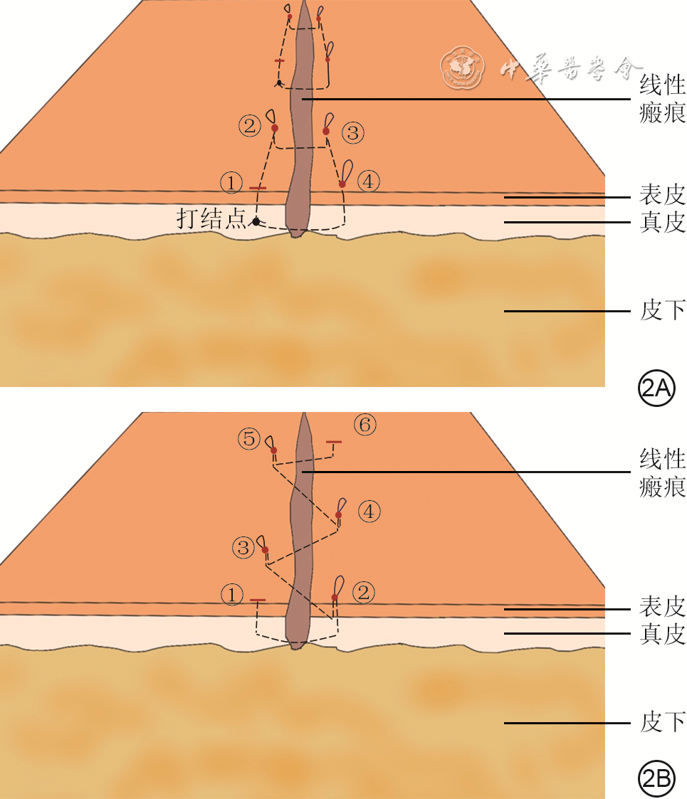
图 2 瘢痕减张方案中的慢吸收光滑缝线的矩形缝合和慢吸收倒刺缝线的W形缝合模式图。2A.矩形缝合;2B.W形缝合
注:图2A中①为进针点,也是最后的出针点,②~④均为缝合过程中回针点;图2B中①为进针点,②~⑤均为缝合过程中回针点,⑥为出针点
-
 线性瘢痕水平埋没皮内缝合操作示例.mp4

-

 下载:
下载:


图(5)
计量
- 文章访问数: 1282
- HTML全文浏览量: 743
- PDF下载量: 121
- 被引次数: 0
