





A prospective study on the dynamic changes of inflammatory and immune indicators after injury and their predictive value for 21-day mortality risk in patients with severe burns
-
摘要:
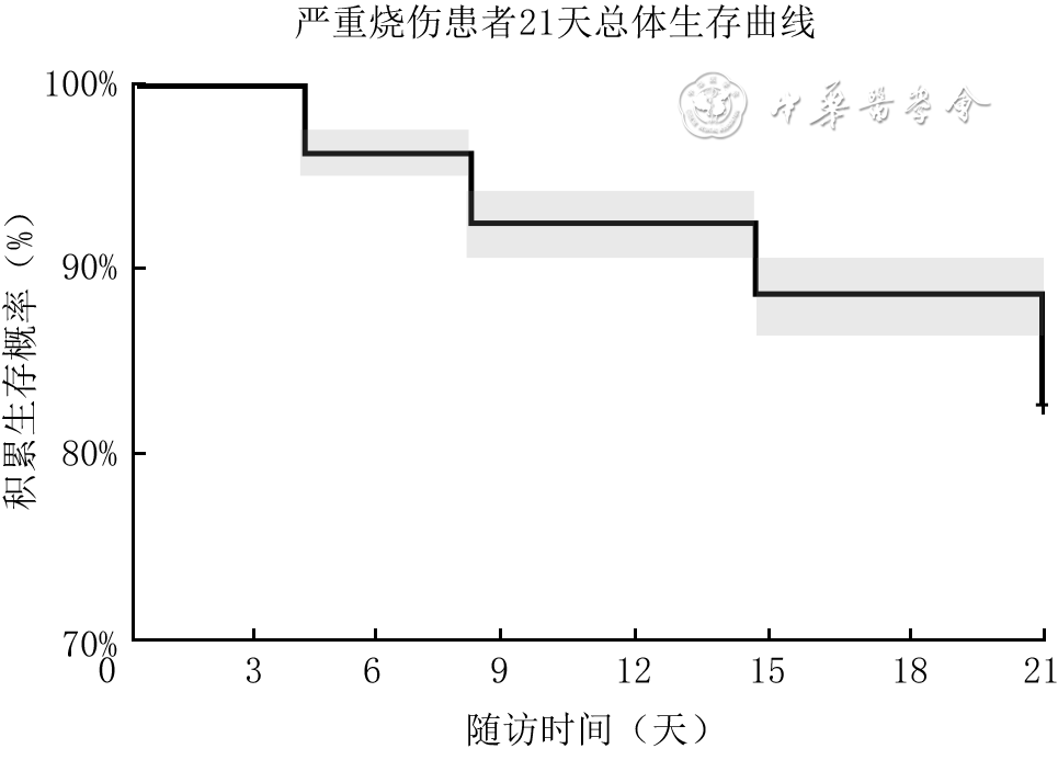
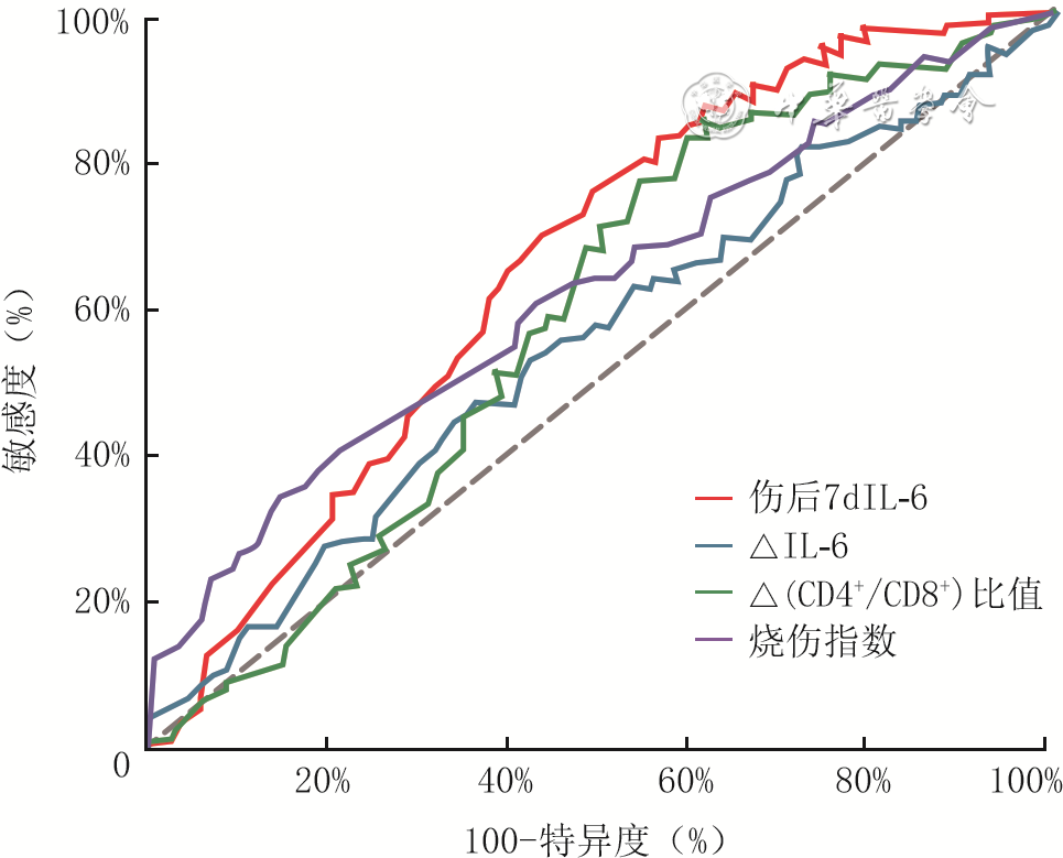
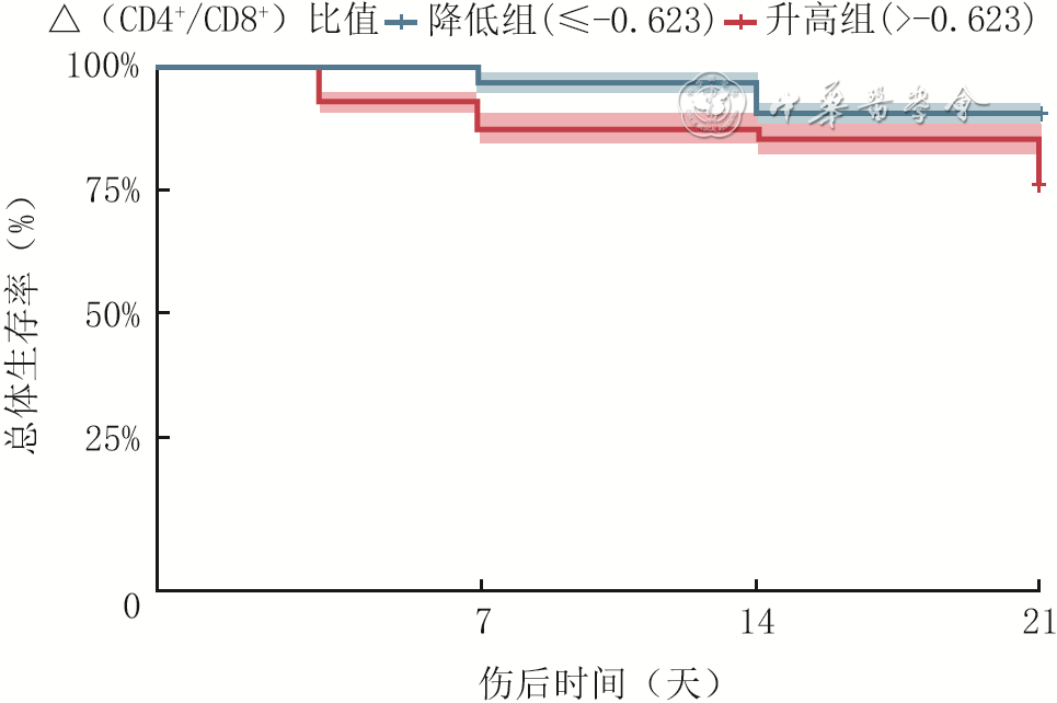
目的 探讨严重烧伤患者伤后炎症免疫指标动态变化及其对21 d死亡风险预测价值。 方法 本研究为前瞻性队列研究。2018年1月—2024年12月,宁波市第二医院烧伤科收治760例符合入选标准的严重烧伤患者。以伤后21 d结局为依据,将患者分为死亡组(139例,男106例、女33例,年龄18~78岁)和存活组(621例,男467例、女154例,年龄18~78岁)。统计两组患者临床资料(如性别、年龄、烧伤指数等),并于伤后1、3、7、14、21 d监测IL-6及CD4⁺/CD8⁺比值;同时计算伤后7 d IL-6、CD4⁺/CD8⁺比值与伤后1 d对应值的差值,即ΔIL-6及Δ(CD4⁺/CD8⁺)比值。分析患者伤后IL-6及CD4+/CD8+比值的纵向变化轨迹(样本数为760)。构建基线Cox比例风险回归模型(以下简称Cox模型)分析烧伤指数及伤后1 d的IL-6、CD4+/CD8+比值等与21 d死亡风险的独立关联性(样本数为753),构建动态Cox模型分析烧伤指数及Δ(CD4+/CD8+)比值等与21 d死亡风险的独立关联性(样本数为746)。采用受试者操作特征曲线(ROC)评估伤后7 d IL-6水平、ΔIL-6、Δ(CD4⁺/CD8⁺)比值及烧伤指数对21 d死亡风险的预测价值,并记录Δ(CD4+/CD8+)的最佳截断值。根据Δ(CD4⁺/CD8⁺)比值是否高于截断值,将患者分为升高组(男363例、女118例,年龄18~78岁)和降低组(男207例、女68例,年龄18~78岁)。绘制前述2组患者的Kaplan-Meier生存曲线并进行比较。采用时间依赖ROC分析,比较基于伤后7 d IL-6、ΔIL-6及Δ(CD4⁺/CD8⁺)比值分别构建的Cox模型(样本数分别为760、746、746),以及基线Cox模型与动态Cox模型,对患者伤后7、14、20 d死亡风险的预测能力。 结果 患者IL-6水平随伤后时间呈下降趋势(估计值为-4.52,标准误为0.10,t=-45.61,P<0.05),死亡组患者IL-6水平高于存活组(估计值为35.64,标准误为4.86,t=7.33,P<0.05),存活组与死亡组患者IL-6水平的纵向变化轨迹比较,差异有统计学意义(估计值为-0.879,标准误为0.23,t=-3.78,P<0.05);患者CD4+/CD8+比值随伤后时间呈升高趋势(估计值为0.02,标准误为<0.01,t=21.48,P<0.05),死亡组患者CD4+/CD8+比值低于存活组(估计值为-0.11,标准误为0.03,t=-4.04,P<0.05),两组患者CD4+/CD8+比值的纵向变化轨迹比较,差异有统计学意义(估计值为<0.01,标准误为<0.01,t=3.64,P<0.05)。基线Cox模型分析结果显示,烧伤指数为21 d死亡风险的独立预测因素(P<0.05),伤后1 d的IL-6、CD4+/CD8+比值为21 d死亡风险的潜在独立预测因素(P值分别为0.053、0.070)。动态Cox模型分析结果显示,烧伤指数为21 d死亡风险的独立预测因素(P<0.05),Δ(CD4+/CD8+)比值为21 d死亡风险的潜在独立预测因素(P=0.058)。伤后7 d IL-6、Δ(CD4+/CD8+)比值、ΔIL-6、及烧伤指数预测21 d死亡风险的AUC分别为0.66(95%CI 0.61~0.70)、0.58(95%CI 0.54~0.63)、0.55(95%CI 0.50~0.61)、0.62(95%CI 0.57~0.68)。Δ(CD4+/CD8+)比值的最佳截断值为-0.623。升高组患者伤后21 d生存率低于降低组(χ²=26.20,P<0.05)。伤后7 d IL-6 Cox模型在伤后7、14、20 d的AUC为71.71%(95%CI 66.03%~77.39%)、61.38%(95%CI 55.42%~67.33%)、61.38%(95%CI 55.42%~67.33%),均高于ΔIL-6 Cox模型的59.38%(95%CI 50.62%~68.14%)、56.70%(95%CI 50.01%~63.38%)、56.70%(95%CI 50.01%~63.38%),Δ(CD4+/CD8+)比值Cox模型的64.92%(95%CI 57.53%~72.31%)、57.19%(95%CI 50.65%~63.72%)、57.19%(95%CI 50.65%~63.72%),基线Cox模型的70.02%(95%CI 63.39%~76.66%)、59.71%(95%CI 53.43%~66.00%)、59.71%(95%CI 53.43%~66.00%),以及动态Cox模型的65.51%(95%CI 58.19%~72.83%)、57.12%(95%CI 50.58%~63.66%)、57.12%(95%CI 50.58%~63.66%)。 结论 严重烧伤患者伤后IL-6和CD4⁺/CD8⁺比值呈动态变化,死亡组患者早期炎症反应更强、免疫抑制更重。伤后7 d IL-6水平对21 d死亡风险有一定预测价值。 Abstract:Objective To investigate the dynamic changes of post-burn inflammatory and immune indicators in patients with severe burns and their predictive value for 21-day mortality risk. Methods This was a prospective cohort study. From January 2018 to December 2024, 760 patients with severe burns who met the inclusion criteria were admitted to the Department of Burns of Ningbo No. 2 Hospital. Based on the outcomes at 21 days post-burn, the patients were divided into death group (139 cases, 106 males and 33 females, aged 18-78 years) and survival group (621 cases, 467 males and 154 females, aged 18-78 years). Clinical data of patients in the two groups (such as sex, age, and burn index) were collected, and IL-6 and CD4+/CD8+ ratio were monitored on days 1, 3, 7, 14, and 21 post-burn. Meanwhile, the differences between IL-6 and CD4+/CD8+ ratio on day 7 post-burn and the corresponding values on day 1 post-burn were calculated, namely ΔIL-6 and Δ(CD4+/CD8+) ratio. The longitudinal trajectories of IL-6 and CD4+/CD8+ ratio post-burn were analyzed (n=760). A baseline Cox proportional hazards regression model (hereinafter referred to as the Cox model) was constructed to analyze the independent associations of age, sex, burn index, and IL-6 and CD4+/CD8+ ratio on day 1 post-burn with 21-day mortality risk (n=753). A dynamic Cox model was constructed to analyze the independent associations of age, sex, burn index and Δ(CD4+/CD8+) ratio with 21-day mortality risk (n=746). Receiver operating characteristic (ROC) curves were used to evaluate the predictive value of IL-6 level on day 7 post-burn, ΔIL-6, Δ(CD4+/CD8+) ratio, and burn index for 21-day mortality risk, and the optimal cut-off value of Δ(CD4+/CD8++) ratio was recorded. According to whether the Δ(CD4+/CD8+) ratio was higher than the cut-off value, patients were divided into increased group (363 males and 118 females, aged 18-78 years) and decreased group (207 males and 68 females, aged 18-78 years), and Kaplan-Meier survival curves were plotted for the two groups and compared. Time-dependent ROC analysis was used to compare the predictive abilities of the Cox models constructed based on IL-6 on day 7 post-burn, ΔIL-6, and Δ(CD4+/CD8+) ratio (with sample sizes of 760, 746, and 746, respectively), as well as the baseline Cox model and the dynamic Cox model, for mortality risk at 7, 14, and 20 days post-burn. Results Patients' IL-6 levels exhibited a downward trend over time post-burn (with an estimate of -4.52, a standard error of 0.10, t=-45.61, P<0.05), and IL-6 levels in death group were higher than those in survival group (with an estimate of 35.64, a standard error of 4.86, t=7.33, P<0.05); the longitudinal trajectories of IL-6 levels between survival group and death group were significantly different (with an estimate of -0.879, a standard error of 0.23, t=-3.78, P<0.05). Patients' CD4+/CD8+ ratio exhibited an upward trend over time post-burn (with an estimate of 0.02, a standard error of <0.01, t=21.48, P<0.05), and the CD4+/CD8+ ratio in death group was lower than that in survival group (with an estimate of -0.11, a standard error of 0.03, t=-4.04, P<0.05); the longitudinal trajectories of CD4+/CD8+ ratio between the two groups were significantly different (with an estimate of <0.01, a standard error of <0.01, t=3.64, P<0.05). The baseline Cox model showed that burn index was an independent predictor of 21-day mortality risk (P<0.05), while IL-6 and CD4+/CD8+ ratio on day 1 post-burn were potential independent predictors of 21-day mortality risk (with P values of 0.053 and 0.070, respectively). The dynamic Cox model showed that burn index was an independent predictor of 21-day mortality risk (P<0.05), while Δ(CD4+/CD8+) ratio was a potential independent predictor of 21-day mortality risk (P=0.058). The AUCs of IL-6 on day 7 post-burn, Δ(CD4+/CD8+) ratio, ΔIL-6, and burn index for predicting 21-day mortality risk were 0.66 (with a 95%CI of 0.61-0.70), 0.58 (with a 95%CI of 0.54-0.63), 0.55 (with a 95%CI of 0.50-0.61), and 0.62 (with a 95%CI of 0.57-0.68), respectively. The optimal cut-off value of Δ(CD4+/CD8+) ratio was -0.623. The 21-day survival rate of patients in increased group was lower than that in decreased group (χ²=26.20, P<0.05). The AUCs of the IL-6 Cox model on day 7 post-burn at 7, 14, and 20 days post-burn were 71.71% (with a 95%CI of 66.03%-77.39%), 61.38% (with a 95%CI of 55.42%-67.33%), and 61.38% (with a 95%CI of 55.42%-67.33%), respectively, all higher than those of the ΔIL-6 Cox model (59.38% (with a 95%CI of 50.62%-68.14%), 56.70% (with a 95%CI of 50.01%-63.38%), and 56.70% (with a 95%CI of 50.01%-63.38%)), the Δ(CD4+/CD8+) ratio Cox model (64.92% (with a 95%CI of 57.53%-72.31%), 57.19% (with a 95%CI of 50.65%-63.72%), and 57.19% (with a 95%CI of 50.65%-63.72%)), the baseline Cox model (70.02% (with a 95%CI of 63.39%-76.66%), 59.71% (with a 95%CI of 53.43%-66.00%), and 59.71% (with a 95%CI of 53.43%-66.00%)), and the dynamic Cox model (65.51% (with a 95%CI of 58.19%-72.83%), 57.12% (with a 95%CI of 50.58%-63.66%), and 57.12% (with a 95%CI of 50.58%-63.66%)). Conclusions In patients with severe burns, IL-6 and CD4+/CD8+ ratio exhibit dynamic changes post-burn; patients in the death group have stronger early inflammatory responses and more severe immunosuppression. IL-6 level on day 7 post-burn has certain predictive value for 21-day mortality. -
Key words:
- Burns /
- Immune dysfunction /
- Dynamic trajectory /
- Biomarkers /
- Interleukin-6 /
- CD4+/CD8+ ratio
-
参考文献
(40) [1] YeJ, HuX, WangZ, et al. The role of mtDAMPs in the trauma-induced systemic inflammatory response syndrome[J]. Front Immunol, 2023,14:1164187. DOI: 10.3389/fimmu.2023.1164187. [2] XiangY, PanBH, ZhangJ, et al. Suppression of overactivated immunity in the early stage is the key to improve the prognosis in severe burns[J]. Front Immunol,2024,15:1455899. DOI: 10.3389/fimmu.2024.1455899. [3] GoossensE, LiJ, CallensC, et al. Acute endotoxemia-induced respiratory and intestinal dysbiosis[J]. Int J Mol Sci, 2022,23(19):11602. DOI: 10.3390/ijms231911602. [4] HoVP, KaafaraniH, RattanR, et al. Sepsis 2019: what surgeons need to know[J]. Surg Infect (Larchmt), 2020,21(3):195-204. DOI: 10.1089/sur.2019.126. [5] JorgensenAM, NelsonRA, HickersonD, et al. Silica-based fiber universal combat matrix reduces inflammation, supporting cellular migration and extracellular matrix remodeling in porcine full-thickness burns[J]. Mil Med, 2025,190(Suppl 2):S521-S527. DOI: 10.1093/milmed/usaf256. [6] ThiagarajanAB, NarlochK, ChernyakM, et al. Probiotic and prebiotic interventions in burn patients: a systematic review[J]. Burns, 2026,52(3):107912. DOI: 10.1016/j.burns.2026.107912. [7] LouJ, KongH, LiJ, et al. Longitudinal biomarker trajectories and their prognostic utility for 21-day mortality in burn patients with sepsis: a retrospective cohort study[J]. Inflamm Res, 2026,75(1):20. DOI: 10.1007/s00011-025-02178-2. [8] XuP, LouJ, KongH, et al. Dynamic biomarker profiling and phenotyping in burn sepsis: a retrospective cohort study using growth mixture modeling[J]. Front Cell Infect Microbiol, 2026,16:1710916. DOI: 10.3389/fcimb.2026.1710916. [9] CaiJ, JinY, LouJ, et al. The V-shaped association between the ratio of neutrophil counts to prognostic nutritional index and 30-, 60-, and 90-day mortality in elderly critically ill patients aged 65 and older with sepsis: a retrospective study based on the MIMIC database[J]. Front Nutr, 2025,12:1602016. DOI: 10.3389/fnut.2025.1602016. [10] MulderPPG, BoekemaBKHL, van der VliesCH, et al. Targeting immune dysregulation after burn injury for improved healing and outcomes[J]. Biomolecules, 2026,16(6):806. DOI: 10.3390/biom16060806. [11] BoonsongsermP, WatcharanurakP, MeevassanaJ, et al. HMGB1 release initiates burn wound healing delay via HMGB1-produced DNA gap reduction, promoting DNA damage-hypomethylation-senescence cascade[J]. Burns, 2026,52(8):108096. DOI: 10.1016/j.burns.2026.108096. [12] LiuH, WangS, LiD, et al. Characteristics and significance of blister fluid cell-free mitochondrial DNA in pediatric small area intermediate-depth burn wounds[J]. Burns, 2026,52(1):107780. DOI: 10.1016/j.burns.2025.107780. [13] SchwachaMG, ScrogginsSR, MontgomeryRK, et al. Burn injury is associated with an infiltration of the wound site with myeloid-derived suppressor cells[J]. Cell Immunol, 2019,338:21-26. DOI: 10.1016/j.cellimm.2019.03.001. [14] WeiJ, ZhangK, PanW, et al. C3AR1 regulates intestinal T-cell immune response after burn by activating the NF-κB signaling pathway[J]. Immunol Cell Biol, 2026,104(4):345-357. DOI: 10.1111/imcb.70099. [15] MulderPPG, VligM, ElgersmaA, et al. Monocytes and T cells incorporated in full skin equivalents to study innate or adaptive immune reactions after burn injury[J]. Front Immunol, 2023,14:1264716. DOI: 10.3389/fimmu.2023.1264716. [16] MoniruzzamanM, ShahA, FairlieT, et al. Immune activation in primary sclerosing cholangitis: a systematic review and comparative analysis with inflammatory bowel diseases[J]. United European Gastroenterol J, 2025,13(9):1740-1753. DOI: 10.1002/ueg2.70115. [17] SayyadioskoieSR, SchwachaMG. Myeloid-derived suppressor cells (MDSCs) and the immunoinflammatory response to injury (Mini Review)[J]. Shock, 2021,56(5):658-666. DOI: 10.1097/SHK.0000000000001795. [18] Morales-PrimoA U, BeckerI, Zamora-ChimalJ. Neutrophil extracellular trap-associated molecules: a review on their immunophysiological and inflammatory roles[J]. Int Rev Immunol, 2022,41(2):253-274. DOI: 10.1080/08830185.2021.1921174. [19] LangleyD, ZimmermannK, KrenskeE, et al. Unremitting pro-inflammatory T-cell phenotypes, and macrophage activity, following paediatric burn injury[J]. Clin Transl Immunology, 2024,13(3):e1496. DOI: 10.1002/cti2.1496. [20] BonoV, MarchettiG C. Immunological recovery with BIC/FTC/TAF: CD4 T-cell count and CD4/CD8 ratio as markers of response[J]. New Microbiologica, 2025,48(1):1-4. [21] ZaluckyA, DumasG, LeligdowiczA, et al. Sepsis and the Immunocompromised Host[J]. Crit Care Clin, 2026,42(4):821-837. DOI: 10.1016/j.ccc.2026.05.007. [22] WuD, ChenY. Lipids for CD8+ TILs: Beneficial or harmful?[J]. Front Immunol, 2022,13:1020422. DOI: 10.3389/fimmu.2022.1020422. [23] MulderP, KoenenH, VligM, et al. Burn-induced local and systemic immune response: systematic review and meta-analysis of animal studies[J]. J Invest Dermatol, 2022,142(11):3093-3109.e15. DOI: 10.1016/j.jid.2022.05.004. [24] YuanY, LeiS, DuanA, et al. Effects on mortality of different blood purification techniques in sepsis patients: an umbrella review of systematic reviews and meta-analyses[J]. Ren Fail, 2026,48(1):2698155. DOI: 10.1080/0886022X.2026.2698155. [25] PengS J, FengY, LiX, et al. Thymopentin (TP-5) prevents lipopolysaccharide-induced neuroinflammation and dopaminergic neuron injury by inhibiting the NF-κB/NLRP3 signaling pathway[J]. Int Immunopharmacol, 2023,119:110109. DOI: 10.1016/j.intimp.2023.110109. [26] MaY, ZhangL, LiuW. Immunosuppressive tumor microenvironment and advance in immunotherapy in melanoma bone metastasis[J]. Front Immunol, 2025,16:1608215. DOI: 10.3389/fimmu.2025.1608215. [27] LiS, LuL, XiongY, et al. Nanomedicine-based immunotherapy for tissue regeneration[J/OL]. Burns Trauma, 2025,13:tkaf015[2025-11-21]. https://pubmed.ncbi.nlm.nih.gov/40568375/. DOI: 10.1093/burnst/tkaf015. [28] 娄家祺, 李琦, 崔庆伟, 等. 肠内免疫营养支持治疗对具有营养风险的成年烧伤患者疗效的前瞻性随机对照研究[J].中华烧伤与创面修复杂志,2022,38(8):722-734. DOI: 10.3760/cma.j.cn501225-20220327-00094. [29] JinY, HuangY, WangJ, et al. From injury to integrity: tissue engineering solutions in combined bone repair and regeneration and muscle repair[J/OL]. Burns Trauma, 2025,13:tkaf052[2025-11-21]. https://pubmed.ncbi.nlm.nih.gov/40979903/. DOI: 10.1093/burnst/tkaf052. [30] ZhangT, YangY, JiangJ, et al. The role of glucose metabolism in wound healing: an overview[J/OL]. Burns Trauma, 2025,13:tkaf053[2025-11-21]. https://pubmed.ncbi.nlm.nih.gov/40979902/. DOI: 10.1093/burnst/tkaf053. [31] LegrandM, HoffmannC, RoquillyA, et al. Early surgery and outcomes in severe burns: a prospective multicenter observational study[J/OL]. Ann Surg, 2026(2026-08-18)[ [2025-11-21]]. https://pubmed.ncbi.nlm.nih.gov/42608728/.DOI: 10.1097/SLA.0000000000007201. [published online ahead of print]. [32] HonoréPM, BlackmanS, PerriensE, et al. Early detection, diagnosis, prevention, and treatment of infection to avoid sepsis and septic shock in severely burned patients: a narrative review[J]. Eur Burn J, 2025,6(1):6. DOI: 10.3390/ebj6010006. [33] MuellerSW, TranNM, BetthauserKD, et al. Vasoactive agents in burn patients: perspectives on angiotensin-II[J]. J Burn Care Res, 2025,46(3):515-525. DOI: 10.1093/jbcr/irae208. [34] YanamaladoddiV, D'CunhaH, CharleyE, et al. Early enteral nutrition in critically-ill patients[J]. World J Crit Care Med, 2025,14(3):102834. DOI: 10.5492/wjccm.v14.i3.102834. [35] KraljR, BarčotZ, KurtanjekM, et al. Prognostic value of serum concentrations of interleukin-6 and interleukin-8 in dermal burn injuries among pediatric patients[J]. J Burn Care Res, 2026,47(1):198-206. DOI: 10.1093/jbcr/iraf136. [36] 贾赤宇, 尹斌, 张泽鑫. 重度烧伤的转录组学研究:价值与机遇[J].中华烧伤与创面修复杂志,2024,40(6):514-520. DOI: 10.3760/cma.j.cn501225-20231026-00135. [37] Ibrahim YoussoufA, BenaissaE, BelouadEM, et al. Epidemiological profile of bacterial infections in burn patients over a five-year period[J]. Cureus, 2024,16(11):e74848. DOI: 10.7759/cureus.74848. [38] YangG, TanL, YaoH, et al. Long-term effects of severe burns on the kidneys: research advances and potential therapeutic approaches[J]. J Inflamm Res, 2023,16:1905-1921. DOI: 10.2147/JIR.S404983. [39] LouJ, FanY, CuiS, et al. Development and validation of a nomogram to predict hypothermia in adult burn patients during escharectomy under general anesthesia[J]. Burns, 2024,50(1):93-105. DOI: 10.1016/j.burns.2023.06.010. [40] 丁泓帆, 郑兴锋, 夏照帆. 代谢组学在危重烧创伤救治中的应用研究进展[J].中华烧伤杂志,2019,35(6):467-471. DOI: 10.3760/cma.j.issn.1009-2587.2019.06.014. -
Table 1. 存活组与死亡组严重烧伤患者临床资料比较
组别 例数 年龄(岁, 性别(例) 烧伤总面积(%TBSA, Ⅲ度烧伤面积(%TBSA, Ⅱ度烧伤面积(%TBSA, 烧伤指数( 男 女 存活组 621 40±14 467 154 41±13 8(0,22) 29±13 25(17,34) 死亡组 139 40±11 106 33 50±19 15(5,32) 26±16 33(24,46) 统计值 t=0.31 χ²=0.08 t=6.73 Z=5.89 t=1.93 Z=7.45 P值 0.758 0.775 <0.001 <0.001 0.054 <0.001 注:ΔIL-6、Δ(CD4⁺/CD8⁺)比值为伤后7 d的IL-6、CD4⁺/CD8⁺比值与伤后1 d对应值的差值;APACHEⅡ为急性生理学与慢性健康状况评分系统Ⅱ;慢性病包括高血压、糖尿病、慢性心肺疾病等  下载: 导出CSV
Table 2. 753例严重烧伤患者各指标预测21 d死亡风险的基线Cox比例风险回归模型分析结果
变量 VIF HR 95%CI P值 IL-6(pg/mL) 1.12 1.00 1.00~1.01 0.053 CD4+/CD8+比值 1.21 0.55 0.29~1.05 0.070 年龄(岁) 1.08 1.01 0.99~1.02 0.370 性别 1.05 0.92 0.66~1.30 0.642 烧伤指数 1.34 1.02 1.01~1.03 <0.001 注:IL-6、CD4+/CD8+比值均为患者伤后1 d的检测值;VIF为方差膨胀因子
下载: 导出CSV
Table 3. 746例严重烧伤患者各指标预测21 d死亡风险的动态Cox比例风险回归模型分析结果
变量 VIF HR 95%CI P值 ΔIL-6(pg/mL) 1.15 0.999 0.996~1.002 0.656 Δ(CD4+/CD8+)比值 1.18 1.407 0.988~2.003 0.058 年龄(岁) 1.09 1.001 0.987~1.015 0.894 性别 1.06 0.947 0.674~1.332 0.756 烧伤指数 1.32 1.023 1.015~1.030 <0.001 注:ΔIL-6、Δ(CD4+/CD8+)比值伤后7 d的IL-6、CD4⁺/CD8⁺比值与伤后1 d对应值的差值;VIF为方差膨胀因子
下载: 导出CSV
Table 4. 不同Cox比例风险回归模型的时间依赖ROC分析[AUC(95%CI)]
模型种类 样本量 伤后7 d 伤后14 d 伤后20 d 基线Cox比例风险回归模型 753 70.02(63.39~76.66) 59.71(53.43~66.00) 59.71(53.43~66.00) 伤后7 d的IL-6 Cox比例风险回归模型 760 71.71(66.03~77.39) 61.38(55.42~67.33) 61.38(55.42~67.33) ΔIL-6 Cox比例风险回归模型 746 59.38(50.62~68.14) 56.70(50.01~63.38) 56.70(50.01~63.38) Δ(CD4+/CD8+)比值Cox比例风险回归模型 746 64.92(57.53~72.31) 57.19(50.65~63.72) 57.19(50.65~63.72) 动态Cox比例风险回归模型 746 65.51(58.19~72.83) 57.12(50.58~63.66) 57.12(50.58~63.66) 注:各模型校正变量均为年龄、性别及烧伤指数;5种模型的事件数分别为137、139、136、136、136
下载: 导出CSV
-

 下载:


图(4) / 表(4)
计量
- 文章访问数: 1
- HTML全文浏览量: 0
- PDF下载量: 0
- 被引次数: 0
