


Mechanism of actions and reflections of Masquelet induced membrane technique in diabetic wound
-
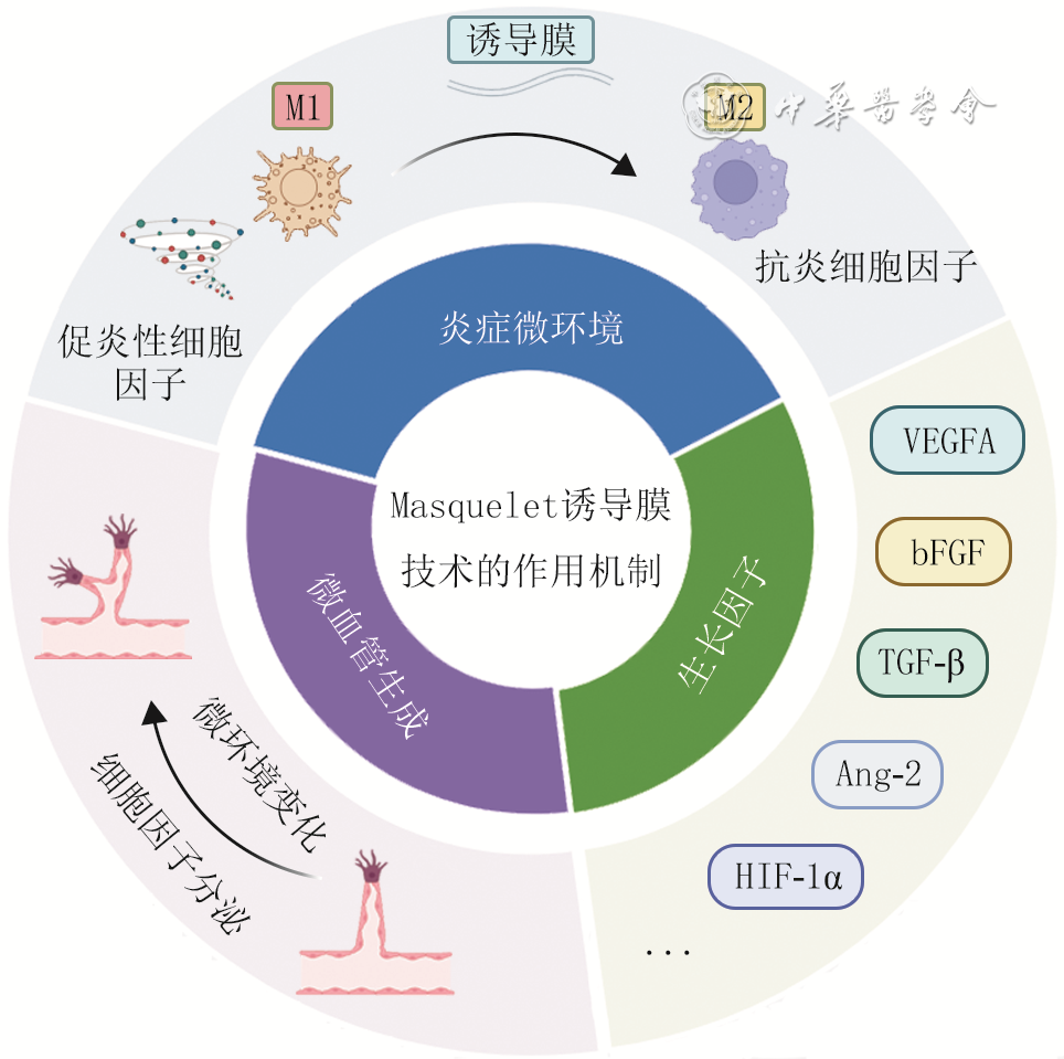
摘要: 糖尿病创面因高糖微环境所致慢性炎症、血管新生障碍及生长因子表达异常等因素,导致愈合过程受阻。Masquelet诱导膜技术(以下简称诱导膜技术)通过在创面置入骨水泥从而在局部形成一个由多种细胞成分组成的富含生长因子及微血管的生物活性膜。该膜主要通过重塑炎症微环境、促进功能性血管新生、产生内源性生长因子3种途径促进创面愈合。该文对诱导膜技术改善糖尿病创面愈合的机制进行了简要阐述,并对其在糖尿病创面中的临床应用进行了梳理,以期为临床治疗糖尿病难愈创面提供新的思路和有效手段。未来研究需聚焦应用诱导膜技术治疗的关键分子通路及新型膜诱导材料的开发,为糖尿病创面治疗提供新的策略。Abstract: Diabetic wounds are hindered in the healing process due to chronic inflammation , vasculari-zation disorder, and abnormal expression of growth factors caused by the high glucose microenvironment. Masquelet induced membrane technique (hereinafter referred to as the induced membrane technique) creates a bioactive membrane composed of various cellular components and rich in growth factors and microvessels by inserting bone cement into the wound. The membrane mainly promotes wound healing by remodeling the inflammatory microenvironment, promoting functional angiogenesis, and producing endogenous growth factors. This article briefly explains the mechanism by which the induced membrane technique improves wound healing and summarizes its clinical application in diabetic wounds. It is expected to provide new ideas and effective means for the clinical treatment of difficult-to-heal diabetic wounds. Future research needs to focus on the key molecular pathways for the application of the induced membrane technique in therapy and the development of new membrane induction materials, so as to provide new strategies for the treatment of diabetic wounds.
-
Key words:
- Diabetic foot /
- Wound healing /
- Diabetic wounds /
- Masquelet /
- Induced membrane technique /
- Angiogenesis
-
参考文献
(62) [1] ArmstrongDG, TanTW, BoultonAJM, et al. Diabetic foot ulcers: a review[J]. JAMA, 2023, 330(1): 62-75. DOI: 10.1001/jama.2023.10578. [2] FuXL, DingH, MiaoWW, et al. Global recurrence rates in diabetic foot ulcers: a systematic review and meta-analysis[J]. Diabetes Metab Res Rev, 2019, 35(6): e3160. DOI: 10.1002/dmrr.3160. [3] MaL, ChenJ, SunY, et al. The perceptions of living with diabetic foot ulcers: a systematic review and meta-synthesis of qualitative studies[J]. J Tissue Viability, 2023, 32(1): 39-50. DOI: 10.1016/j.jtv.2022.11.005. [4] DengH, LiB, ShenQ, et al. Mechanisms of diabetic foot ulceration: a review[J]. J Diabetes, 2023, 15(4): 299-312. DOI: 10.1111/1753-0407.13372. [5] DuY, WangJ, FanW, et al. Preclinical study of diabetic foot ulcers: from pathogenesis to vivo/vitro models and clinical therapeutic transformation[J]. Int Wound J, 2023, 20(10): 4394-4409. DOI: 10.1111/iwj.14311. [6] MohsinF, JavaidS, TariqM, et al. Molecular immunological mechanisms of impaired wound healing in diabetic foot ulcers (DFU), current therapeutic strategies and future directions[J]. Int Immunopharmacol, 2024, 139:112713. DOI: 10.1016/j.intimp.2024.112713. [7] LiuH, WangX, GaoH, et al. Physiological and pathological characteristics of vascular endothelial injury in diabetes and the regulatory mechanism of autophagy[J]. Front Endocrinol (Lausanne), 2023, 14:1191426. DOI: 10.3389/fendo.2023.1191426. [8] LiuJ, PanS, WangX, et al. Role of advanced glycation end products in diabetic vascular injury: molecular mechanisms and therapeutic perspectives[J]. Eur J Med Res, 2023, 28(1):553. DOI: 10.1186/s40001-023-01431-w. [9] LuY, LiuX, ZhaoJ, et al. Single-cell profiling reveals transcriptomic signatures of vascular endothelial cells in non-healing diabetic foot ulcers[J]. Front Endocrinol (Lausanne), 2023, 14:1275612. DOI: 10.3389/fendo.2023.1275612. [10] 陶克,曹涛,郝彤. 糖尿病足溃疡血管形成障碍的机制及干预策略[J]. 中华烧伤与创面修复杂志,2025,41(2):120-126.DOI: 10.3760/cma.j.cn501225-20241204-00474. [11] VillaF, MarchandinH, LavigneJ-P, et al. Anaerobes in diabetic foot infections: pathophysiology, epidemiology, virulence, and management[J]. Clin Microbiol Rev, 2024, 37(3): e0014323. DOI: 10.1128/cmr.00143-23. [12] ZhangN, LiuY, YanW, et al. The effect of negative pressure wound therapy on the outcome of diabetic foot ulcers: a meta-analysis[J]. Int Wound J, 2024, 21(4): e14886. DOI: 10.1111/iwj.14886. [13] BardillJR, LaughterMR, StagerM, et al. Topical gel-based biomaterials for the treatment of diabetic foot ulcers[J]. Acta Biomater, 2022, 138: 73-91. DOI: 10.1016/j.actbio.2021.10.045. [14] AlfordAI, NicolaouD, HakeM, et al. Masquelet's induced membrane technique: review of current concepts and future directions[J]. J Orthop Res, 2021, 39(4): 707-718. DOI: 10.1002/jor.24978. [15] LiuX, MinHS, ChaiY, et al. Masquelet technique with radical debridement and alternative fixation in treatment of infected bone nonunion[J]. Front Surg, 2022, 9:1000340. DOI: 10.3389/fsurg.2022.1000340. [16] PederivaD, De LucaL, FaldiniC, et al. Masquelet's induced membrane technique in the upper limb: a systematic review of the current outcomes[J]. J Orthop Traumatol, 2025, 26(1):4. DOI: 10.1186/s10195-024-00815-w. [17] AllesinaL, Alessio-MazzolaM, BelluatiA, et al. Surgical treatment of critical size bone defects with Masquelet technique versus bone transport: a systematic review and meta-analysis of comparative studies[J]. Arch Orthop Trauma Surg, 2023, 143(12): 7081-7096. DOI: 10.1007/s00402-023-05049-9. [18] KleinC, MonetM, BarbierV, et al. The Masquelet technique: current concepts, animal models, and perspectives[J]. J Tissue Eng Regen Med, 2020, 14(9):1349-1359. DOI: 10.1002/term.3097. [19] MathieuL, MourtialonR, DurandM, et al. Masquelet technique in military practice: specificities and future directions for combat-related bone defect reconstruction[J]. Mil Med Res, 2022, 9(1): 48. DOI: 10.1186/s40779-022-00411-1. [20] ChenH, YaoL, ZhouY, et al. Evaluation of antibiotic-loaded bone cement in treatment of infected diabetic foot: systematic review and meta-analysis[J]. Diabetes Metab Res Rev, 2024, 40(8): e70002. DOI: 10.1002/dmrr.70002. [21] 杨成兰,陈静,闫雪萍,等.Masquelet诱导膜技术联合多种治疗策略在糖尿病足创面中的临床应用进展[J].中华糖尿病杂志,2025,17(8):1057-1061.DOI: 10.3760/cma.j.cn115791-20241102-00655. [22] DurandM, MathieuL, VenantJ, et al. Engineering the bone reconstruction surgery: the case of the Masquelet-induced membrane technique[J]. Eur J Trauma Emerg Surg, 2025, 51(1):138. DOI: 10.1007/s00068-025-02815-9. [23] KlopfleischR, JungF. The pathology of the foreign body reaction against biomaterials[J]. J Biomed Mater Res A, 2017, 105(3): 927-940. DOI: 10.1002/jbm.a.35958. [24] LuW, ZhaoR, FanX, et al. Time-varying characteristics of the induced membrane and its effects on bone defect repair[J]. Injury, 2023, 54(2): 318-328. DOI: 10.1016/j.injury.2022.12.026. [25] WuJH, BaoQW, WangSK, et al. Mechanisms of the Masquelet technique to promote bone defect repair and its influencing factors[J]. Chin J Traumatol, 2025, 28(3):157-163. DOI: 10.1016/j.cjtee.2024.04.003. [26] SheikhZ, BrooksPJ, BarzilayO, et al. Macrophages, foreign body giant cells and their response to implantable biomaterials[J]. Materials (Basel), 2015, 8(9): 5671-5701. DOI: 10.3390/ma8095269. [27] KanekoY, MineharaH, SonobeT, et al. Differences in macrophage expression in induced membranes by fixation method - Masquelet technique using a mouse's femur critical-sized bone defect model[J]. Injury, 2024, 55(6):111135. DOI: 10.1016/j.injury.2023.111135. [28] GindrauxF, RondotT, de BillyB, et al. Similarities between induced membrane and amniotic membrane: novelty for bone repair[J]. Placenta, 2017, 59:116-123. DOI: 10.1016/j.placenta.2017.06.340. [29] AhoOM, LehenkariP, RistiniemiJ, et al. The mechanism of action of induced membranes in bone repair[J]. J Bone Joint Surg Am, 2013, 95(7): 597-604. DOI: 10.2106/JBJS.L.00310. [30] HenrichD, SeebachC, NauC, et al. Establishment and characterization of the Masquelet induced membrane technique in a rat femur critical-sized defect model[J]. J Tissue Eng Regen Med, 2016, 10(10): E382-E396. DOI: 10.1002/term.1826. [31] TarchalaM, HarveyEJ, BarraletJ. Biomaterial-stabilized soft tissue healing for healing of critical-sized bone defects: the Masquelet technique[J]. Adv Healthc Mater, 2016, 5(6): 630-640. DOI: 10.1002/adhm.201500793. [32] TothZ, RoiM, EvansE, et al. Masquelet technique: effects of spacer material and micro-topography on factor expression and bone regeneration[J]. Ann Biomed Eng, 2019, 47(1):174-189. DOI: 10.1007/s10439-018-02137-5. [33] YinQ, ChenX, DaiB, et al. Varying degrees of spontaneous osteogenesis of Masquelet's induced membrane: experimental and clinical observations[J]. BMC Musculoskelet Disord, 2023, 24(1): 384. DOI: 10.1186/s12891-023-06498-4. [34] DawiJ, TumanyanK, TomasK, et al. Diabetic foot ulcers: pathophysiology, immune dysregulation, and emerging therapeutic strategies[J]. Biomedicines, 2025, 13(5):1076. DOI: 10.3390/biomedicines13051076. [35] JiangN, XuC, XuY, et al. Comprehensive transcriptomic analysis of immune-related genes in diabetic foot ulcers: new insights into mechanisms and therapeutic targets[J]. Int Immunopharmacol, 2024, 139:112638. DOI: 10.1016/j.intimp.2024.112638. [36] ⅠPastar, BalukoffNC, MarjanovicJ, et al. Molecular pathophysiology of chronic wounds: current state and future directions[J]. Cold Spring Harb Perspect Biol, 2023, 15(4):a041243. DOI: 10.1101/cshperspect.a041243. [37] ZhangY, JiaF, LiM, et al. Engineering macrophage phenotype switching via nucleotide-binding oligomerization domain-like receptor protein 3 inflammasome inhibition: a translational approach using antibiotic cement for diabetic foot ulcers[J]. Bioeng Transl Med, 2025, 10(6): e70073. DOI: 10.1002/btm2.70073. [38] 曹涛, 计鹏, 张智, 等. 抗生素骨水泥治疗糖尿病足溃疡的前瞻性随机对照研究[J]. 中华烧伤与创面修复杂志, 2023, 39(4): 311-318. DOI: 10.3760/cma.j.cn501225-20221111-00485. [39] ZhongM, GuoJ, QaharM, et al. Combination therapy of negative pressure wound therapy and antibiotic-loaded bone cement for accelerating diabetic foot ulcer healing: a prospective randomised controlled trial[J]. Int Wound J, 2024, 21(10): e70089. DOI: 10.1111/iwj.70089. [40] RoyR, ZayasJ, MohamedMF, et al. IL-10 dysregulation underlies chemokine insufficiency, delayed macrophage response, and impaired healing in diabetic wounds[J]. J Invest Dermatol, 2022, 142(3Pt A): 692-704.e14. DOI: 10.1016/j.jid.2021.08.428. [41] LiuC, YouJX, ChenYX, et al. Effect of induced membrane formation followed by polymethylmethacrylate implantation on diabetic foot ulcer healing when revascularization is not feasible[J]. J Diabetes Res, 2019, 2019: 2429136. DOI: 10.1155/2019/2429136. [42] MartinDC, SempleJL, SeftonMV. Poly(methacrylic acid-co-methyl methacrylate) beads promote vascularization and wound repair in diabetic mice[J]. J Biomed Mater Res A, 2010, 93(2): 484-492. DOI: 10.1002/jbm.a.32528. [43] NiikuraT, OdaT, JimboN, et al. Immunohistochemical analysis revealed the expression of bone morphogenetic proteins-4, 6, 7, and 9 in human induced membrane samples treated with the Masquelet technique[J]. J Orthop Surg Res, 2022, 17(1): 29. DOI: 10.1186/s13018-022-02922-y. [44] HoitG, KainMS, SparkmanJW, et al. The induced membrane technique for bone defects: basic science, clinical evidence, and technical tips[J]. OTA Int, 2021, 4(2Suppl):Se106(1-5). DOI: 10.1097/OI9.0000000000000106. [45] OkonkwoUA, DiPietroLA. Diabetes and wound angiogenesis[J]. Int J Mol Sci, 2017, 18(7):1419. DOI: 10.3390/ijms18071419. [46] OkonkwoUA, ChenL, MaD, et al. Compromised angiogenesis and vascular Integrity in impaired diabetic wound healing[J]. PLoS One, 2020, 15(4): e0231962. DOI: 10.1371/journal.pone.0231962. [47] TangQ, JinH, TongM, et al. Inhibition of Dll4/Notch1 pathway promotes angiogenesis of Masquelet's induced membrane in rats[J]. Exp Mol Med, 2018, 50(4):1-15. DOI: 10.1038/s12276-018-0062-9. [48] PelissierP, MasqueletAC, BareilleR, et al. Induced membranes secrete growth factors including vascular and osteoinductive factors and could stimulate bone regeneration[J]. J Orthop Res, 2004, 22(1): 73-79. DOI: 10.1016/S0736-0266(03)00165-7. [49] WangW, ZuoR, LongH, et al. Advances in the Masquelet technique: myeloid-derived suppressor cells promote angiogenesis in PMMA-induced membranes[J]. Acta Biomater, 2020, 108: 223-236. DOI: 10.1016/j.actbio.2020.03.010. [50] WuH, TanJ, SunD, et al. Discovery of multipotent progenitor cells from human induced membrane: equivalent to periosteum-derived stem cells in bone regeneration[J]. J Orthop Translat, 2023, 42: 82-93. DOI: 10.1016/j.jot.2023.07.004. [51] QiM, ZhouQ, ZengW, et al. Growth factors in the pathogenesis of diabetic foot ulcers[J]. Front Biosci (Landmark Ed), 2018, 23(2): 310-317. DOI: 10.2741/4593. [52] ZubairM, AhmadJ. Role of growth factors and cytokines in diabetic foot ulcer healing: a detailed review[J]. Rev Endocr Metab Disord, 2019, 20(2): 207-217. DOI: 10.1007/s11154-019-09492-1. [53] GessmannJ, RosteiusT, BaeckerH, et al. Is the bioactivity of induced membranes time dependent?[J]. Eur J Trauma Emerg Surg, 2022, 48(4): 3051-3061. DOI: 10.1007/s00068-021-01844-4. [54] WangX, WeiF, LuoF, et al. Induction of granulation tissue for the secretion of growth factors and the promotion of bone defect repair[J]. J Orthop Surg Res, 2015, 10:147. DOI: 10.1186/s13018-015-0287-4. [55] 赵晨兵,张会峰.载抗生素骨水泥治疗糖尿病足感染创面的研究进展[J].中华糖尿病杂志,2022,14(7):724-729.DOI: 10.3760/cma.j.cn115791-20211213-00660. [56] DaiJ, ZhouY, MeiS, et al. Application of antibiotic bone cement in the treatment of infected diabetic foot ulcers in type 2 diabetes[J]. BMC Musculoskelet Disord, 2023, 24(1):135. DOI: 10.1186/s12891-023-06244-w. [57] DongT, HuangQ, SunZ. Antibiotic-laden bone cement for diabetic foot infected wounds: a systematic review and meta-analysis[J]. Front Endocrinol (Lausanne), 2023, 14:1134318. DOI: 10.3389/fendo.2023.1134318. [58] JianY, LiL, ChenW, et al. Comparative analysis of the therapeutic effect of antibiotic bone cement on Wagner grade 3 or 4 diabetic foot ulcer in heel and non-heel areas: a retrospective cohort study[J]. Front Endocrinol (Lausanne), 2025, 16:1662731. DOI: 10.3389/fendo.2025.1662731. [59] SunYW, LiL, ZhangZH. Antibiotic-loaded bone cement combined with vacuum-assisted closure facilitating wound healing in Wagner 3-4 diabetic foot ulcers[J]. Int J Low Extrem Wounds, 2025, 24(3): 672-677. DOI: 10.1177/15347346221109045. [60] 钟宇惠, 崔旭, 周思拓, 等. 抗生素骨水泥与VSD联合游离股前外侧穿支嵌合肌皮瓣序贯治疗糖尿病性跟骨骨髓炎创面的临床效果[J]. 中华烧伤与创面修复杂志, 2026, 42(3): 225-233. DOI: 10.3760/cma.j.cn501225-20251129-00493. [61] DingX, YuanY, LuH, et al. Analysis of the effect of antibiotic bone cement in the treatment of diabetic foot ulcer through tibia transverse transport[J]. Orthop Surg, 2022, 14(9): 2141-2149. DOI: 10.1111/os.13412. [62] 徐林刚,杨冠龙,刘磊,等.抗生素骨水泥联合自体富血小板血浆治疗糖尿病足[J].河南科技大学学报(医学版),2019,37(1):26-29.DOI: 10.15926/j.cnki.issn1672-688x.2019.01.007. -

 下载:
下载:

图(2)
计量
- 文章访问数: 1612
- HTML全文浏览量: 334
- PDF下载量: 70
- 被引次数: 0
