





Performance of phenylboronic acid-modified GelMA hydrogel loaded with copper-doped cerium oxide nanozyme and its effect on healing of infected burn wounds in mice
-
摘要:
目的 探讨负载含铜氧化铈纳米酶的苯硼酸修饰的甲基丙烯酸酐明胶(GelMA)水凝胶(以下简称复合水凝胶)的性能及其对小鼠烧伤感染性创面愈合的影响。 方法 该研究为包含成组设计与重复测量设计的实验研究。制备具有三维疏松多孔网状结构的苯硼酸修饰的GelMA水凝胶(以下简称单纯水凝胶),采用水热法制备呈均匀球形结构的含铜氧化铈纳米酶。筛选出终质量浓度为100 μg/mL的含铜氧化铈纳米酶对人脐静脉内皮细胞活力无明显影响且具有较高的类超氧化物歧化酶和过氧化氢酶活性,将其加入单纯水凝胶中,制备复合水凝胶。取复合水凝胶,用磷酸盐缓冲液(PBS)或过氧化氢溶液浸泡24、48、72 h后,计算复合水凝胶中含铜氧化铈纳米酶的释放百分比,样本数均为3。取30只8周龄雄性BALB/c小鼠,于脊柱两侧对称位置各造成1个烧伤创面后,滴加耐甲氧西林金黄色葡萄球菌(MRSA)菌液,构建烧伤感染性创面模型。按随机数字表法(分组方法后同)将小鼠分为对照组、单纯水凝胶组和复合水凝胶组,每组10只小鼠(20个创面)。对照组小鼠创面不行药物干预,单纯水凝胶组和复合水凝胶组小鼠创面分别滴加单纯水凝胶、复合水凝胶。于伤后3、7、14 d,测量剩余创面面积并计算剩余创面面积百分比。于伤后3 d,采用平板计数法测定创面中MRSA浓度。于伤后5 d,采用激光散斑血流成像系统评估创面血流灌注量。另取6只8周龄雄性BALB/c小鼠,分为PBS组和复合水凝胶埋置组,每组3只,分别于小鼠背部中央皮下注射PBS和皮下埋置复合水凝胶。于处理14 d后,采集小鼠腹腔全血,检测血常规指标和血生化指标。 结果 在PBS和过氧化氢溶液中分别浸泡24、48、72 h后,复合水凝胶中含铜氧化铈纳米酶的释放百分比总体比较,差异均有统计学意义(F值分别为30.29、65.16,P<0.05)。伤后3、7、14 d,与对照组比较,单纯水凝胶组和复合水凝胶组小鼠剩余创面面积百分比均显著降低(P<0.05);复合水凝胶组小鼠剩余创面面积百分比均明显低于单纯水凝胶组(P<0.05)。伤后3 d,与对照组比较,单纯水凝胶组和复合水凝胶组小鼠创面中MRSA浓度均明显降低(P<0.05);复合水凝胶组小鼠创面中MRSA浓度明显低于单纯水凝胶组(P<0.05)。伤后5 d,与对照组[(221±25)灌注单位]比较,单纯水凝胶组和复合水凝胶组小鼠创面血流灌注量[分别为(313±53)、(535±104)灌注单位]均明显升高(P<0.05);复合水凝胶组小鼠创面血流灌注量明显高于单纯水凝胶组(P<0.05)。处理14 d后,复合水凝胶埋置组和PBS组小鼠血常规指标和血生化指标均维持在正常生理范围内,组间比较,差异均无统计学意义(P>0.05)。 结论 复合水凝胶具有良好的生物相容性、活性氧响应性和优异的抗菌性,可通过早期消除小鼠烧伤感染性创面MRSA和促进血流恢复,加速创面愈合。 Abstract:Objective To explore the performance of phenylboronic acid-modified gelatin methacryloyl (GelMA) hydrogel loaded with copper-doped cerium oxide nanozyme (hereinafter referred to as composite hydrogel) and its effects on healing of infected burn wounds in mice. Methods This study was an experimental study including group design and repeated measures design. The phenylboronic acid-modified GelMA hydrogel (hereinafter referred to as simple hydrogel) with a three-dimensional loose porous network structure was prepared. The copper-doped cerium oxide nanozyme with a uniform spherical structure was prepared by a hydrothermal method. The copper-doped cerium oxide nanozyme with a final mass concentration of 100 μg/mL, which had no obvious effect on the viability of human umbilical vein endothelial cells and possessed high superoxide dismutase-like and catalase-like activities, was screened out and added into the simple hydrogel to prepare the composite hydrogel. After the composite hydrogels were soaked in phosphate buffer saline (PBS) or hydrogen peroxide solution for 24, 48, and 72 h, the release percentages of copper-doped cerium oxide nanozymes in the composite hydrogel were calculated, with a sample size of 3. Thirty 8-week-old male BALB/c mice were taken to establish a burn wound infection model by creating symmetrical burn wounds on both sides of the spine, and then dropping methicillin-resistant Staphylococcus aureus (MRSA) bacterial suspension to the wounds. According to the random number table method (the same grouping method below), the mice were divided into control group, simple hydrogel group, and composite hydrogel group, with 10 mice in each group (20 wounds). No drug intervention was administered to the wounds of mice in control group, the simple hydrogel and composite hydrogel were dropped on the wounds of mice in simple hydrogel group and composite hydrogel group, respectively. At post injury day (PID) 3, 7, and 14, the residual wound area was measured and the percentage of residual wound area was calculated. At PID 3, the MRSA concentration in the wound was measured using the plate counting method. At PID 5, the wound blood perfusion volume was evaluated by a laser speckle blood flow imaging system. Another six 8-week-old male BALB/c mice were taken and divided into PBS group and composite hydrogel implantation group, with 3 mice in each group, and PBS was subcutaneously injected and composite hydrogels were subcutaneously implanted into the center of the mice's backs, respectively. At 14 d after treatment, abdominal whole blood of mice was collected, then the blood routine test indexes and blood biochemical test indexes were detected. Results After immersion in PBS or hydrogen peroxide solution for 24, 48, and 72 h, respectively, the overall differences in the release percentages of copper-doped cerium oxide nanozymes in the composite hydrogel were statistically significant (with F values of 30.29 and 65.16, respectively, P<0.05). At PID 3, 7, and 14, compared with those in control group, the percentages of residual wound area of mice in simple hydrogel group and composite hydrogel group were all significantly reduced (P<0.05); the percentages of residual wound area of mice in composite hydrogel group were significantly lower than those in simple hydrogel group (P<0.05). At PID 3, compared with that in control group, the MRSA concentrations in the wounds of mice in simple hydrogel group and composite hydrogel group were both significantly decreased (P<0.05); the MRSA concentration in the wounds of mice in composite hydrogel group was significantly lower than that in simple hydrogel group (P<0.05). At PID 5, compared with (221±25) perfusion units in control group, the wound blood perfusion volumes of mice in simple hydrogel group and composite hydrogel group ((313±53) and (535±104) perfusion units, respectively) were both significantly increased (P<0.05); the wound blood perfusion volume of mice in composite hydrogel group was significantly higher than that in simple hydrogel group (P<0.05). At 14 d after treatment, the blood routine test indexes and blood biochemical test indexes of mice in composite hydrogel implantation group and PBS group were all maintained within normal physiological ranges, with no statistically significant differences between the two groups (P>0.05). Conclusions The composite hydrogel possesses good biocompatibility, reactive oxygen species responsiveness, and excellent antibacterial properties. It can accelerate wound healing by eliminating MRSA from the infected burn wounds in mice at early stage and promoting blood flow recovery. -
参考文献
(38) [1] MaJ, LiS, ZhangL, et al. Oxidativestress-scavenging thermo-activated MXene hydrogel for rapid repair of MRSA impaired wounds and burn wounds[J]. Mater Today, 2024, 80: 139-155. DOI: 10.1016/j.mattod.2024.08.010. [2] XuM, KhanA, WangT, et al. Mussel-inspired hydrogel with potent in vivo contact-active antimicrobial and wound healing promoting activities[J]. ACS Appl Bio Mater, 2019, 2(8): 3329-3340. DOI: 10.1021/acsabm.9b00353. [3] LiM, HouL, ZhangZ, et al. Pomegranate peel extracellular vesicle-incorporated GelMA hydrogel targets Staphylococcus aureus infection and accelerates wound healing[J]. ACS Appl Bio Mater, 2026, 9(3): 1522-1532. DOI: 10.1021/acsabm.5c02091. [4] LiuY,YangQ,ZhangL,et al.An ROS-responsive antioxidant hydrogel with immunomodulatory activity for promoting diabetic wound healing[J].Biomater Sci,2026,14(5):1292-1308.DOI: 10.1039/d6bm00018e. [5] ZhuZ,DingJ,QinM,et al.Enhanced ·OH-scavenging activity of Cu-CeOx nanozyme via resurrecting macrophage Nrf2 transcriptional activity facilitates diabetic wound healing[J].Adv Healthc Mater,2024,13(12):e2303229.DOI: 10.1002/adhm.202303229. [6] LiX,LiangH,HuangY,et al.Near-infrared light-responsive copper-cerium bimetallic oxide nanozyme with antibacterial and antioxidant abilities for periodontitis therapy[J].Colloids Surf B Biointerfaces,2025,252:114685.DOI: 10.1016/j.colsurfb.2025.114685. [7] LuoD, DengM, NiP, et al. Nanoparticle-enhanced injectable GelMA composite hydrogel for gastrointestinal wound healing[J]. ACS Biomater Sci Eng, 2026, 12(1): 588-599. DOI: 10.1021/acsbiomaterials.5c01231. [8] YueK,Trujillo-de SantiagoG,AlvarezMM,et al.Synthesis, properties, and biomedical applications of gelatin methacryloyl (GelMA) hydrogels[J].Biomaterials,2015,73:254-271.DOI: 10.1016/j.biomaterials.2015.08.045. [9] RenL, ZhouX, FengL, et al. ROS-scavenging and oxygen-generating MgMn-LDH integrated smart injectable hydrogel for microenvironment-reprogrammable spinal cord injury repair[J]. Mater Adv, 2026,7(7): 3586-3607. DOI: 10.1039/d6ma00108d. [10] ZhangX,NanK,ZhangY,et al.A novel injectable hydrogel prepared from phenylboronic acid modified gelatin and oxidized-dextran for bone tissue engineering[J].Int J Biol Macromol,2024,261(Pt 1):129666.DOI: 10.1016/j.ijbiomac.2024.129666. [11] ZhuY,XiuZ,JiangX,et al.Injectable hydrogels with ROS-triggered drug release enable the co-delivery of antibacterial agent and anti-inflammatory nanoparticle for periodontitis treatment[J].J Nanobiotechnology,2025,23(1):205.DOI: 10.1186/s12951-025-03275-4. [12] WangKY,JinXY,MaYH,et al.Injectable stress relaxation gelatin-based hydrogels with positive surface charge for adsorption of aggrecan and facile cartilage tissue regeneration[J].J Nanobiotechnology,2021,19(1):214.DOI: 10.1186/s12951-021-00950-0. [13] GengX, LiuK, WangJ, et al. Preparation of ultra-small copper nanoparticles-loaded self-healing hydrogels with antibacterial, inflammation-suppressing and angiogenesis-enhancing properties for promoting diabetic wound healing[J]. Int J Nanomed, 2023, 18: 3339-3358. DOI: 10.2147/IJN.S399933. [14] BorkowG, MelamedE. The journey of copper-impregnated dressings in wound healing: from a medical hypothesis to clinical practice[J]. Biomedicines, 2025, 13(3): 562. DOI: 10.3390/biomedicines13030562. [15] 顾雅男, 徐翔昊, 王彦平, 等. 氧化铈纳米酶-甲基丙烯酸酐化明胶水凝胶在小鼠全层皮肤缺损感染创面修复中的作用[J]. 中华烧伤与创面修复杂志, 2024, 40(2): 131-140. DOI: 10.3760/cma.j.cn501225-20231120-00201. [16] GuoY, ZhangX, BanX, et al. Photothermally sterilizable hydrogel patch for accelerated burn wound healing[J]. Int J Biol Macromol, 2026,363:152156. DOI: 10.1016/j.ijbiomac.2026.152156. [17] HoEC, OlsonKE, ButlerM, et al. Clinical impact of pleural fluid Streptococcus pneumoniae polymerase chain reaction testing in children with complicated pneumonia[J]. Clin Infect Dis, 2024,79(6):1487-1494. DOI: 10.1093/cid/ciae439. [18] TangY, ZhaoR, YiM, et al. Multifunctional hydrogel enhances inflammatory control, antimicrobial activity, and oxygenation to promote healing in infectious wounds[J]. Biomacromolecules, 2024, 25(4): 2423-2437. DOI: 10.1021/acs.biomac.3c01386. [19] HanY, YinZ, WangY, et al. Photopolymerizable and antibacterial hydrogels loaded with metabolites from lacticaseibacillus rhamnosusGG for infected wound healing[J]. Biomacromolecules, 2024, 25(4): 2587-2596. DOI: 10.1021/acs.biomac.4c00124. [20] AbbasA, LiN, NaseerS, et al. Development of curcumin/ADP-loaded gelatin methacrylate hydrogel for enhanced wound healing with hemostatic, anti-inflammatory, and antibacterial properties[J]. Gels, 2026,12(6):456. DOI: 10.3390/gels12060456. [21] TangY,PengF,LiC,et al.Nicotinamide mononucleotide modified CeO2 hydrogels promote diabetic wound healing by managing exudate and reducing inflammation[J].iScience,2026,29(2):114247.DOI: 10.1016/j.isci.2025.114247. [22] FuZ, ChenL, MoL, et al. Cerium oxide nanoparticles for enhanced healing of diabetic foot ulcers[J]. Front Bioeng Biotechnol, 2026,13: 1695821. DOI: 10.3389/fbioe.2025.1695821. [23] ZhangP,LiY,TangY,et al.Copper-based metal-organic framework as a controllable nitric oxide-releasing vehicle for enhanced diabetic wound healing[J].ACS Appl Mater Interfaces,2020,12(16):18319-18331.DOI: 10.1021/acsami.0c01792. [24] LiuJ,ZhuY,FanY,et al.The pH-dependent multiple nanozyme activities of copper-cerium dioxide and its application in regulating intracellular oxygen and hydrogen peroxide levels[J].J Colloid Interface Sci,2024,654(Pt B):1054-1062.DOI: 10.1016/j.jcis.2023.10.050. [25] JinM, PengL, HuangY, et al. On-demand switching of a pH/ROS-responsive Ce single-atom nanozyme spray for infected wounds[J]. Nano Lett, 2026, 26(17): 5934-5944. DOI: 10.1021/acs.nanolett.6c00541. [26] LiM, DingQ, HuS, et al. A triple-responsive nanozyme platform of AgAu-CeO2 heterojunction integrated with probiotics for precision antibacterial therapy in periodontitis[J]. Biomaterials, 2026,327:123731. DOI: 10.1016/j.biomaterials.2025.123731. [27] WangW, CuiY, WeiX, et al. CuCo2O4 nanoflowers with multiple enzyme activities for treating bacterium-infected wounds via cuproptosis-like death[J]. ACS Nano, 2024, 18(24): 15845-15863. DOI: 10.1021/acsnano.4c02825. [28] XiangC,PuC,ZhongX,et al.Functional hydrogels promote chronic infectious wound healing by re-rousing macrophage M1 and inducing bacterial copper-like death[J].Mater Today Bio,2025,31:101571.DOI: 10.1016/j.mtbio.2025.101571. [29] KrishnamoorthiR,HsiaoCY,ChouYP,et al.Green synthesis of a bacitracin@Ag-CeO2 nanocomposite@hydrogel for dual antibiofilm and anti-inflammatory therapy against MRSA wound infections[J].J Mater Chem B,2026,14(13):4125-4143.DOI: 10.1039/d5tb02286j. [30] HouQ, LiuK, LianC, et al. A gelatin-based composite hydrogel with a "one stone, two birds" strategy for photothermal antibacterial and vascularization of infected wounds[J]. Biomacromolecules, 2023, 24(7): 3397-3410. DOI: 10.1021/acs.biomac.3c00471. [31] TerriacL,HelesbeuxJJ,MaugarsY,et al.Boronate ester hydrogels for biomedical applications: challenges and opportunities[J]. Chem Mater,2024,36(14):6674-6695.DOI: 10.1021/acs.chemmater.4c00507. [32] LiQ,SongH,LiS,et al.Macrophage metabolism reprogramming EGCG-Cu coordination capsules delivered in polyzwitterionic hydrogel for burn wound healing and regeneration[J].Bioact Mater,2023,29:251-264.DOI: 10.1016/j.bioactmat.2023.07.011. [33] ZhaoM,KangM,WangJ,et al.Stem cell-derived nanovesicles embedded in dual-layered hydrogel for programmed ROS regulation and comprehensive tissue regeneration in burn wound healing[J].Adv Mater,2024,36(32):e2401369.DOI: 10.1002/adma.202401369. [34] Shirvani-FilAbadiM,ReiisiS,ShirianS.Enhancement of wound healing through chitosan/gelatin/polyvinyl alcohol-based nanocomposites containing chrysin-loaded cerium oxide nanoparticles: a synergistic approach to tissue regeneration[J].J Biomater Sci Polym Ed,2026:1-25.DOI: 10.1080/09205063.2026.2627417. [35] ZhaoS, LingJ, WangN, et al. Cerium dioxide nanozyme doped hybrid hydrogel with antioxidant and antibacterial abilities for promoting diabetic wound healing[J]. Chem Eng J, 2024, 497: 154517. DOI: 10.1016/j.cej.2024.154517. [36] SaravanakumarG,KimJ,KimWJ.Reactive-oxygen-species-responsive drug delivery systems: promises and challenges[J].Adv Sci (Weinh),2016,4(1):1600124.DOI: 10.1002/advs.201600124. [37] QiaoB,WangJ,QiaoL,et al.ROS-responsive hydrogels with spatiotemporally sequential delivery of antibacterial and anti-inflammatory drugs for the repair of MRSA-infected wounds[J].Regen Biomater,2024,11:rbad110.DOI: 10.1093/rb/rbad110. [38] TuC,LuH,ZhouT,et al.Promoting the healing of infected diabetic wound by an anti-bacterial and nano-enzyme-containing hydrogel with inflammation-suppressing, ROS-scavenging, oxygen and nitric oxide-generating properties[J].Biomaterials,2022,286:121597.DOI: 10.1016/j.biomaterials.2022.121597. -

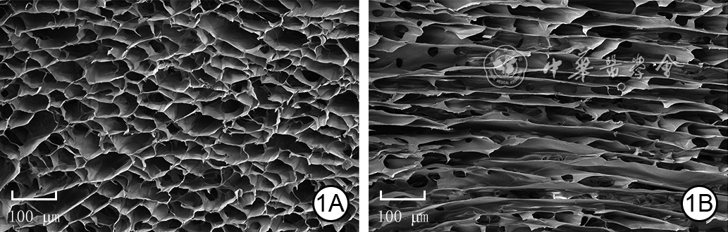
图 1 单纯水凝胶的微观形貌 扫描电子显微镜 ×100。1A、1B.分别为单纯水凝胶的纵切面、横切面,均呈多孔疏松的网状空间结构
注:单纯水凝胶为苯硼酸修饰的甲基丙烯酸酐明胶水凝胶

图 2 含铜氧化铈纳米酶的表征。2A.含铜氧化铈纳米酶呈均匀的球形 扫描电子显微镜 ×600 000;2B.含铜氧化铈纳米酶球形表面略粗糙 透射电子显微镜 ×1 000 000;2C、2D、2E.分别为X射线能谱仪分析的含铜氧化铈纳米酶中铈、铜、氧元素的分布图

图 3 3组烧伤感染性创面小鼠伤后各时间点创面愈合情况。3A、3B、3C.分别为对照组伤后3、7、14 d的创面情况;3D、3E、3F.分别为单纯水凝胶组伤后3、7、14 d的创面情况;3G、3H、3I.分别为复合水凝胶组伤后3、7、14 d的创面情况,图3I中剩余创面面积明显小于图3C、3F
注:单纯水凝胶为苯硼酸修饰的甲基丙烯酸酐明胶水凝胶,复合水凝胶为负载含铜氧化铈纳米酶的苯硼酸修饰的甲基丙烯酸酐明胶水凝胶;对照组小鼠创面仅用无菌透明手术敷贴封闭;单纯水凝胶组和复合水凝胶组小鼠创面先分别滴加单纯水凝胶和复合水凝胶,再用无菌透明手术敷贴封闭;图中绿圈直径为10 mm

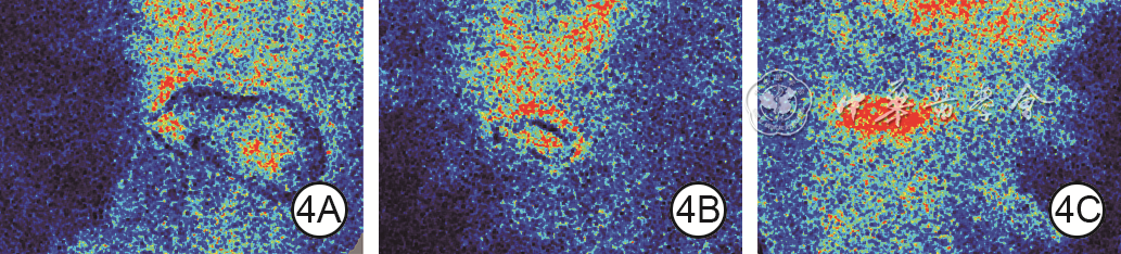
图 4 3组烧伤感染性创面小鼠伤后5 d创面血流灌注情况。4A、4B、4C.分别为对照组、单纯水凝胶组、复合水凝胶组,图4C创面血流灌注量明显多于图4A、4B
注:单纯水凝胶为苯硼酸修饰的甲基丙烯酸酐明胶水凝胶,复合水凝胶为负载含铜氧化铈纳米酶的苯硼酸修饰的甲基丙烯酸酐明胶水凝胶;对照组小鼠创面仅用无菌透明手术敷贴封闭;单纯水凝胶组和复合水凝胶组小鼠创面先分别滴加单纯水凝胶和复合水凝胶,再用无菌透明手术敷贴封闭;黑色表示无血流灌注,蓝色表示血流灌注很少,绿色表示血流灌注一般,红色表示血流灌注充足

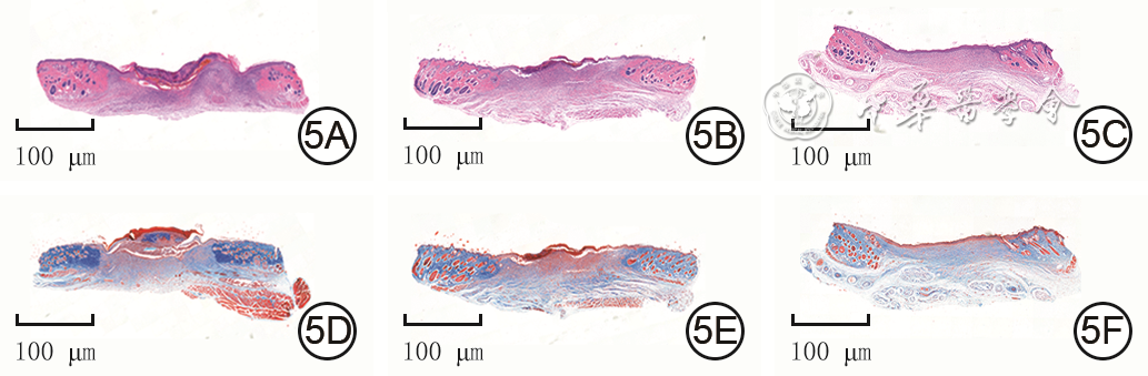
图 5 3组烧伤感染性创面小鼠伤后14 d创面新生肉芽组织、上皮化程度和胶原纤维沉积情况。5A、5B、5C.分别为对照组、单纯水凝胶组和复合水凝胶组新生肉芽组织和上皮化情况,图5C新生肉芽组织和上皮化情况优于图5A、5B 苏木精-伊红 ×5;5D、5E、5F.分别为对照组、单纯水凝胶组和复合水凝胶组胶原纤维沉积情况,图5F较图5D、5E胶原纤维分布更加有序整齐 Masson ×5
注:单纯水凝胶为苯硼酸修饰的甲基丙烯酸酐明胶水凝胶,复合水凝胶为负载含铜氧化铈纳米酶的苯硼酸修饰的甲基丙烯酸酐明胶水凝胶;对照组小鼠创面仅用无菌透明手术敷贴封闭;单纯水凝胶组和复合水凝胶组小鼠创面先分别滴加单纯水凝胶和复合水凝胶,再用无菌透明手术敷贴封闭
Table 1. 3组烧伤感染性创面小鼠伤后各时间点剩余创面面积百分比比较(%,
组别 样本数 3 d 7 d 14 d 对照组 4 63.7±3.0 45.8±1.7 28.8±3.8 单纯水凝胶组 4 57.8±3.9 40.0±1.6 18.5±1.3 复合水凝胶组 4 53.5±1.9 34.3±1.0 11.0±2.2 F值 11.11 31.74 46.29 P值 0.004 <0.001 <0.001 P1值 0.011 0.015 <0.001 P2值 <0.001 <0.001 <0.001 P3值 0.037 0.015 0.002 注:单纯水凝胶为苯硼酸修饰的甲基丙烯酸酐明胶水凝胶,复合水凝胶为负载含铜氧化铈纳米酶的苯硼酸修饰的甲基丙烯酸酐明胶水凝胶;对照组小鼠创面仅用无菌透明手术敷贴封闭;单纯水凝胶组和复合水凝胶组小鼠创面先分别滴加单纯水凝胶和复合水凝胶,再用无菌透明手术敷贴封闭;处理因素主效应,F=70.80,P<0.001;时间因素主效应,F=7 089.13,P<0.001;两者交互作用,F=26.72,P<0.001;F值、P值为3组间各时间点总体比较所得;P1值为单纯水凝胶组与对照组比较所得,P2值为复合水凝胶组与对照组比较所得,P3值为复合水凝胶组与单纯水凝胶组比较所得  下载: 导出CSV
Table 2. 2组小鼠处理14 d后血常规指标和血生化指标比较(
组别 样本数 血常规指标 白细胞计数(×109/L) 淋巴细胞计数(×109/L) 单核细胞计数(×109/L) 中性粒细胞计数(×109/L) 红细胞计数(×109/L) 血红蛋白水平(g/L) 血小板计数(×109/L) PBS组 3 5.6±0.4 4.30±0.15 0.16±0.04 1.46±0.27 8.6±0.6 134±7 957±231 复合水凝胶埋置组 3 5.8±0.8 5.10±0.42 0.15±0.06 1.54±0.33 8.5±0.9 140±9 1 007±215 t值 0.24 3.11 0.24 0.32 0.20 0.90 0.27 P值 0.818 0.069 0.821 0.762 0.847 0.416 0.796 注:PBS为磷酸盐缓冲液,ALT为丙氨酸氨基转移酶,AST为天冬氨酸氨基转移酶;复合水凝胶为负载含铜氧化铈纳米酶的苯硼酸修饰的甲基丙烯酸酐明胶水凝胶;PBS组小鼠于背部皮下注射PBS,复合水凝胶埋置组小鼠于背部皮下埋置复合水凝胶
下载: 导出CSV
-

 下载:
下载:




计量
- 文章访问数: 3365
- HTML全文浏览量: 134
- PDF下载量: 23
- 被引次数: 0
