2022 Vol. 38, No. 12

Expert Forums
Burn injury should be considered as a chronic disease
Luo Gaoxing, Li Haisheng
2022, 38(12): 1101-1104. doi: 10.3760/cma.j.cn501225-20221010-00444
Abstract:
Burn is a special type of trauma, which initiates with skin injury and subsequently causes damages to various systems of the whole body. Burn was regarded as an acute injury in the past, while burn is also considered as a disease now. In recent years, more and more recent evidences have demonstrated that burn injury has a continuous and long-term influence on the life, work, and health status of burn survivors. Therefore, we proposed that burn should be considered as a chronic disease and briefly summarized the long-term effects of burns on mortality, psychology, metabolism, immune system, cardiovascular system, and occurrence of infection and tumor in discharged burn victims. We hope this work can improve the understanding of the long-term effects of burns among medical staff, burn patients and their relatives, and social workers.
A long way for the popularization and improvement of burn rehabilitation
Xie Weiguo
2022, 38(12): 1105-1109. doi: 10.3760/cma.j.cn501225-20221111-00484
Abstract:
In more than half a century, burn rehabilitation in China has experienced three periods: initial exploration in the latter half of the 20th century, gradual development in the first decade of the 21st century, and the accelerating popularization in the recent decade. Looking back at the historical development of burn rehabilitation, we can see not only great progresses, but also many problems and deficiencies. In China, many units still do not even provide burn rehabilitation treatment, and many units where rehabilitation is also in practice, are commonly experiencing problems such as lack of enough therapists, insufficient team construction and training, incomplete treatment programs and limited curative effects, unstable multi-disciplinary cooperation mode, and lack of high-level rehabilitation research and technological innovation. Based on this, this paper puts forward some suggestions for further development and improvement, hoping to push the burn rehabilitation in China from popularization towards a comprehensive high level.
Original Articles·Burn Rehabilitation
Comparison of the effects of self-made three-dimensional printed assistant tableware and traditional universal cuff assistant tableware in patients with upper limb dysfunction after burn injury
Luo Hao, Chen Pei, Xie Weiguo
2022, 38(12): 1110-1116. doi: 10.3760/cma.j.cn501120-20220103-00004
Abstract:
  Objective  To compare the effects of self-made three-dimensional (3D) printed assistant tableware (hereinafter referred to as 3D assistive device) and traditional universal cuff assistant tableware (hereinafter referred to as universal cuff) on the voluntary eating function of patients with upper limb dysfunction after burns.  Methods  The prospective self-control study was conducted. From March 2020 to June 2021, 18 patients with upper limb dysfunction after moderate to severe burns who met the inclusion criteria were admitted to Tongren Hospital of Wuhan University & Wuhan Third Hospital, including 15 males and 3 females, aged 21-58 (42±11) years. After using the 3D software and 3D printer to customize 3D assistive devices for patients, the patients were instructed to eat with the 3D assistive devices and the universal cuff on alternate days for 14 consecutive days, each for 7 days. During this time, the patients could also be fed by someone else without using the assistive device. The number of times the patients ate regular meals with each assistive device and the proportion of them to the total number of regular meals ate during the 7 days to which they belonged were counted. After the total use of two assistive devices for 14 days (hereinafter referred to as after use of 14 days), the amount and time of transferring liquid using the two assistive devices of patients were measured; the patients' feeding items in the modified Barthel index (MBI) were scored respectively when eating without the assistive device, with the 3D assistive device, or with the universal cuff; the satisfaction degree of feeding when eating without the assistive device, with the 3D assistive device, or with the universal cuff was evaluated using the 5-grade Likert scale. Data were statistically analyzed with paired sample t test, Wilcoxon signed rank sum test, and chi-square test.  Results  The number of the patients ate regular meals with the 3D assistive device during the 7 days was (18.1±2.0) times, which was significantly more than (4.0±2.0) times with the universal cuff (t=53.72, P<0.01). The proportion of the number of the patients ate regular meals with the 3D assistive device to the total number of regular meals ate during the 7 days to which it belonged was 72.4% (325/449), which was significantly higher than 16.7% (72/431) with the universal cuff (χ2=257.36, P<0.01). After use of 14 days, the patients' time and amount of transferring liquid using the 3D assistive device were respectively significantly shorter and more than using the universal cuff (with t values of 2.49 and 7.52, respectively, P<0.05 or P<0.01). The patients' MBI feeding scores when eating with the 3D assistive device and with the universal cuff were close (P>0.05), which were both significantly higher than the score when eating without the assistive device (with Z values of 3.90 and 3.86, respectively, P<0.01).The patients' satisfaction scores of feeding when eating with the 3D assistive device and with the universal cuff were close (P>0.05), which were both significantly higher than the score when eating without the assistive device (with Z values of 3.61 and 3.00, respectively, P<0.01).  Conclusions  Both the 3D assistive device and the universal cuff can compensate the limb function of patients with upper limb dysfunction after burns and improve their self-feeding ability, but the 3D assistive device has more advantages in improving patients' willingness to eat and the efficiency of food transfer.
A prospective randomized controlled study on the effects of progressive core muscle group training combined with lower limb intelligent rehabilitation training for burn patients with lower limb dysfunction
Zhao Haiyang, Liu Jiaqi, Han Juntao, Zhu Chan, Zhou Qin, Xu Jing, Liang Min, Zhang Bowen, Qi Zongshi
2022, 38(12): 1117-1125. doi: 10.3760/cma.j.cn501225-20220616-00236
Abstract:
  Objective  To observe the effect of progressive core muscle group training combined with lower limb intelligent rehabilitation training on burn patients with lower limb dysfunction.  Methods  A prospective randomized controlled study was conducted. From March 2017 to May 2020, 60 patients with motor and balance dysfunction after deep partial-thickness burns or full-thickness burns of both lower extremities who met the inclusion criteria were admitted to the First Affiliated Hospital of Air Force Military Medical University. They were divided into simple intelligent rehabilitation group (30 cases, 20 males and 10 females, aged (40±3) years) and combined rehabilitation group (30 cases, 16 males and 14 females, aged (39±3) years) according to the random number table method. The patients in both groups started red light treatment after the wound healing or when the scattered residual wound area was less than 5% total body surface area. After 2 weeks of red light treatment, patients in the combined rehabilitation group started progressive core muscle group training on the basis of lower limb intelligent rehabilitation training in simple intelligent rehabilitation group, and the training time was 6 weeks. Before and after 6 weeks of training, the lower limb motor function was evaluated with the simple Fugl-Meyer scale, the balance capacity was evaluated with the Berg balance scale, and the walking capacity was evaluated with the Holden walking ability rating scale. After 6 weeks of training, a self-designed questionnaire was used to investigate patients' satisfaction for the treatment effect. The patients were followed up for 6 months after the treatment to observe the balance stability of standing on one foot in the flexion position and their participation in activities of daily life. Data were statistically analyzed with independent sample t test, paired sample t test, and chi-square test.  Results  Before training, the lower limb motor function score of patients in simple intelligent rehabilitation group was 24.9±2.7, which was close to 23.9±2.3 in combined rehabilitation group (P>0.05). After 6 weeks of training, the lower limb motor function score of patients in combined rehabilitation group was 29.6±3.9, which was significantly higher than 27.3±3.8 in simple rehabilitation group (t=-2.28, P<0.05). The lower limb motor function scores of patients in combined rehabilitation group and simple intelligent rehabilitation group after 6 weeks of training were significantly higher than those before training (with t values of -6.50 and -3.21, respectively, P<0.01). After 6 weeks of training, the balance capacity score of patients in combined rehabilitation group was 41±7, which was significantly higher than 36±5 in simple intelligent rehabilitation group (t=-2.68, P<0.05); the balance capacity scores of patients in combined rehabilitation group and simple intelligent rehabilitation group after 6 weeks of training were significantly higher than those before training (with t values of -8.72 and -8.09, respectively, P<0.01). After 6 weeks of training, the walking capacity grading of patients in combined rehabilitation group was significantly improved compared with that in simple intelligent rehabilitation group (χ2=-2.14, P<0.05), and the walking capacity grading of patients in simple intelligent rehabilitation group and combined rehabilitation group after 6 weeks of training was significantly improved compared with that before treatment (with χ2 values of -4.94 and -5.26, respectively, P<0.01). After 6 weeks of training, the satisfaction score for the treatment effect of patients in combined rehabilitation group was 13.7±1.2, which was significantly higher than 7.8±1.4 in simple intelligent rehabilitation group (t=22.84, P<0.01). The patients in both groups could stand on one foot to maintain balance in the flexion position of lower limb, and their activities of daily life were not affected 6 months after treatment.  Conclusions  On the basis of conventional rehabilitation therapy, the combination of progressive core muscle group training and lower limb intelligent rehabilitation training can significantly promote the recovery of lower limb motor and balance function of burn patients.
Effects and mechanism of pressure treatment on hemodynamic changes in patients with hypertrophic scar secondary to extensive burns
Song Chunhong, Li Jingbo, Lan Wei, Chen Shangqing, Liu Yanzhi, Ji Xueliang, Yi Xianfeng, Lin Yueqing, Sun Tianbao
2022, 38(12): 1126-1132. doi: 10.3760/cma.j.cn501225-20220616-00235
Abstract:
  Objective  To investigate the hemodynamic changes of the main arteries and veins of the extremities and the heart in patients with hypertrophic scar secondary to extensive burns after pressure treatment, and to analyze the relevant mechanisms.  Methods  A retrospective before-after self-control study was conducted. From January 2017 to February 2022, 37 patients with hypertrophic scar secondary to extensive burns who met the inclusion criteria were hospitalized in the Burn Rehabilitation Department of Guangdong Industrial Injury Rehabilitation Hospital, including 25 males and 12 females, aged 23-52 years. The patients were admitted to the hospital within 12 weeks after wound healing, and within one week after admission, rehabilitation therapists, occupational therapists, and tailors custom-made pressure products such as full-body pressure garment, pressure pants, vests, split finger gloves, split finger socks, hoods, and plastic collars, with the pressure at each part maintained at 2.67-4.00 kPa when wearing. Before the first treatment with pressure products (hereinafter referred to as before pressure treatment) and at 1 h of the first treatment with pressure products (hereinafter referred to as 1 h of pressure treatment), color Doppler ultrasonography was performed to check the pulse rate of the axillary artery, the lumen diameter, peak systolic velocity (PSV), and resistance index of the axillary artery and femoral artery on the left side, the lumen diameter, cross-sectional area, and average blood flow velocity of the axillary vein and femoral vein, and the mitral valve E peak, mitral valve A peak, tricuspid valve E peak, aortic valve PSV, and pulmonary valve PSV of the heart; an optical chromatographic skin detector was used to detect the red color, red pigment, and surface brightness of the scar on the back of the hand to reflect the filling and distribution of the scar microvessels. Data were statistically analyzed with paired sample t test.  Results  Compared with those before pressure treatment, the PSV of the axillary artery of patients was significantly slowed down at 1 h of pressure treatment (t=55.42, P<0.01); the average blood flow velocity of the axillary vein was significantly accelerated (t=-60.50, P<0.01); the pulse rate, lumen diameter, and resistance index of the axillary artery, as well as the lumen diameter and cross-sectional area of the axillary vein did not change obviously (P>0.05); the average blood flow velocity of the femoral vein was significantly accelerated (t=-80.52, P<0.01); the lumen diameter, PSV, and resistance index of the femoral artery, as well as the lumen diameter and cross-sectional area of the femoral vein had no significant change (P>0.05); the mitral valve E peak and mitral valve A peak of the heart decreased significantly (with t values of 10.71 and 21.96, respectively, P<0.01); the tricuspid valve E peak of the heart increased significantly (t=7.57, P<0.01); the PSV of the aortic valve and pulmonary valve of the heart did not change obviously (P>0.05). At 1 h of pressure treatment, the red color and red pigment values of the scar on the back of the hand of patients were 15.3±1.1 and 16.8±1.2, respectively, which were significantly lower than 24.5±1.3 and 23.8±1.2 before pressure treatment (with t values of 8.32 and 8.04, respectively, P<0.01). The brightness value of the scar surface on the back of the hand of patients at 1 h of pressure treatment was similar to that before pressure treatment (P>0.05).  Conclusions  After pressure treatment for the hypertrophic scar in patients secondary to extensive burn, the average blood flow velocity of the axillary vein and femoral vein in patients are obviously accelerated, the PSV of the axillary artery is significantly slowed down, the peak values of mitral valve E and mitral valve A of the heart are significantly decreased, and the tricuspid valve E peak is significantly increased. These hemodynamic changes may be related to the reduction of microvascular blood flow in the local area of scar after systemic pressure treatment.
Original Articles
Anatomical characteristics and clinical application of anterolateral thigh perforator flap pedicled with oblique branch trunk of lateral circumflex femoral artery in the muscular septum
Yang Lin, Liu Zhijin, Cheng Junnan, Gao Qinfeng, Yang Chengpeng, Liu Shengzhe, Zhang Tao, Sun Fengwen, Huang Yongtao, Ju Jihui
2022, 38(12): 1133-1139. doi: 10.3760/cma.j.cn501225-20220227-00039
Abstract:
  Objective  To explore the anatomical characteristics of the anterolateral thigh perforator flap pedicled with oblique branch trunk of lateral circumflex femoral artery in the muscular septum and the clinical effect of this flap in repairing skin and soft tissue defects in the extremities.  Methods  A retrospective observational study was conducted. From December 2020 to April 2021, 59 patients with skin and soft tissue defects in the extremities admitted to the Department of Hand Surgery of Suzhou Ruihua Orthopaedic Hospital met the inclusion criteria, including 46 males and 13 females, aged 20 to 81 years. The wound area after debridement ranged from 8 cm×5 cm to 38 cm×20 cm. According to the condition of the wound, the perforators in the anterolateral femoral region on the unaffected side were located by color Doppler ultrasound. The anterolateral thigh perforator flap with the oblique branch of lateral circumflex femoral artery as the source artery was designed, and the wound was repaired by unilateral flap or series combination of bilateral flaps (with the area of unilateral flap ranging from 7 cm×5 cm to 37 cm×11 cm). The wound of the donor site was sutured directly. The following items were recorded, including the number of perforators in the anterolateral femoral region marked before operation, the course characteristics of oblique branch trunk of lateral circumflex femoral artery and its perforators, and the number, origin, and type of perforators observed during operation, the flap repair mode and the flap harvest time. After operation, the survival condition of the flap, the wound healing time in the recipient site, and the suture healing in the donor area were observed, and the recovery of the donor and recipient areas was followed up. At the last follow-up, the sensation function evaluated by sensory rating scale of British Medical Research Association and two-point discrimination of the area transplanted with flap were recorded, and the improved comprehensive curative effect evaluation scale of flap was used to evaluate the repair effect of the flap.  Results  A total of 156 perforators were marked in the anterolateral femoral region before operation, and 144 perforators were observed during the operation, of which 98 came from the oblique branch of the lateral circumflex femoral artery, and the first perforator of the oblique branch was the intermuscular septal perforator. Once formed, most oblique branch trunk of lateral circumflex femoral artery in the muscular septum could be divided into the deep branch and the superficial branch at the middle and upper 1/3 junction of the line between the anterior superior iliac spine and the lateral edge of the patella, the deep branch mainly ran in the muscle of vastus intermedius and vastus lateralis, and rarely developed the skin perforators, while the superficial branch mainly ran in the muscular septum between the rectus femoris and the vastus lateralis, and grew out the perforators to the proximal skin of the anterolateral femoral region. Fifty-six patients were repaired with unilateral flap and 3 patients with bilateral flap in series combination, with the harvesting time of the unilateral flap ranged from 9 to 99 min. Three patients developed arterial crisis within 48 hours after operation and survived after timely exploration; the flap of 1 patient developed necrosis 11 days after operation and was repaired by abdominal split-thickness skin graft; the other flaps survived smoothly. The wound healing time in the recipient area was 10 to 42 days after operation, and the sutures in the donor area healed well. During the follow-up of 6 to 10 months, 8 patients underwent thinning and plastic surgery 6 months after operation because of bloating in the flap transplantation area, while the other patients had a good shape of the flap transplantation area, with no deep tissue infection such as osteomyelitis, with soft texture, good elasticity, no pain, and good blood circulation; all the donor areas were left with linear scars, normal blood circulation in the distal extremities, no limitation of knee joint motion or quadriceps muscle strength. At the last follow-up, the sensation function was restored in the flap transplantation area of the affected limb, including 14 cases of S1 grade and 45 cases of S2 grade; there was only one point of two-point discrimination; the curative effect evaluation of flap repair included excellent in 24 cases, good in 35 cases, and general in 3 cases.  Conclusions  The anterolateral thigh perforator flap pedicled with oblique branch trunk of lateral circumflex femoral artery in the muscular septum has the advantages of relatively concealed donor area, large cutting width, and simple operation during surgery. It protects the donor area to the greatest extent while repairing skin and soft tissue defects in the extremities, and is a useful supplement to the classic anterolateral thigh perforator flap.
Plasmids carried by carbapenems-resistant Klebsiella pneumoniae in burn patients and its correlation with strain transmission
Huang Siyi, Gong Yali, Zhou Dapeng, Jiang Lingli, Xiang Lijuan, Chen Zehui, Wang Dali, Huang Guangtao
2022, 38(12): 1140-1147. doi: 10.3760/cma.j.cn501120-20210930-00343
Abstract:
  Objective  To explore the carrier status of carbapenems-resistant Klebsiella pneumoniae (CRKP) plasmids in burn patients and analyze the correlation of these plasmids with the transmission of CRKP.  Methods  A retrospective observational study was conducted. A total of 26 CRKP strains, which were isolated from the clinic-related samples of 22 burn patients (with 20 males and 2 females, aged (42±16) years) admitted to the First Affiliated Hospital of Army Medical University (the Third Military Medical University) from January to December 2017, were collected and individually numbered. The plasmids of the strains were extracted by alkali lysis. After determination of the plasmid concentration by a nucleic acid concentration detector, the agarose gel electrophoresis was used to visualize the bands, and rough plasmids typing was performed. The plasmid of the smallest numbered CRKP in each plasmid type was transformed into competent Escherichia coli (E. coli) strain Top10 (hereinafter referred to as TOP10 strain). The growth of each transformed strains and a Top10 strain cultivated in ampicillin containing Luria-Bertani (LB) agar medium overnight was observed, and the proportion of successful transformation was calculated. The plasmids from the smallest numbered plasmid carrying CRKP strain of successfully transformed Top10 strains (hereinafter referred to as the smallest successfully transformed strain) and correspondingly numbered CRKP were extracted, and then, the agarose gel electrophoresis was used to visualize the bands. Aforementioned successfully transformed strains and a TOP10 strain were used for the antimicrobial susceptibility testing with 17 antibiotics commonly used in clinic. The plasmid from the smallest successfully transformed strain was sequenced using the next-generation sequencing technology. Bioinformatics analyses such as protein-coding gene prediction and protein sequence alignment were performed successively. The sequence was subsequently named pKP03-NDM1 according to the carrying of drug resistance gene. According to the whole genome sequence of the plasmid carried by the smallest successfully transformed strain, the polymerase chain reaction, agarose gel electrophoresis, and gene sequencing were used to detect the New Delhi metallo-beta lactamase-1 (blaNDM-1) of plasmids in the remaining 25 strains of CRKP. The ST typing in multilocus sequence typing of 26 strains of CRKP was analyzed based on the literature.  Results  Plasmids were successfully extracted from 26 CRKP, with mass concentrations ranging from 19.3 to 189.8 ng/μL. Each of the 26 CRKP carrying plasmids showed at least one band longer than 2 500 bp in the agarose gel electrophoresis, which were roughly divided into 6 patterns of A, B, C, D, E, and F. After overnight cultivation, no growth of strains was observed in LB agar medium containing ampicillin inoculated with the TOP10 strain or TOP10 strains transformed by the plasmid of CRKP patterning A, B, D, or E. In contrast, TOP10 strains transformed by the pattern C plasmid from NO.3 CRKP and the pattern F plasmid from NO.15 CRKP resulted in numerous colony growths, and those transformed strains were named as TOP10-pKP03 and TOP10-pKP15, respectively. The proportion of successful transformation was 1/3. The plasmid carried by TOP10-pKP03 showed a single band in the agarose gel electrophoresis, which was the same size as the largest band of the plasmid from NO.3 CRKP. The TOP10 strain was sensitive to the 17 antibiotics commonly used in clinic. TOP10-pKP03 and TOP10-pKP15 were resistant to penicillins, cephalosporins, and carbapenems but remained sensitive to monocyclic β-lactam, aminoglycosides, quinolones and tigecycline. The full length of the plasmid carried by TOP10-pKP03 was 41 190 bp. In addition to blaNDM-1, this plasmid carried bleMBL, T4SS, bleomycin resistance gene, conjugation transfer elements, and relaxase, etc. The plasmid showed 99% nucleotide identity similarity and the same length to the plasmid pJN24NDM1 extracted from an E. coli isolate JN24. Totally 16 (61.5%) CRKP were confirmed to carrying blaNDM-1 gene, among the ST typing of the 16 strains, 11 strains were ST11, while ST215, ST260, ST395, ST2230, and new ST had 1 strain each. Among the ST typing of 10 blaNDM-1-negative CRKP, 8 strains were ST11, while ST395 and ST2230 had 1 strain each.  Conclusions  A blaNDM-1 gene carrying plasmid pKP03-NDM1 was extracted and sequenced from CRKP isolated from burn patients, with a high plasmid carrying rate. Meanwhile, this plasmid may mediate inter-CRKP and CRKP-E. coli horizontal transfer of blaNDM-1, leading to transmission of antimicrobial resistance.
Clinical effects of free descending branch tissue flap of lateral circumflex femoral artery in repairing Wagner grade 3 or 4 diabetic foot wounds
Yang Li, Li Kelin, Pang Yuanxiang, Zhou Wenchang
2022, 38(12): 1148-1155. doi: 10.3760/cma.j.cn501225-20220321-00076
Abstract:
  Objective  To investigate the clinical effects of free descending branch tissue flap of lateral circumflex femoral artery in repairing Wagner grade 3 or 4 diabetic foot wounds.  Methods  A retrospective observational study was conducted. From October 2019 to January 2022, 12 patients (10 males and 2 females, aged 38-66 years, with an average of 52 years) with diabetic foot wounds who met the inclusion criteria were admitted to the First People's Hospital of Yulin, of whom 5 cases being Wagner grade 3 and 7 cases being Wagner grade 4. Wound debridement and vacuum sealing drainage were performed to control infection, and the wound area after debridement was between 13 cm×6 cm and 28 cm×11 cm. The wounds were repaired with free descending branch tissue flaps of lateral circumflex femoral artery. Among them, the wounds of 4 cases were repaired with single flap of musculocutaneous branch or intermuscular branch of descending branch tissue flap of lateral circumflex femoral artery, 1 case with lobulated flap, 1 case with bilateral tandem flap, and 6 cases with chimeric lateral femoral muscle flap. The area of resected tissue flap was 9 cm×7 cm to 21 cm×10 cm; end-to-end anastomosis was selected between vessels in the donor sites and the recipient sites, and pressurized treatment should be performed if necessary. The donor site wounds of 10 patients were treated with cosmetic tension-relieving suture, and the donor site wounds of 2 patients were repaired with split-thickness skin graft from head. After operation, the condition of wound repair was recorded. After wound healing, the level of 2-hour postprandial blood glucose of patients was measured. During follow-up, the wound healing of the recipient site and scar formation of the donor site were observed. Before and 6 months after operation, computer tomography angiography was used to detect and compare the blood perfusion of the affected limb. At the last follow-up, the walking ability of the patients was recorded.  Results  The wounds of 9 patients healed well after operation; the flaps in the recipient site of 2 patients were infected on postoperative day 5 after surgery and were repaired by suturing in stage Ⅱ after open drainage and inflammation control; the distal end of the grafted tandem flap in 1 patient gradually developed purple necrosis on postoperative day 4 and was repaired with a skin graft after debridement. After wound healing, the 2-hour postprandial blood glucose value of all patients was controlled in the range of 8-12 mmol/L. During Follow-up of 6 months to 1 year, 3 patients had partial ulceration in the plantar compression area, which healed after decompression combined with dressing change or flap repair, while the other 9 patients had no ulceration in the recipient area; the appearance of the affected foot was plump after wound healing, the transplanted flaps had good blood supply and good fit with the surrounding tissue, and were wear-resistant, but insensitive. During follow-up, only linear scar remained in the donor area of direct suture in 10 cases, and the skin grafts in the donor area were completely survived in 2 cases. Compared with the patency of the main blood vessels of the ankle joint segment of the affected limb before operation, the vascular network of the flap transplantation area in the affected foot was formed 6 months after operation. At the last follow-up, 8 patients could walk independently, and 4 patients could walk with crutches.  Conclusions  The free descending branch tissue flap of lateral circumflex femoral artery is effective in repairing Wagner grade 3 or 4 diabetic foot wounds, which can shorten the course of disease and improve local blood flow.
Influence factors of poor efficacy after flap repair operation in patients with pressure ulcers
Pan Nanfang, Liang Zunhong, Lin Ping, Wang Xin, Lin Shishuai, Huang Yangjun, Chen Xianxian, Pan Yunchuan
2022, 38(12): 1156-1161. doi: 10.3760/cma.j.cn501225-20220228-00041
Abstract:
  Objective  To investigate the influence factors of poor efficacy after flap repair operation in patients with pressure ulcers.  Methods  The retrospective case series study was conducted. From January 2011 to June 2021, 125 patients with stage Ⅲ and Ⅳ pressure ulcers treated in Hainan General Hospital met the inclusion criteria. There were 82 males and 43 females, aged 15-90 (57±20) years. According to the postoperative effects, the patients were divided into poor efficacy group (47 cases) and good efficacy group (78 cases). The clinical data of patients in the two groups were collected, including the age, gender, location, stage, size, and bone exposure of pressure ulcers, preoperative microorganism culture results of wound exudate sample, whether combined with osteomyelitis, diabetes, lower limb paroxysmal myospasm, and gatism or not, the number of surgical debridement combined with negative-pressure wound therapy, type of surgical flap, postoperative position, and preoperative albumin, leukocyte, C-reactive protein (CRP), and hemoglobin. Data were statistically analyzed with independent sample t test, Mann-Whitney U test, and chi-square test. The binary multivariate logistic regression analysis was conducted to screen the independent risk factors influencing the poor efficacy after flap repair operation in 125 patients with stage Ⅲ and Ⅳ pressure ulcers.  Results  The ratio of patients with lower limb paroxysmal myospasm in poor efficacy group was 22/47, which was significantly higher than 3/78 in good efficacy group (χ2=33.83, P<0.01). The preoperative hemoglobin level of patients in poor efficacy group was (102±17) g/L, which was significantly lower than (113±20) g/L in good efficacy group (t=-3.24, P<0.01). The preoperative CRP level of patients was 39.1 (14.1, 91.6) mg/L in poor efficacy group, which was significantly higher than 15.3 (6.6, 42.0) mg/L in good efficacy group (Z=-3.04, P<0.01). There were no statistically significant differences in other indexes between patients in the two groups (P>0.05). Multivariate logistic regression analysis showed that age, lower limb paroxysmal myospasm, and preoperative hemoglobin level were the independent risk factors for poor efficacy after flap repair operation in patients with pressure ulcers (with odds ratios of 1.03, 40.69, and 0.97, 95% confidence intervals of 1.00-1.06, 9.18-180.39, and 0.95-1.00, respectively, P<0.05 or P<0.01).  Conclusions  Poor efficacy after flap repair operation in patients with pressure ulcers is affected by many factors, among which the age, lower limb paroxysmal myospasm, and preoperative hemoglobin level are the independent risk factors.
Effects of mechanical tension on the formation of hypertrophic scars in rabbit ears and transforming growth factor-β1/Smad signaling pathway
Cao Peng, Wang Yunwei, Guan Hao, Yang Yunshu, Li Shaohui, Chen Yang, Zhu Chan, Wan Yu, Ren Liying, Yao Ming
2022, 38(12): 1162-1169. doi: 10.3760/cma.j.cn501120-20211213-00412
Abstract:
  Objective  To explore the effects of mechanical tension on the formation of hypertrophic scars in rabbit ears and transforming growth factor-β1 (TGF-β1)/Smad signaling pathway.  Methods  The experimental research method was adopted. Six New Zealand white rabbits, male or female, aged 3-5 months were used and 5 full-thickness skin defect wounds were made on the ventral surface of each rabbit ear. The appearance of all rabbit ear wounds was observed on post surgery day (PSD) 0 (immediately), 7, 14, 21, and 28. On PSD 28, the scar formation rate was calculated. Three mature scars in the left ear of each rabbit were included in tension group and the arch was continuously expanded with a spiral expander. Three mature scars in the right ear of each rabbit were included in sham tension group and only the spiral expander was sutured without expansion. There were 18 scars in each group. After mechanical tension treatment (hereinafter referred to as treatment) for 40 days, the color and texture of scar tissue in the two groups were observed. On treatment day 40, the scar elevation index (SEI) was observed and calculated; the histology was observed after hematoxylin eosin staining, and the collagen morphology was observed after Masson staining; mRNA expressions of TGF-β1, Smad3, collagen Ⅰ, collagen Ⅲ, and α-smooth muscle actin (α-SMA) in scar tissue were detected by real-time fluorescence quantitative reverse transcription polymerase chain reaction; and the protein expressions of TGF-β1, collagen Ⅰ, collagen Ⅲ, and α-SMA, and phosphorylation level of Smad3 in scar tissue were detected by Western blotting. The number of samples of each group in the experiments was 3. Data were statistically analyzed with independent sample t test.  Results  On PSD 0, 5 fresh wounds were formed on all the rabbit ears; on PSD 7, the wounds were scabbed; on PSD 14, most of the wounds were epithelialized; on PSD 21, all the wounds were epithelialized; on PSD 28, obvious hypertrophic scars were formed. The scar formation rate was 75% (45/60) on PSD 28. On treatment day 40, the scar tissue of rabbit ears in tension group was more prominent than that in sham tension group, the scar tissue was harder and the color was more ruddy; the SEI of the scar tissue of rabbit ears in tension group (2.02±0.08) was significantly higher than 1.70±0.08 in sham tension group (t=5.07, P<0.01). On treatment day 40, compared with those in sham tension group, the stratum corneum of scar tissue became thicker, and a large number of new capillaries, inflammatory cells, and fibroblasts were observed in the dermis, and collagen was more disordered, with nodular or swirling distribution in the scar tissue of rabbit ears in tension group. On treatment day 40, the mRNA expressions of TGF-β1, Smad3, collagen Ⅰ, collagen Ⅲ, and α-SMA in the scar tissue of rabbit ears in tension group were respectively 1.81±0.25, 5.71±0.82, 7.86±0.56, 4.35±0.28, and 5.89±0.47, which were significantly higher than 1.00±0.08, 1.00±0.12, 1.00±0.13, 1.00±0.14, and 1.00±0.14 in sham tension group (with t values of 5.36, 9.82, 20.60, 18.26, and 17.13, respectively, all P<0.01); the protein expressions of TGF-β1, collagen Ⅰ, collagen Ⅲ, and α-SMA, and phosphorylation level of Smad3 in the scar tissue of rabbit ears in tension group were respectively 0.865±0.050, 0.895±0.042, 0.972±0.027, 1.012±0.057, and 0.968±0.087, which were significantly higher than 0.657±0.050, 0.271±0.029, 0.631±0.027, 0.418±0.023, and 0.511±0.035 in sham tension group (with t values of 5.08, 21.27, 15.55, 16.70, and 8.40, respectively, all P<0.01).  Conclusions  Mechanical tension can inhibit the regression of hypertrophic scars in rabbit ears through stimulating the hyperplasia of scars, inhibiting the normal arrangement of dermal collagen fibers, and intensifying the deposition of collagen fibers, and the mechanism may be related to the activation of TGF-β1/Smad signaling pathway by mechanical tension.
Original Article·Nursing Column
Epidemiological characteristics and the establishment and evaluation of a risk prediction model for nosocomial infection in burn patients
Wang Hua, Zhao Peng, Sun Dan, Wu Xing, Yuan Qinfang, Wang Kewei
2022, 38(12): 1170-1178. doi: 10.3760/cma.j.cn501225-20220214-00025
Abstract:
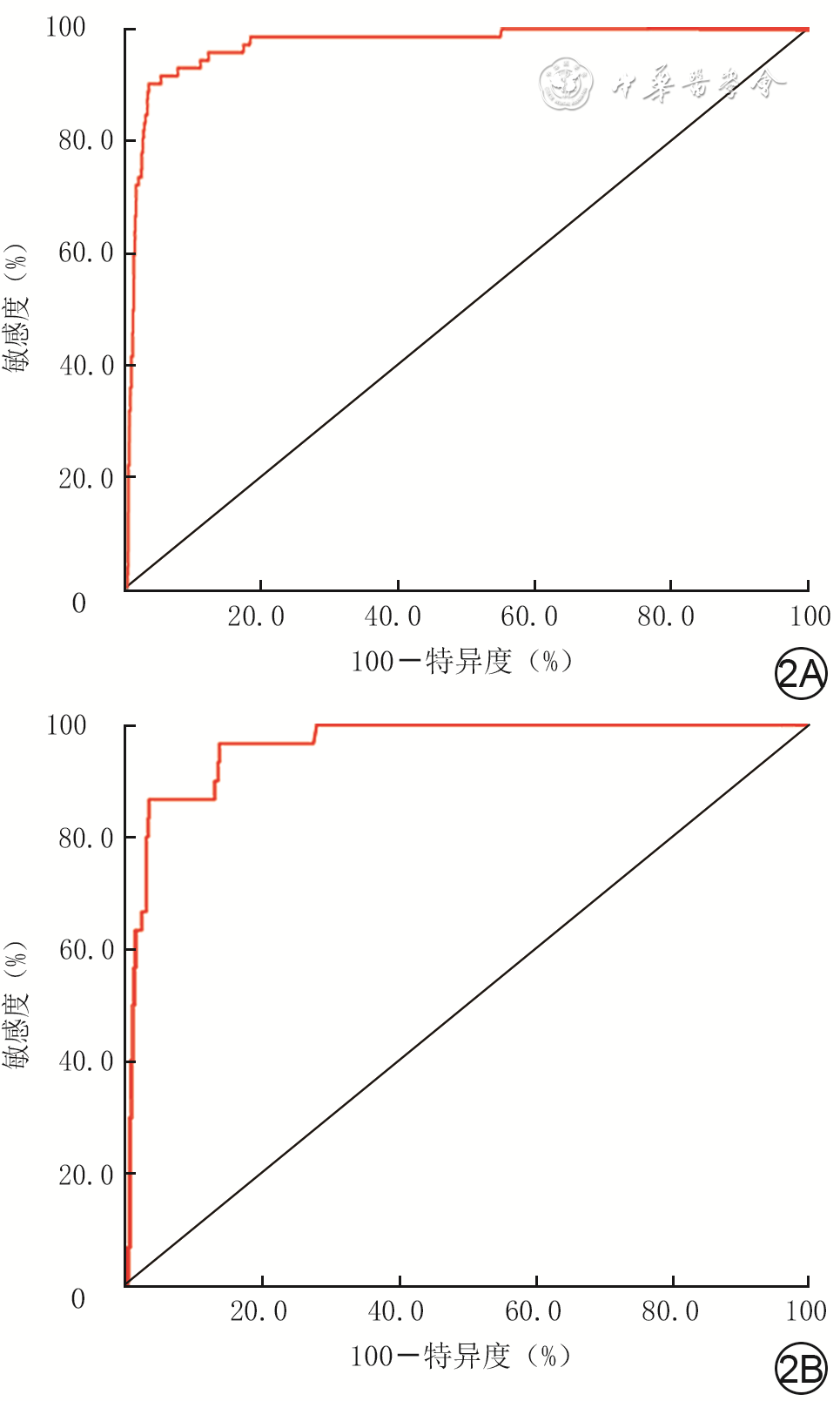
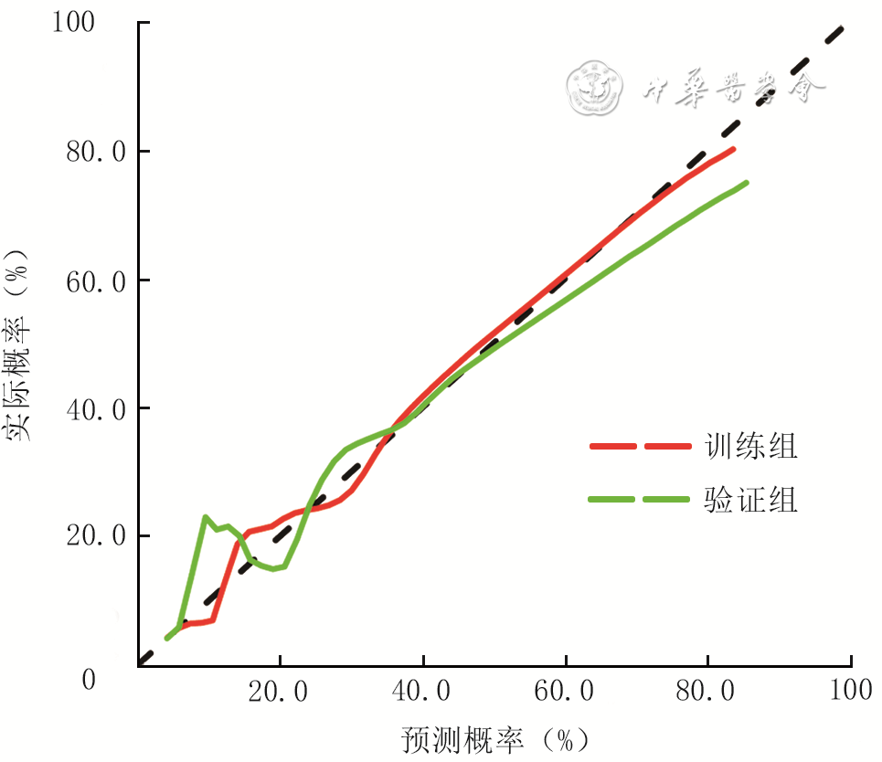
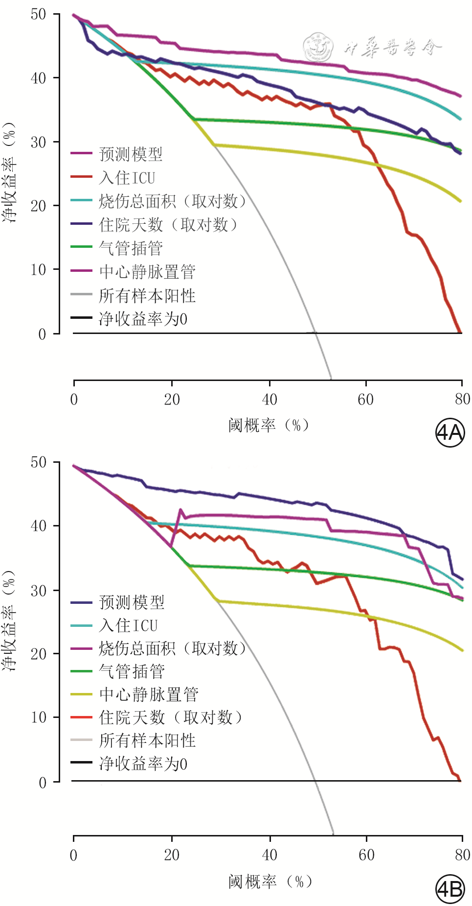
  Objective  To find the epidemiological characteristics of nosocomial infection in burn patients, to establish a risk prediction model for nosocomial infection in burn patients based on the screened independent risk factors of the infection, and to analyze its predictive value.  Methods  A retrospective case series study was conducted. From May 2016 to December 2019, 3 475 burn patients who were admitted to the Department of Burns of Affiliated Hospital of Jiangnan University met the inclusion criteria, including 2 290 males and 1 185 females, aged from 1 to 94 years. The incidence of nosocomial infection, the detection site and specific composition of pathogenic bacteria were counted. The patients were randomly divided into training group (2 434 cases) and verification group (1 041 cases) in R 4.1.3 statistic software with a ratio of about 7∶3. Factors including gender, age, total burn area, combination of full-thickness burn/inhalation injury/shock/diabetes on admission, admission to intensive care unit (ICU), status of central venous catheterization/endotracheal intubation/urethral catheter indwelling/surgery, nosocomial infection status, days of antibiotic use, and days of hospital stay of patients were compared between the two groups. According to the occurrence of nosocomial infection, the patients were divided into nosocomial infection group (102 cases) and non-nosocomial infection group (3 373 cases), and in addition to the aforementioned data, non-nosocomial infection related data, the season of admission and types of antibiotics used were compared between the two groups. The above-mentioned data were statistically analyzed with one-way analysis of independent sample t test, chi-square test, and Mann-Whitney U test, and the indicators with statistically significant differences between nosocomial infection group and non-nosocomial infection group were included as variables in multivariate logistic regression analysis to screen independent risk factors for the development of nosocomial infection in 3 475 burn patients. On the basis of independent risk factors and important clinical characteristics, a nomogram prediction model was constructed for the risk of developing nosocomial infection of burn patients in training group. In both training group and verification group, receiver operating characteristic (ROC) curves for prediction of nosocomial infection by the prediction model were plotted, and the area under the ROC curve was calculated; calibration curves were plotted to evaluate the conformity between the predicted results of the prediction model and the actual situation; clinical decision curves were plotted to evaluate the clinical utility of the prediction model.  Results  The incidence of nosocomial infection of patients included in this study was 2.94% (102/3 475); pathogens were detected from 212 specimens, mainly wound (78 cases, accounting for 36.79%) and blood (64 cases, accounting for 30.19%) specimens; 250 strains of pathogenic bacteria were detected, mainly gram-negative bacteria (153 strains, accounting for 61.20%). All clinical characteristics of patients between training group and verification group were similar (P>0.05). There were statistically significant differences between patients in nosocomial infection group and non-nosocomial infection group in the aspects of age, total burn area, days of antibiotic use, antibiotic use type, days of hospital stay, combination of full-thickness burn, combination of inhalation injury, combination of shock, ICU admission status, central venous catheterization status, endotracheal intubation status, urethral catheter indwelling status, surgery status (with Z values of 4.41, 14.95, 15.70, 650.32, and 13.73, χ2 values of 151.09, 508.30, 771.20, 955.79, 522.67, 967.40, 732.11, and 225.35, respectively, P<0.01). ICU admission, endotracheal intubation, urethral catheter indwelling, and days of hospital stay were independent risk factors for developing nosocomial infection by 3 475 burn patients (with odds ratios of 5.99, 3.39, 9.32, and 6.21, 95% confidence intervals of 2.25-15.99, 1.56-7.39, 2.77-31.31, and 2.48-15.92, respectively, P<0.01). In training group and verification group, the area under ROC curves of the nosocomial infection prediction model based on independent risk factors, total burn area, and central vein catheterization were both 0.97 (with both 95% confidence intervals being 0.95-0.99); the calibration curve analysis showed that the prediction results of the prediction model were in good agreement with the actual situation; the clinical decision curve analysis showed that the prediction model had good clinical utility.  Conclusions  The nosocomial infection in burn patients is mainly caused by gram-negative bacteria, with wound as the main infection site, and the independent risk factors including ICU admission, endotracheal intubation, urethral catheter indwelling, and days of hospital stay. Based on independent risk factors and important clinical features, the risk prediction model for nosocomial infection has a good ability to predict nosocomial infection in burn patients.
Reviews
Research advances on pharmacological interventions for hypertrophic scar
Shi Liangliang, Liu Mingzhuo, Jiang Zhengying, Yu Xiaoting, Li Jiaqi, Guo Guanghua
2022, 38(12): 1179-1184. doi: 10.3760/cma.j.cn501120-20211118-00388
Abstract:
Hypertrophic scar is an abnormal fibrous proliferative disease that occurs after deep cutaneous injury, which not only affects aesthetics and function but also has negative psychological effects on the patients. However, the mechanism of hypertrophic scar formation has not been fully elucidated, and its clinical treatment is complex with a high rate of recurrence and no radicle cure. Intervention based on molecular targets will likely be the future direction for the prevention and treatment of hypertrophic scar. In this article, we review the specific roles and mechanisms of drug-targeted interventions in hypertrophic scar formation, including general drugs, cytokines, immunomodulators, herbal extracts, exosomes, and nanomaterials, in the context of advances in both basic and clinical research at home and abroad.
Research advances on the treatment of diabetic foot ulcers with autologous platelet-rich fibrin
Yu Xiaoyuan, Zhao Mingyu, Zhang Ying, Xu Gang
2022, 38(12): 1185-1189. doi: 10.3760/cma.j.cn501225-20220110-00001
Abstract:
Diabetic foot is one of the serious complications of diabetic patients, which makes the society and public health bear a huge economic burden. In recent years, more and more studies at home and abroad have been conducted on the treatment of chronic wounds with autologous platelet-rich fibrin, and the therapeutic concepts and methods have been updated constantly. In this paper, we reviewed the general situation of autologous platelet-rich fibrin, the mechanism of autologous platelet-rich fibrin in promoting the healing of diabetic foot ulcers and the new progress in its application, so as to provide a new strategy for the repair of diabetic foot ulcers.
Progress on the mechanism and application of adipose-derived stem cells in promoting wound repair
Sun Tianyi, Lu Feng, Gao Jianhua, Zhou Cheng, Dong Ziqing, Li Bin
2022, 38(12): 1190-1195. doi: 10.3760/cma.j.cn501225-20220729-00320
Abstract:
Chronic refractory wounds and scars caused by abnormal wound repair seriously damage the health of patients and affect their quality of life. At present, there is a lack of simple but effective and economical treatment methods. Adipose-derived stem cells (ASCs), as a kind of mesenchymal stem cells with multi-directional differentiation potential, have been confirmed by several in vivo and in vitro studies to promote wound healing by promoting epithelialization, angiogenesis, immunoregulation, antioxidant properties, and other mechanisms. ASCs and their derivatives have been used in the treatment of refractory wounds caused by burns, diabetic, and radiation injuries with good results achieved. Their potential to become new materials for wound repair has also been confirmed. This paper reviewed the mechanism and clinical application of ASCs in promoting wound repair, and looked into its research direction and prospects.
Interpretation of Guidelines
Interpretation of 2020 edition of Japanese "Guidelines for the management of connective tissue disease/vasculitis-associated skin ulcers"
Jia Rufu, Cao Mingcong
2022, 38(12): 1196-1200. doi: 10.3760/cma.j.cn501225-20220320-00073
Abstract:
Patients with connective tissue diseases and vasculitis often develop refractory skin ulcers in various parts of the body due to abnormal autoimmune reactions, and the skin ulcers caused by different types of diseases have strong heterogeneity. In 2020, The Journal of Dermatology published the Japanese "Guidelines for the management of connective tissue disease/vasculitis-associated skin ulcers", which is an updated version of its 2016 edition, to guide clinical management of related diseases in a more scientific and standard way. By interpreting the guidelines, this paper aims to help domestic clinicians clarify the diagnosis and treatment objectives of connective tissue disease and vasculitis-associated skin ulcers, accelerate skin wound healing, and improve healing quality.