2022 Vol. 38, No. 11

Expert Forum
To strengthen the basic and translational research of mesenchymal stem cell-based therapy for refractory wounds
Shi Chunmeng
2022, 38(11): 999-1003. doi: 10.3760/cma.j.cn501225-20220913-00405
Abstract:
In recent years, the application of cell-based therapy in the field of refractory wound repair has shown broad prospects, among which the mesenchymal stem cell is the most concerned and widely studied cell type. Despite the rapid development of clinical translational research, the therapeutic effect of cell-based therapy is not consistent, and most clinical trials have not achieved the desired results. Further studies have found that heterogeneity is an important issue that restricts the further development of cell-based therapy and urgently needs to be studied. Based on the research progress of mesenchymal stem cells, in the review, we discuss the current status and challenges of cell-based therapy strategies for refractory wounds.
Original Articles · Wound Repair and Cell Therapy
Effects of exosomes from hepatocyte growth factor-modified human adipose mesenchymal stem cells on full-thickness skin defect in diabetic mice
Cao Tao, Xiao Dan, Ji Peng, Zhang Zhi, Cai Weixia, Han Chao, Li Wen, Tao Ke
2022, 38(11): 1004-1013. doi: 10.3760/cma.j.cn501225-20220731-00330
Abstract:
  Objective  To investigate the effects and mechanism of exosomes from hepatocyte growth factor (HGF)-modified human adipose mesenchymal stem cells (ADSCs) on full-thickness skin defect wounds in diabetic mice.  Methods  The experimental study method was adopted. Discarded adipose tissue of 3 healthy females (10-25 years old) who underwent abdominal surgery in the Department of Plastic Surgery of First Affiliated Hospital of Air Force Medical University from February to May 2021 was collected, and primary ADSCs were obtained by collagenase digestion method and cultured for 7 days. Cell morphology was observed by inverted phase contrast microscope. The ADSCs of third passage were transfected with HGF lentivirus and cultured for 5 days, and then the fluorescence of cells was observed by imaging system and the transfection rate was calculated. The exosomes of ADSCs of the third to sixth passages and the HGF transfected ADSCs of the third to sixth passages were extracted by density gradient centrifugation, respectively, and named, ADSC exosomes and HGF-ADSC exosomes. The microscopic morphology of exosomes was observed by transmission electron microscopy, and the positive expressions of CD9, CD63, and CD81 of exosomes were detected by flow cytometry, respectively. Twenty-four 6-week-old male Kunming mice were selected to make the diabetic models, and full-thickness skin defect wounds were made on the backs of mice. According to the random number table method, the mice were divided into phosphate buffer solution (PBS) group, HGF alone group, ADSC exosome alone group, and HGF-ADSC exosome group, with 6 mice in each group, and treated accordingly. On post injury day (PID) 3, 7, 10, and 14, the wounds were observed and the wound healing rate was calculated; the blood flow intensity of wound base was detected by Doppler flowmeter and the ratio of relative blood flow intensity on PID 10 was calculated. On PID 10, the number of Ki67 positive cells in wounds was detected by immunofluorescence method, and the number of new-vascularity of CD31 positive staining and tubular neovascularization in the wounds was detected by immunohistochemistry method; the protein expressions of protein endothelial nitric oxide synthase (eNOS), phosphatidylinositol 3-kinase (PI3K), phosphorylated PI3K (p-PI3K), protein kinase B (Akt) and phosphorylated Akt (p-Akt) in wounds were detected by Western blotting, and the ratios of p-PI3K to PI3K and p-Akt to Akt were calculated. On PID 14, the defect length and collagen regeneration of wound skin tissue were detected by hematoxylin and eosin staining and Masson staining, respectively, and the collagen volume fraction (CVF) was calculated. The number of samples is 3 in all cases. Data were statistically analyzed with repeated measurement analysis of variance, one-way analysis of variance, and Tukey test.  Results  After 7 days of culture, the primary ADSCs were spindle shaped and arranged in vortex shape after dense growth. After 5 days of culture, HGF transfected ADSCs of the third passage carried green fluorescence, and the transfection rate was 85%. The ADSC exosomes and HGF-ADSC exosomes were similar in microscopic morphology, showing vesicular structures with an average particle size of 103 nm and 98 nm respectively, and both were CD9, CD63, and CD81 positive. On PID 3, the wounds of mice in the 4 groups were all red and swollen, with a small amount of exudate. On PID 7, the wounds of HGF-ADSC exosome group were gradually reduced, while the wounds of the other three groups were not significantly reduced. On PID 10, the wounds in the 4 groups were all reduced and scabbed. On PID 14, the wounds in HGF-ADSC exosome group were basically healed, while the residual wounds were found in the other three groups. On PID 3, the healing rates of wounds in the four groups were similar (P>0.05); On PID 7 and 10, the wound healing rates in HGF-ADSC exosome group were significantly higher than those in PBS group, HGF alone group, and ADSC exosome alone group, respectively (with q values of 13.11, 13.11, 11.89, 12.85, 11.28, and 7.74, respectively, all P<0.01); on PID 14, the wound healing rate in HGF-ADSC exosome group was significantly higher than that in PBS group, HGF alone group, and ADSC exosome alone group (with q values of 15.50, 11.64, and 6.36, respectively, all P<0.01). On PID 3, there was no obvious blood supply in wound base of mice in the 4 groups. On PID 7, microvessels began to form in the wound base of HGF-ADSC exosome group, while the wound base of the other three groups was only congested at the wound edge. On PID 10, microvessel formation in wound base was observed in the other 3 groups except in PBS group, which had no obvious blood supply. On PID 14, the blood flow intensity of wound base in HGF-ADSC exosome group was stronger than that in the other 3 groups, and the distribution was uniform. On PID 10, the ratio of wound base relative blood flow intensity in HGF-ADSC exosome group was significantly higher than that in PBS group, HGF alone group, and ADSC exosome alone group (with q values of 23.73, 19.32, and 9.48, respectively, all P<0.01); The numbers of Ki67-positive cells and new-vascularity of wounds in HGF-ADSC exosome group were significantly higher than those in PBS group, HGF alone group, and ADSC exosome alone group, respectively (with q values of 19.58, 18.20, 11.04, 20.68, 13.79, and 8.12, respectively, P<0.01). On PID 10, the protein expression level of eNOS of wounds in HGF-ADSC exosome group was higher than that in PBS group, HGF alone group, and ADSC exosome alone group (with q values of 53.23, 42.54, and 26.54, respectively, all P<0.01); the ratio of p-PI3K to PI3K and the ratio of p-Akt to Akt of wounds in HGF-ADSC exosome group were significantly higher than those in PBS group, HGF alone group, and ADSC exosome alone group, respectively (with q values of 16.11, 11.78, 6.08, 65.54, 31.63, and 37.86, respectively, P<0.01). On PID 14, the length of skin tissue defect in the wounds of HGF-ADSC exosome group was shorter than that in PBS group, HGF alone group, and ADSC exosome alone group (with q values of 20.51, 18.50, and 11.99, respectively, all P<0.01); the CVF of wounds in HGF-ADSC exosome group was significantly higher than that in PBS group, HGF alone group and ADSC exosome alone group (with q values of 31.31, 28.52, and 12.35, respectively, all P<0.01).  Conclusions  Human HGF-ADSC exosomes can significantly promote wound healing in diabetic mice by increasing neovascularization in wound tissue, and the mechanism may be related to the increased expression of eNOS in wounds by activating PI3K/Akt signaling pathway.
Effect of P62 on the migration and motility of human epidermal cell line HaCaT in high glucose microenvironment and its mechanism
Zhang Yuping, Zhang Qiong, Deng Fang, Chen Bing, Zhang Junhui, Hu Jiongyu
2022, 38(11): 1014-1022. doi: 10.3760/cma.j.cn501225-20220630-00272
Abstract:
  Objective  To investigate the effect of P62 on the migration and motility of human epidermal cell line HaCaT in high glucose microenvironment and its possible molecular mechanism, so as to explore the mechanism of refractory diabetic foot wound healing.  Methods  The method of experimental research was used. HaCaT cells in logarithmic growth phase was taken for experiment. The cells were collected and divided into normal control group (culture solution containing glucose with final molarity of 5.5 mmol/L) and high glucose (culture solution containing glucose with final molarity of 30.0 mmol/L) 24 h group, high glucose 48 h group, and high glucose 72 h group according to the random number table (the same grouping method below). The cells in normal control group were routinely cultured for 72 h, cells in high glucose 72 h group were cultured with high glucose for 72 h, cells in high glucose 48 h group were routinely cultured for 24 h then cultured with high glucose for 48 h, cells in high glucose 24 h group were routinely cultured for 48 h then cultured with high glucose for 24 h. Then the protein expression of P62 was detected by Western blotting. The cells were collected and divided into normal control group and high glucose group. After being correspondingly cultured for 48 h as before, the protein expression of P62 was detected by immunofluorescence method (indicated as green fluorescence). The cells were collected and divided into negative control small interfering RNA (siRNA) group, P62-siRNA-1 group, P62-siRNA-2 group, and P62-siRNA-3 group, and transfected with the corresponding reagents. At post transfection hour (PTH) 72, the protein expression of P62 was detected by Western blotting. The cells were collected and divided into normal glucose+negative control siRNA group, normal glucose+P62-siRNA group, high glucose+negative control siRNA group, and high glucose+P62-siRNA group. After the corresponding treatment, the protein expression of P62 was detected by Western blotting at PTH 72 h, the cell migration rate was detected and calculated at 24 h after scratching by scratch test, with the number of samples being 9; and the range of cell movement was observed and the trajectory velocity was calculated within 3 h under the living cell workstation, with the number of samples being 76, 75, 80, and 79 in normal glucose+negative control siRNA group, normal glucose+P62-siRNA group, high glucose+negative control siRNA group, and high glucose+P62-siRNA group, respectively. The cells were collected and divided into normal glucose+phosphate buffered solution (PBS) group, high glucose+PBS group, and high glucose+N-acetylcysteine (NAC) group. After the corresponding treatment, the protein expression of P62 at 48 h of culture was detected by Western blotting and immunofluorescence method, respectively. Except for scratch test and cell motility experiment, the number of samples was all 3 in the rest experiments. Data were statistically analyzed with one-way analysis of variance and least significant difference test.  Results  Compared with the protein expression in normal control group, the protein expressions of P62 of cells in high glucose 24 h group, high glucose 48 h group, and high glucose 72 h group were significantly increased (P<0.01). At 48 h of culture, the green fluorescence of P62 of cells in high glucose group was stronger than that in normal control group. At PTH 72, compared with the protein expression in negative control siRNA group, the protein expressions of P62 of cells in P62-siRNA-1 group, P62-siRNA-2 group, and P62-siRNA-3 group were significantly decreased (P<0.01). At PTH 72, compared with the protein expression in normal glucose+negative control siRNA group, the protein expression of P62 of cells in normal glucose+P62-siRNA group was significantly decreased (P<0.01), while the protein expression of P62 of cells in high glucose+negative control siRNA group was significantly increased (P<0.01); compared with the protein expression in high glucose+negative control siRNA group, the protein expression of P62 of cells in high glucose+P62-siRNA group was significantly decreased (P<0.01). At 24 h after scratching, compared with (55±7)% in normal glucose+negative control siRNA group, the cell migration rate in normal glucose+P62-siRNA group was significantly increased ((72±14)%, P<0.01), while the cell migration rate in high glucose+negative control siRNA group was significantly decreased ((37±7)%, P<0.01); compared with that in high glucose+negative control siRNA group, the cell migration rate in high glucose+P62-siRNA group was significantly increased ((54±10)%, P<0.01). Within 3 h of observation, the cell movement range in high glucose+negative control siRNA group was smaller than that in normal glucose+negative control siRNA group, while the cell movement range in normal glucose+P62-siRNA group was larger than that in normal glucose+negative control siRNA group, and the cell movement range in high glucose+P62-siRNA group was larger than that in high glucose+negative control siRNA group. Compared with that in normal glucose+negative control siRNA group, the cell trajectory speed in normal glucose+P62-siRNA group was significantly increased (P<0.01), while the cell trajectory speed in high glucose+negative control siRNA group was significantly decreased (P<0.01); compared with that in high glucose+negative control siRNA group, the cell trajectory speed in high glucose+P62-siRNA group was significantly increased (P<0.01). At 48 h of culture, compared with that in normal glucose+PBS group, the protein expression of P62 of cells in high glucose+PBS group was significantly increased (P<0.01); compared with that in high glucose+PBS group, the protein expression of P62 of cells in high glucose+NAC group was significantly decreased (P<0.01). At 48 h of culture, the green fluorescence of P62 of cells in high glucose+PBS group was stronger than that in normal glucose+PBS group, while the green fluorescence of P62 of cells in high glucose+NAC group was weaker than that in high glucose+PBS group.  Conclusions  In HaCaT cells, high glucose microenvironment can promote the protein expression of P62; knockdown of P62 protein can promote the migration and increase the mobility of HaCaT cells; and the increase of reactive oxygen species in high glucose microenvironment may be the underlying mechanism for the increase of P62 expression.
Effects and mechanism of human umbilical vein endothelial cells-derived exosomes on wound healing in diabetic rabbits
Yi Jiarong, Li Zenan, Xie Huiqing, Chen Shuyue, Jiang Bimei, Qian Li, Xu Lixin, Li Haihong, Lei Shaorong, Chen Zhizhao, Zhou Jianda
2022, 38(11): 1023-1033. doi: 10.3760/cma.j.cn501225-20220622-00254
Abstract:
  Objective  The investigate the effects and mechanism of exosomes derived from human umbilical vein endothelial cells (HUVECs) on wound healing in diabetes rabbits.  Methods  The experimental research methods were used. The primary vascular endothelial cells (VECs) and human skin fibroblasts (HSFs) were extracted from skin tissue around ulcer by surgical excision of two patients with diabetic ulcer (the male aged 49 years and the female aged 58 years) admitted to Xiangya Third Hospital of Central South University in June 2019. The cells were successfully identified through morphological observation and flow cytometry. The HUVEC exosomes were extracted by ultracentrifugation and identified successfully by morphological observation, particle size detection, and Western blotting detection. Twenty female 3-month-old New Zealand rabbits were taken to create one type 2 diabetic full-thickness skin defect wound respectively on both sides of the back. The wounds were divided into exosomes group and phosphate buffer solution (PBS) group and treated accordingly, with 20 wounds in each group, the time of complete tissue coverage of wound was recorded. On PID 14, hematoxylin-eosin staining or Masson staining was performed to observe angiogenesis or collagen fiber hyperplasia (n=20). The VECs and HSFs were co-cultured with HUVEC exosomes for 24 h to observe the uptake of HUVEC exosomes by the two kinds of cells. The VECs and HSFs were divided to exosome group treated with HUVEC exosomes and PBS group treated with PBS to detect the cell proliferation on 4 d of culture with cell count kit 8, to detect and calculate the cell migration rate at 24 and 48 h after scratch by scratch test, to detect the cell migration number at 24 h of culture with Transwell test, and to detect the mRNA expressions of nuclear factor-erythroid 2-related factor 2 (NRF2) and transcription activating factor 3 (ATF3) by real time fluorescence quantitative reverse transcription polymerase chain reaction. Besides, the number of vascular branches and vascular length were observed in the tube forming experiment after 12 h of culture of VECs (n=3). The VECs and HSFs were taken and divided into PBS group and exosome group treated as before, and NRF2 interference group, ATF3 interference group, and no-load interference group with corresponding gene interference. The proliferation and migration of the two kinds of cells, and angiogenesis of VECs were detected as before (n=3). Data were statistically analyzed with analysis of variance for repeated measurement, one-way analysis of variance, independent sample t test, and least significant difference test.  Results  The time of complete tissue coverage of wound in exosome group was (17.9±1.9) d, which was significantly shorter than (25.2±2.3) d in PBS group (t=4.54, P<0.05). On PID14, the vascular density of wound in PBS group was significantly lower than that in exosome group (t=10.12, P<0.01), and the collagen fiber hyperplasia was less than that in exosome group. After 24 h of culture, HUVEC exosomes were successfully absorbed by VECs and HSFs. The proliferative activity of HSFs and VECs in exosome group was significantly higher than that in PBS group after 4 d of culture (with t values of 54.73 and 7.05, respectively, P<0.01). At 24 and 48 h after scratch, the migration rates of HSFs (with t values of 3.42 and 11.87, respectively, P<0.05 or P<0.01) and VECs (with t values of 21.42 and 5.49, respectively, P<0.05 or P<0.01) in exosome group were significantly higher than those in PBS group. After 24 h of culture, the migration numbers of VECs and HSFs in exosome group were significantly higher than those in PBS group (with t values of 12.31 and 16.78, respectively, P<0.01). After 12 h of culture, the mRNA expressions of NRF2 in HSFs and VECs in exosome group were significantly higher than those in PBS group (with t values of 7.52 and 5.78, respectively, P<0.05 or P<0.01), and the mRNA expressions of ATF3 were significantly lower than those in PBS group (with t values of 13.44 and 8.99, respectively, P<0.01). After 12 h of culture, the number of vascular branches of VECs in exosome group was significantly more than that in PBS group (t=17.60, P<0.01), and the vascular length was significantly longer than that in PBS group (t=77.30, P<0.01). After 4 d of culture, the proliferation activity of HSFs and VECs in NRF2 interference group was significantly lower than that in PBS group and exosome group (P<0.05 or P<0.01); the proliferation activity of HSFs and VECs in ATF3 interference group was significantly higher than that in PBS group (P<0.05 or P<0.01) and significantly lower than that in exosome group (P<0.05 or P<0.01). At 24 and 48 h after scratch, the migration rates of HSFs and VECs in ATF3 interference group were significantly higher than those in PBS group (P<0.05 or P<0.01) and significantly lower than those in exosome group (P<0.05 or P<0.01). At 24 and 48 h after scratch, the migration rates of HSFs and VECs in NRF2 interference group were significantly lower than those in PBS group and exosome group (P<0.05 or P<0.01). After 24 h of culture, the migration numbers of VECs and HSFs in ATF3 interference group were significantly more than those in PBS group (P<0.05) and significantly less than those in exosome group (P<0.05 or P<0.01); the migration numbers of VECs and HSFs in NRF2 interference group were significantly less than those in PBS group and exosome group (P<0.01). After 12 h of culture, the vascular length and number of branches of VECs in NRF2 interference group were significantly decreased compared with those in PBS group and exosome group (P<0.01); the vascular length and number of branches of VECs in ATF3 interference group were significantly increased compared with those in PBS group (P<0.01) and were significantly decreased compared with those in exosome group (P<0.01).  Conclusions  HUVEC exosomes can promote the wound healing of diabetic rabbits by promoting the proliferation and migration of VECs and HSFs, and NRF2 and ATF3 are obviously affected by exosomes in this process, which are the possible targets of exosome action.
Original Articles
Application and clinical efficacy of ultrasound debridement method in residual burn wounds
He Zeliang, Li Jin, Sui Zhenyang, Zhang Julei, An Liang'en, Liu Lingling, Zhang Chengliang, Yao Yuanyuan, Qiu Shulin, Li Xiaodong
2022, 38(11): 1034-1039. doi: 10.3760/cma.j.cn501120-20211123-00396
Abstract:
  Objective  To investigate the application and clinical efficacy of ultrasound debridement method in residual burn wounds.  Methods  A retrospective cohort study was conducted. From August 2017 to August 2021, 64 patients with residual burn wounds who met the inclusion criteria were admitted to the 980th Hospital of the Joint Logistic Support Force of PLA. According to the debridement method adopted for the residual wounds, the patients were divided into ultrasound debridement group (34 cases, 22 males and 12 females, aged (31±13) years) and traditional debridement group (30 cases, 19 males and 11 females, aged (32±13) years). After the corresponding debridement, the wounds of patients in the two groups were selected for stamp skin grafting or large skin grafting according to the wound site and skin donor status. For unhealed wounds after stage Ⅰ surgery, secondary debridement and skin grafting were be performed, with the wound debridement methods in the 2 groups being the same as those of stage Ⅰ, respectively. On postoperative day 3, drug-sensitive test was used to detect the bacteria in the wound and the positive rate of bacteria was calculate. On postoperative day 7, the survival rate of skin slices in wound and the incidence of subcutaneous hematoma were calculated. At discharge, wound healing time and debridement times of patients were counted, and the secondary debridement rate was calculated. Data were statistically analyzed with independent sample t test or chi-square test.  Results  On postoperative day 3, the wounds in ultrasound debridement group were infected with Staphylococcus aureus in 2 cases and Pseudomonas aeruginosa in 2 cases, and the wounds in traditional debridement group were infected with Staphylococcus aureus in 5 cases, Pseudomonas aeruginosa in 3 cases, Acinetobacter baumannii in 1 cases, Klebsiella pneumoniae in 1 cases, and Enterobacter cloacae in 1 cases. The positive rate of bacteria of wound in ultrasound debridement group was significantly lower than that in traditional debridement group (χ2=5.51, P<0.05). On postoperative day 7, the survival rate of skin grafts in ultrasound debridement group was (92±5) %, which was significantly higher than (84±10) % in traditional debridement group (χ2=6.78, P<0.01); the incidence of subcutaneous hematoma in ultrasound debridement group was 17.6% (6/34), which was significantly lower than 40.0%( 12/30) in traditional debridement group, χ2=3.94, P<0.05. At discharge, the wound healing time in ultrasound debridement group was (11.0±2.0) d, which was significantly shorter than (13.0±3.1) d in traditional debridement group (t=3.81, P<0.01); the secondary debridement rate of wounds in ultrasound debridement group was 2.9% (1/34), which was significantly lower than 20.0% (6/30) in traditional debridement group (χ2=4.76, P<0.05).  Conclusions  Ultrasound debridement method can significantly reduce the bacterial load of residual burn wounds, reduce postoperative hematoma formation, and promote the survival of skin grafts to shorten the course of disease of patients.
The regularity of sensory recovery after wound repair on the wrist and back of hand with anterolateral femoral flap without nerve anastomosis
Zhou Yao, Ju Jihui, Tang Linfeng, Wang Kai, Zhou Rong, Guo Liping, Yang Liang
2022, 38(11): 1040-1046. doi: 10.3760/cma.j.cn501120-20211014-00350
Abstract:
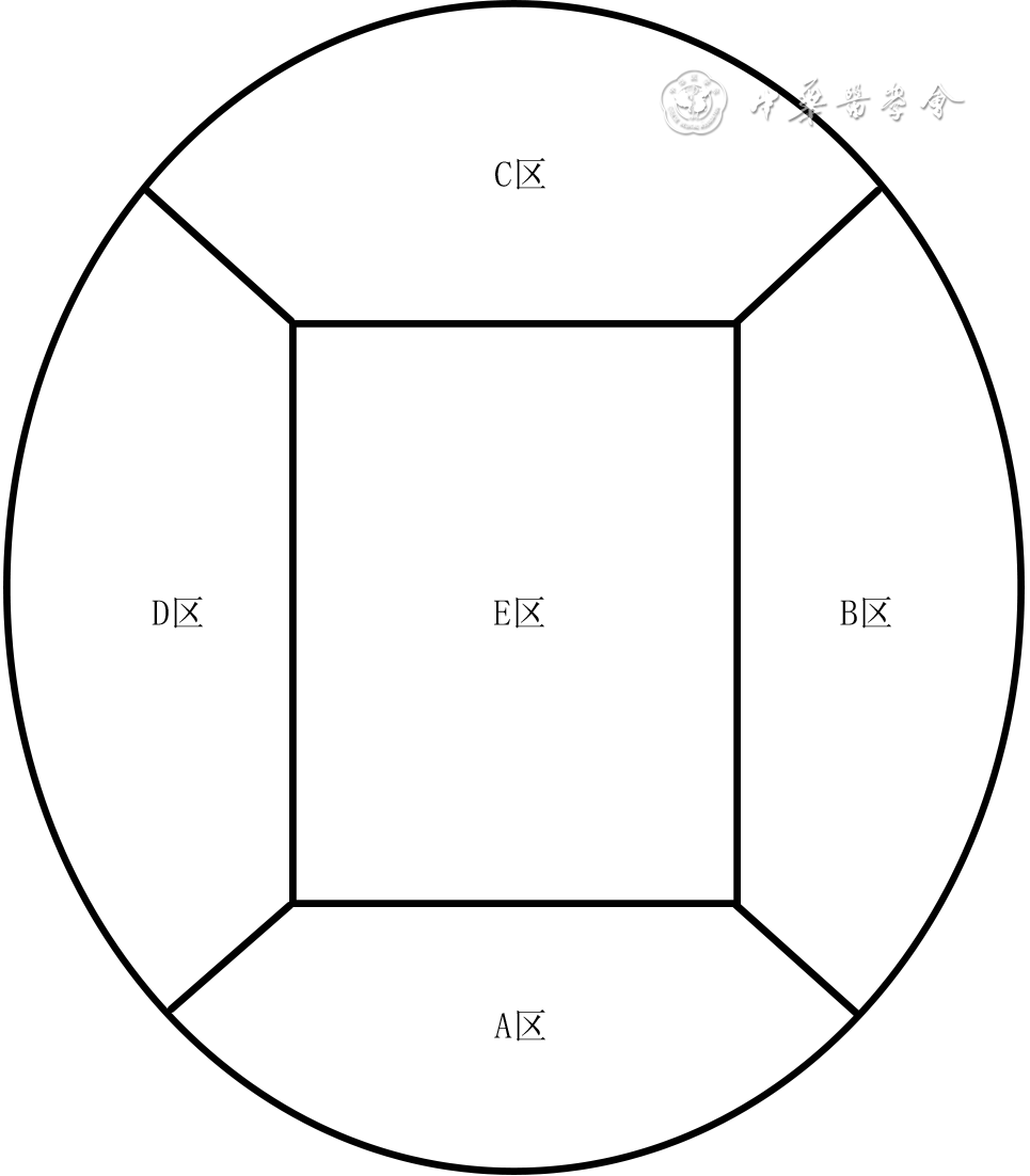
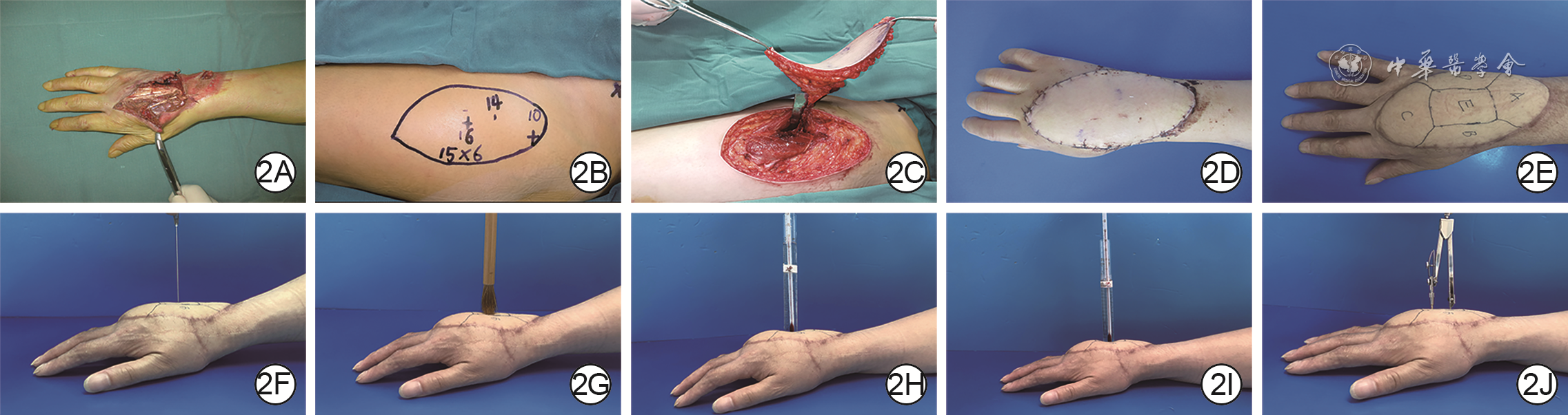
  Objective   To investigate the regularity of sensory recovery after repairing the wounds on the wrist and back of hand with anterolateral femoral flap without nerve anastomosis.   Methods   A cross-sectional study was conducted. From January 2018 to December 2020, patients who underwent free anterolateral femoral flaps without nerve anastomosis to repair wounds on the wrist and back of hand and met the inclusion criteria in Changshu Hai Yu Health Centre and Suzhou Ruihua Orthopedic Hospital were included in this study. Depending on the time interval between the day of the patient's surgery and the day of the cross-sectional survey, 80 patients were divided into 6-month group (15 males and 5 females, aged 22-63 years), 12-month group (16 males and 4 females, aged 21-65 years), 18-month group (15 males and 5 females, aged 25-61 years), and 24-month group (14 males and 6 females, aged 20-65 years), with 20 patients in each group. The area of skin and soft tissue defects after debridement ranged from 6.0 cm×4.5 cm to 18.0 cm×9.0 cm. Anterolateral femoral flaps were cut with areas of 7 cm×5 cm to 20 cm×10 cm and a thickness of 1.0 to 2.5 cm. Each transplanted flap was divided into A (proximal), B/D (bilateral), C (distal), and E (central) regions. The pain sensation, touch sensation, cold sensation, warmth sensation, and two-point discrimination (2-PD) in the aforementioned five regions and the differences in the five senses of the whole flap were tested and compared. Data were statistically analyzed with one-way analysis of variance, Fisher's exact probability test, chi-square test, or McNemar test.   Results   In A region of anterolateral femoral flap without nerve anastomosis, compared with those in 6-month group, the pain sensation, touch sensation, cold sensation, and warmth sensation of flap of patients in 12-month group were significantly recovered (with χ 2 values of 10.10, 14.55, 12.13, and 4.29, respectively, P<0.05 or P<0.01); compared with that in 12-month group, the warmth sensation of flap of patients in 18-month group recovered significantly ( χ 2=5.23, P<0.05). In B region, compared with those in 6-month group, the pain sensation, touch sensation, and cold sensation of flap of patients in 12-month group recovered significantly (with χ 2 values of 5.58, 3.96, and 4.29, respectively, P<0.05); compared with those in 12-month group, the pain sensation, touch sensation, cold sensation, and warmth sensation of flap of patients in 18-month group recovered significantly (with χ 2 values of 5.58, 3.96, 7.03, and 12.38, respectively, P<0.05 or P<0.01). In C region, compared with that in 6-month group, the pain sensation of flap of patients in 12-month group recovered significantly ( χ 2=4.80, P<0.05); Compared with that in 12-month group, the warmth sensation of flap of patients in 18-month group recovered significantly ( χ 2=10.16, P<0.01). In D region, compared with those in 6-month group, the pain sensation, touch sensation, and cold sensation of flap of patients in 12-month group recovered significantly (with χ 2 values of 5.58, 4.29, and 3.96, respectively, P<0.05); compared with those in 12-month group, the pain sensation, touch sensation, cold sensation, and warmth sensation of flap of patients in 18-month group recovered significantly (with χ 2 values of 5.58, 4.29, 3.96, and 10.10, respectively, P<0.05 or P<0.01). In E region, compared with that in 6-month group, the cold sensation of flap of patients in 12-month group recovered significantly ( χ 2=4.80, P<0.05); compared with those in 12-month group, the pain sensation, touch sensation, and warmth sensation of flap of patients in 18-month group recovered significantly (with χ 2 values of 6.47, 4.91, and 9.23, respectively, P<0.05 or P<0.01). The five senses in the 5 regions of flap of patients in 24-month group were similar to those in 18-month group ( P>0.05). The recovery of 2-PD in the 5 regions of flap of patients was similar between the two adjacent groups ( P>0.05). In 12-month group, the recoveries of pain sensation, touch sensation, and cold sensation of flap of patients in A region were better than those in the other 4 regions ( P<0.05 or P<0.01), the recovery of warmth sensation was better than that of B region, C region, and E region ( P<0.05 or P<0.01); in 18-month group, the recovery of pain sensation, touch sensation, cold sensation, and warmth sensation of flap of patients in A region of was better than those in area C region ( P<0.05). Compared with those in 6-month group, the pain sensation, touch sensation, and cold sensation of the whole flap of patients in 12-month group recovered significantly (with χ 2 values of 7.62, 7.03, and 5.58, respectively, P<0.05 or P<0.01). Compared with the 12-month group in which 10, 11, 10, and 4 patients had a recovery of pain, touch sensation, cold sensation, and warmth sensation in the whole flap, the 18-month group had significantly more patients with sensations recovered, which were 17, 17, 16, and 14, respectively (with χ 2 values of 5.58, 4.29, 3.96, and 10.10, respectively, P<0.05 or P<0.01). The five senses of the whole flap of patients in 24-month group were similar to those in 18-month group ( P>0.05).   Conclusions   In the anterolateral femoral flap without nerve anastomosis for repairing wounds on the wrist and back of hand, the sensation gradually recovered from the proximal end to the distal end. The sensation of touch, pain, and cold began to recover from 6 months after operation, and entered the stable recover period at 18 months after operation. Warmth sensation began to recover from 12 months after operation, and entered the stable recovery period at 18 months after operation. The 2-PD of most flaps was still not recovered 2-year after operation.
Effects of Krüppel-like factor 4 on inflammatory response and organ injury in septic mice
Wang Yunwei, Liu Yang, Cao Peng, Zhang Qingyi, Chen Yang, Li Shaohui, Guan Hao
2022, 38(11): 1047-1056. doi: 10.3760/cma.j.cn501225-20220111-00005
Abstract:
  Objective  To explore the expression characteristics and role of Krüppel-like factor 4 (KLF4) in macrophage inflammatory response and its effects on inflammatory response and organ injury in septic mice, so as to lay a theoretical foundation for targeted treatment of burns and trauma sepsis.  Methods  The method of experimental research was used. Mouse RAW264.7 macrophages and primary peritoneal macrophages (PMs) isolated from 10 male C57BL/6J mice aged 6-8 weeks were used for the experiments. RAW264.7 macrophages and PMs were treated with endotoxin/lipopolysaccharide (LPS) for 0 (without treatment), 1, 2, 4, 6, 8, 12, and 24 h, respectively, to establish macrophage inflammatory response model. The mRNA expression of interleukin 1β (IL-1β), IL-6, CC chemokine ligand 2 (CCL2) and tumor necrosis factor-α (TNF-α) were detected by real-time fluorescence quantitative reverse transcription polymerase chain reaction (RT-PCR), and the LPS treatment time was determined for some of the subsequent experiments. RAW264.7 macrophages were treated with LPS for 0 and 8 h, the localization and protein expression of KLF4 were detected by immunofluorescence method, transcriptome sequencing of the cells was performed using the high-throughput sequencing technology platform, and the differently expressed genes (DEGs) between the two time points treated cells were screened by DESeq2 software. RAW264.7 macrophages and PMs were treated with LPS for 0, 1, 2, 4, 6, 8, 12, and 24 h, respectively, and the mRNA and protein expressions of KLF4 were detected by real-time fluorescence quantitative RT-PCR and Western blotting, respectively. RAW264.7 macrophages were divided into negative control (NC) group and KLF4-overexpression group according to the random number table, which were treated with LPS for 0 and 8 h respectively after transfection of corresponding plasmid. The mRNA expressions of KLF4, IL-1β, IL-6, CCL2, and TNF-α were detected by real-time fluorescence quantitative RT-PCR, while the protein expression of KLF4 was detected by Western blotting. The number of samples in aforementioned experiments was all 3. Forty male C57BL/6J mice aged 6-8 weeks were divided into KLF4-overexpression group and NC group (with 20 mice in each group) according to the random number table, and the sepsis model of cecal ligation perforation was established after the corresponding transfection injection was injected respectively. Twelve mice were selected from each of the two groups according to the random number table, and the survival status within 72 hours after modeling was observed. Eight hours after modeling, the remaining 8 mice in each of the two groups were selected, the eyeball blood samples were collected to detect the levels of IL-1β and IL-6 in serum by enzyme-linked immunosorbent assay, and the levels of alanine aminotransferase (ALT) and aspartate aminotransferase (AST) in serum by dry chemical method. Subsequently, the heart, lung, and liver tissue was collected, and the injury was observed after hematoxylin-eosin staining. Data were statistically analyzed with independent sample t test, Cochran & Cox approximate t test, one-way analysis of variance, Dunnett test, Brown-Forsythe and Welch one-way analysis of variance, Dunnett T3 test, log-rank (Mantel-Cox) test.  Results  Compared with that of LPS treatment for 0 h, the mRNA expressions of IL-1β in RAW264.7 macrophages treated with LPS for 6 h and 8 h, the mRNA expressions of IL-6 in RAW264.7 macrophages treated with LPS for 4-12 h, the mRNA expressions of CCL2 in RAW264.7 macrophages treated with LPS for 8 h and 12 h, and the mRNA expressions of TNF-α in RAW264.7 macrophages treated with LPS for 4-8 h were significantly up-regulated (P<0.05 or P<0.01), while the mRNA expressions of IL-1β and CCL2 in PMs treated with LPS for 4-8 h, the mRNA expressions of IL-6 in PMs treated with LPS for 2-24 h, and the mRNA expressions of TNF-α in PMs treated with LPS for 2-12 h were significantly up-regulated (P<0.05 or P<0.01). Eight hours was selected as the LPS treatment time for some of the subsequent experiments. KLF4 mainly located in the nucleus of RAW264.7 macrophages. Compared with those of LPS treatment for 0 h, the protein expression of KLF4 in RAW264.7 macrophages treated with LPS for 8 h was obviously decreased, and there were 1 470 statistically differentially expressed DEGs in RAW264.7 macrophages treated with LPS for 8 h, including KLF4 with significantly down-regulated transcriptional expression (false discovery rate<0.05, log2 (fold change)=-2.47). Compared with those of LPS treatment for 0 h, the mRNA expressions of KLF4 in RAW264.7 macrophages treated with LPS for 6-24 h, the protein expressions of KLF4 in RAW264.7 macrophages and PMs treated with LPS for 1-24 h, and the mRNA expressions of KLF4 in PM treated with LPS for 4-24 h were significantly decreased (P<0.05 or P<0.01). Compared with those in NC group, the mRNA (with t' values of 17.03 and 8.61, respectively, P<0.05 or P<0.01) and protein expressions of KLF4 in RAW264.7 macrophages treated with LPS for 0 h and 8 h in KLF4-overexpression group were significantly increased, the mRNA expressions of IL-6 and CCL2 increased significantly in RAW264.7 macrophages treated with LPS for 0 h (with t values of 6.29 and 3.40, respectively, P<0.05 or P<0.01), while the mRNA expressions of IL-1β, IL-6, CCL2, and TNF-α decreased significantly in RAW264.7 macrophages treated with LPS for 8 h (with t values of 10.52, 9.60, 4.58, and 8.58, respectively, P<0.01). The survival proportion of mice within 72 h after modeling in KLF4-overexpression group was significantly higher than that in NC group (χ2=4.01, P<0.05). Eight hours after modeling, the serum levels of IL-1β, IL-6 and ALT, AST of mice in KLF4-overexpression group were (161±63), (476±161) pg/mL and (144±24), (264±93) U/L, respectively, which were significantly lower than (257±58), (654±129) pg/mL and (196±27), (407±84) U/L (with t values of 3.16, 2.44 and 4.04, 3.24, respectively, P<0.05 or P<0.01) in NC group. Eight hours after modeling, compared with those in NC group, the disorder of tissue structure of heart, lung, and liver, inflammatory exudation, and pathological changes of organ parenchyma cells in KLF4-overexpression group were obviously alleviated.  Conclusions  The expression of KLF4 is significantly down-regulated in LPS-induced macrophage inflammatory response, which significantly inhibits the macrophage inflammatory response. KLF4 significantly enhances the survival rate of septic mice and alleviates inflammatory response and sepsis-related organ injury.
Effect of deep dermal tissue dislocation injury on skin fibrosis in pig
Yu Xiaoping, Liu Yingkai, Ma Xian, Tang Jiajun, Niu Yiwen, Zhou Junli, Lu Shuliang
2022, 38(11): 1057-1065. doi: 10.3760/cma.j.cn501120-20210831-00301
Abstract:
  Objective  To explore the effect of deep dermal tissue dislocation injury on skin fibrosis in pig, in order to provide some theoretical basis for burn scar treatment.  Methods  The experimental research method was applied. Six 2-month-old female Duroc pigs were taken. Fifteen operative areas on the right dorsum of pigs on which medium-thick skin grafts and deep dermal tissue slices were cut and re-implanted were included into dermal in situ reimplantation group, and fifteen operative areas on the left dorsum of pigs on which medium-thick skin grafts and deep dermal tissue slices were cut and the deep dermal tissue slice was placed under the fat layer were included into the dermal dislocation group. The hair growth in the operative areas on post-injury day (PID) 7, 14, and 21 and the cross-sectional structure on PID 14 were observed in the two groups. On PID 7, 14, and 21, the skin thickness (the distance from the epidermis to the upper edge of the fat), the dermal thickness (the distance from the lower edge of the epidermis to the upper edge of the fat, excluding the fibrotic tissue thickness between the dermis and the fat), and the fibrosis tissue thickness of the dermis-fat interface (from the lower edge of the deep dermis to the upper edge of the fat in dermal in situ reimplantation group and from the lower edge of the superficial dermis to the upper edge of the fat in dermal dislocation group) in the operative areas were measured and compared between the two groups; the fibrotic tissue thickness at the dermal cutting interface (from the lower edge of the superficial dermis to the upper edge of the deep dermis) in the operative areas in dermal in situ reimplantation group was measured and compared with the fibrotic tissue thickness at the dermal-fat interface. Sirius red staining was performed to observe and compare the type Ⅰ and Ⅲ collagen content in the dermal-fat interface in the operative areas between the 2 groups and between the dermal cutting interface and dermal-fat interface in the operative areas in dermal in situ reimplantation group. Immunohistochemical staining was performed to observe the positive expressions of proliferating cell nuclear antigen (PCNA), transforming growth factor β1 (TGF-β1), fibroblast growth factor 2 (FGF-2), and hepatocyte growth factor (HGF) in the operative areas in the two groups. The sample number was 6. Data were statistically analyzed with independent sample t test.  Results  On PID 7, 14, and 21, the hairs in the operative areas in dermal in situ reimplantation group were denser than those in dermal dislocation group. On PID 14, the skin cross section in the operative areas in dermal dislocation group showed a "sandwich"-like structure, while the skin cross section in the operative areas in dermal in situ reimplantation group had normal structure. On PID 7, 14, and 21, the skin thickness in the operative areas in dermal dislocation group was (4 234±186), (4 688±360), and (4 548±360) μm, respectively, which was close to (4 425±156), (4 714±141), and (4 310±473) μm in dermal in situ reimplantation group (P>0.05); the dermal thickness in the operative areas in dermal dislocation group was significantly thinner than that in dermal in situ reimplantation group (with t values of -9.73, -15.85, and -15.41, respectively, P<0.01); the fibrotic tissue thickness at the dermal-fat interface in the operative areas in dermal dislocation group was significantly thicker than that in dermal in situ reimplantation group (with t values of 14.48, 20.58, and 15.67, respectively, P<0.01); there was no statistically significant difference between the fibrotic tissue thickness at the dermal-fat interface and the dermal cutting interface in the operative areas in dermal in situ reimplantation group (P>0.05). On PID 7, 14, 21, the type Ⅲ collagen content in the dermal-fat interface in the operative areas in dermal dislocation group was increased significantly compared with that in dermal in situ replantation group (with t values of 2.65, 0.61, and 7.39, respectively, P<0.05 or P<0.01), whereas there were no statistically significant differences in the type Ⅰ collagen content at the dermal-fat interface in the operative areas between the 2 groups (P>0.05) and the type Ⅰ and Ⅲ collagen content between the dermal-fat interface and the dermal cutting interface in the operative areas in dermal in situ reimplantation group (P>0.05). On PID 7, 14, and 21, PCNA, TGF-β1, FGF-2, and HGF were positively expressed in the superficial dermis and adipose tissue in the operative areas in dermal dislocation group, while PCNA, TGF-β1, FGF-2, and HGF were positively expressed in the superficial dermis, deep dermis, and adipose tissue in the operative areas in dermal in situ reimplantation group.  Conclusions  Inadequate intrinsic thickness of dermal tissue is the key factor causing fibrosis, and the biological purpose of fibrosis is to "compensate" the intrinsic thickness of the skin. Besides, adipose tissue may also be an important component of fibrotic skin repair.
Regulatory effects of bio-intensity electric field on microtubule acetylation in human epidermal cell line HaCaT
Wu Yating, Zhang Ze, Ji Ran, Zhang Shuhao, Wang Wenping, Wu Chao, Zhang Jiaping, Jiang Xupin, Zhang Hengshu
2022, 38(11): 1066-1072. doi: 10.3760/cma.j.cn501120-20211105-00377
Abstract:
  Objective  To investigate the regulatory effects of bio-intensity electric field on directional migration and microtubule acetylation in human epidermal cell line HaCaT, aiming to provide molecular theoretical basis for the clinical treatment of wound repair.  Methods  The experimental research methods were used. HaCaT cells were collected and divided into simulated electric field group (n=54) placed in the electric field device without electricity for 3 h and electric field treatment group (n=52) treated with 200 mV/mm electric field for 3 h (the same treatment methods below). The cell movement direction was observed in the living cell workstation and the movement velocity, trajectory velocity, and direction of cosθ of cell movement within 3 h of treatment were calculated. HaCaT cells were divided into simulated electric field group and electric field treatment 1 h group, electric field treatment 2 h group, and electric field treatment 3 h group which were treated with 200 mV/mm electric field for corresponding time. HaCaT cells were divided into simulated electric field group and 100 mV/mm electric field group, 200 mV/mm electric field group, and 300 mV/mm electric field group treated with electric field of corresponding intensities for 3 h. The protein expression of acetylated α-tubulin was detected by Western blotting (n=3). HaCaT cells were divided into simulated electric field group and electric field treatment group, and the protein expression of acetylated α-tubulin was detected and located by immunofluorescence method (n=3). Data were statistically analyzed with Kruskal-Wallis H test,Mann-Whitney U test, Bonferroni correction, one-way analysis of variance, least significant difference test, and independent sample t test.  Results  Within 3 h of treatment, compared with that in simulated electric field group, the cells in electric field treatment group had obvious tendency to move directionally, the movement velocity and trajectory velocity were increased significantly (with Z values of -8.53 and -2.05, respectively, P<0.05 or P<0.01), and the directionality was significantly enhanced (Z=-8.65, P<0.01). Compared with (0.80±0.14) in simulated electric field group, the protein expressions of acetylated α-tubulin in electric field treatment 1 h group (1.50±0.08) and electric field treatment 2 h group (1.89±0.06) were not changed obviously (P>0.05), while the protein expression of acetylated α-tubulin of cells in electric field treatment 3 h group (3.37±0.36) was increased significantly (Z=-3.06, P<0.05). After treatment for 3 h, the protein expressions of acetylated α-tubulin of cells in 100 mV/mm electric field group, 200 mV/mm electric field group, and 300 mV/mm electric field group were 1.63±0.05, 2.24±0.08, and 2.00±0.13, respectively, which were significantly more than 0.95±0.27 in simulated electric field group (P<0.01). Compared with that in 100 mV/mm electric field group, the protein expressions of acetylated α-tubulin in 200 mV/mm electric field group and 300 mV/mm electric field group were increased significantly (P<0.01); the protein expression of acetylated α-tubulin of cells in 300 mV/mm electric field group was significantly lower than that in 200 mV/mm electric field group (P<0.05). After treatment for 3 h, compared with that in simulated electric field group, the acetylated α-tubulin of cells had enhanced directional distribution and higher protein expression (t=5.78, P<0.01).  Conclusions  Bio-intensity electric field can induce the directional migration of HaCaT cells and obviously up-regulate the level of α-ubulin acetylation after treatment at 200 mV/mm bio-intensity electric field for 3 h.
Original Article·Nursing Column
A cross-sectional survey and analysis of influencing factors of humanistic of the current status of humanistic care ability of burn specialist nurses
Jiang Qiqi, Zhang Yin, Qiao Liang, Zha Qinghua, Xie Lin, Luo Zhizhen
2022, 38(11): 1073-1078. doi: 10.3760/cma.j.cn501120-20210318-00092
Abstract:
  Objective  To investigate the current status of humanistic care ability of burn specialist nurses and to analyze the influencing factors.  Methods  A single-center cross-sectional research method was conducted. From May to August 2020, 63 burn specialist nurses who met the inclusion criteria in Ruijin Hospital affiliated to Shanghai Jiao Tong University School of Medicine were selected. Self-made general data questionnaire was used to investigate 17 indexes, including gender, age, professional title, working years, whether received humanistic care training, academic qualification, and caring ability inventory (CAI) was used to evaluate their humanistic care ability. After the nurses were classified by the general data, independent sample t test and one-way analysis of variance were performed on the data to analyze the total score of CAI. The CAI total scores and scores of cognition, courage, and patience of the nurses were compared with the international norm. The factors with statistically significant differences in unvariate analysis were selected for multiple linear regression analysis to screen the independent influencing factors of humanistic care ability of burn specialist nurses.  Results  A total of 63 questionnaires were collected in this survey, all of which were valid. Among the 63 nurses, there were 4 males and 59 females, with the age mainly ranging from 20 to 30 years (30 nurses, 47.62%), the professional titles mainly being nurse practitioner (36 nurses, 57.14%), the working years mainly being more than 10 years (28 nurses, 44.44%), 32 nurses not receiving humanistic care training, and academic qualifications mostly being junior college (37 nurses, 58.73%). There were significant differences in the total scores of CAI among nurses with different ages, professional titles, working years, whether received humanistic care training, and academic qualifications (with F values of 53.95, 49.14, and 75.42, t values of 6.08 and -2.82, respectively, P<0.01). The scores of cognition, courage, and patience and the total scores of CAI of nurses in this group were significantly lower than those of international norm (with t values of -2.02, -2.04, -6.19, and -3.89, respectively, P<0.05 or P<0.01). Multiple linear regression analysis showed that age, working years, professional title, and whether received humanistic care training were the independent influencing factors of humanistic care ability of burn specialist nurses (with 95% confidence intervals of 1.91-23.23, 16.25-31.48, 1.05-19.09, and 6.72-31.82, unstandardized coefficient values of 12.57, 23.86, 10.07, and 19.27, respectively, P<0.05 or P<0.01).  Conclusions  The humanistic care ability of burn specialist nurses is relatively weak. Age, professional title, working years, and whether received humanistic care training are the independent influencing factors of humanistic care ability of burn specialist nurses.
Meta-analysis of the interventional effects of music therapy on pain and anxiety of burn patients in wound dressing change
Li Ye, Liu Fangli, Yuan Ju, Li Jing, Liu Ziwei, Guan Ningxiao
2022, 38(11): 1079-1084. doi: 10.3760/cma.j.cn501120-20210716-00252
Abstract:
  Objective  To evaluate the interventional effects of music therapy on pain and anxiety of burn patients in wound dressing change.  Methods  The meta-analysis method was adopted. Databases including China National Knowledge Internet, Wanfang Database, and VIP database were retrieved with the search terms in Chinese version of "音乐, 烧伤, 创面换药/伤口换药, 疼痛/操作性疼痛", and PubMed, Embase, and Cochrane Library were retrieved with the search terms in English version of "music, burn, dressing change/wound dressing, pain/ache/sore" to obtain the publicly published randomized controlled trials on the application of music therapy for wound dressing change in burn patients from the establishment of each database to May 2021. The outcome indexes included pain score/percentage and anxiety score after dressing change. Rev Man 5.4 and Stata 14.0 statistical software were used to conduct a meta-analysis of eligible studies.  Results  A total of 520 burn patients from 7 studies were included, including 260 patients in music therapy group who received music therapy and 260 patients in routine dressing change group who received routine dressing change. The bias risk of all the 7 included studies was uncertain. Compared with those in routine dressing change group, the pain percentages (relative risk=0.06, 95% confidence interval=0.01-0.41, P<0.01) and pain scores after dressing change (standardized mean difference (SMD)=-0.91, 95% confidence interval=-1.61--0.22, P<0.05) of patients in music therapy group were significantly lower. Subgroup analysis showed that music type and timing of intervention might be the source of heterogeneity in pain scores after dressing change. The anxiety scores of patients in music therapy group were significantly lower than those in routine dressing change group (SMD=-0.64, 95% confidence interval=-1.09--0.19, P<0.01). There was no publication bias in pain or anxiety scores after dressing change.  Conclusions  Music therapy can relieve the pain and anxiety of burn patients during dressing change.
Reviews
Research advances on the mechanism of refractory healing of diabetic foot ulcer
Wang Ning, Ju Shang
2022, 38(11): 1085-1089. doi: 10.3760/cma.j.cn501225-20220227-00038
Abstract:
The number of patients with diabetic foot ulcer (DFU) has increased progressively year by year. Refractory DFU has brought great burden to the country and individuals. How to accelerate the healing of DFU has become the main emphasis of research. However currently, the mechanism of its refractory healing is not fully elucidated, and the correlation between the various mechanisms are not high. Therefore, its clinical standardization, and precise clinical diagnosis and treatment still face several challenges. Based on the progress of clinical research and basic research at home and abroad, this paper reviewed the specific mechanisms that lead to refractory DFU, with the focus on chronic inflammation, bacteria biofilm formation, high oxidative stress, growth factor inhibition, impaired microcirculation, and accumulation of advanced glycation end products.
Progress in research and development of soft tissue three-dimensional bioprinting and its supporting equipment
Hu Yanke, Chen Shuying, Zhou Fei, Xiong Yahui, Chen Lei, Qi Shaohai
2022, 38(11): 1090-1095. doi: 10.3760/cma.j.cn501120-20210922-00327
Abstract:
As a cutting-edge technology of tissue engineering, three-dimensional bioprinting can accurately fabricate biomimetic tissue, which has made great progress in the field of hard tissue printing such as bones and teeth. Meanwhile, the research on soft tissue bioprinting is also developing rapidly. This article mainly discussed the development progress in various bioprinting technologies and supporting equipment including printing software, printing hardware, supporting consumables, and bioreactors for soft tissue three-dimensional bioprinting, and made a prospect for the future research and development direction of soft tissue three-dimensional bioprinting.
Research progress of the application of methacrylic anhydride gelatin hydrogel in wound repair
Ding Neng, Fu Xinxin, Wu Haimei, Zhu Lie
2022, 38(11): 1096-1100. doi: 10.3760/cma.j.cn501225-20220308-00056
Abstract:
Wound repair is a common clinical problem, which seriously affects the quality of life of patients and also brings a heavy burden to the society. Hydrogel-based multifunctional dressing has shown strong potential in the treatment of acute and chronic wounds. In addition to its good histocompatibility, cell adhesion, and biodegradability, methacrylic anhydride gelatin (GelMA) hydrogel has also attracted much attention due to its low cost, mild reaction conditions, adjustable physicochemical properties, and wide clinical applications. In this paper, the characteristics of GelMA hydrogel and its research progress in wound repair are introduced, and the future development of multifunctional GelMA hydrogel dressing for wound treatment is prospected.