2023 Vol. 39, No. 3

Guidelines and Consensuses
National expert consensus on clinical diagnosis and treatment of hot crush injury wound (2023 version)
2023, 39(3): 201-208. doi: 10.3760/cma.j.cn501225-20220608-00227
Abstract:
Hot crush injury is a kind of damage with high disability rate. Although some experience has been accumulated in diagnosis and treatment of hot crush injury over the years, its treatment is often not standardized and difficult to achieve a satisfactory therapeutic effect due to insufficient understanding of the particularity of hot crush injury wounds. In order to standardize the treatment of hot crush injury wounds and improve the treatment effect of hot crush injury wounds and the life quality of patients, the Burns and Trauma Branch of Chinese Geriatrics Society, Branch of Burn and Trauma Wound Repair Materials of Chinese Society of Biomaterials, and Wound Repair Professional Committee of Chinese Medical Doctor Association have reached a consensus on the causes, types, wound treatment, and precautions of hot crush injury based on the current situation and clinical experience in the diagnosis and treatment of hot crush injury at home and abroad. It would provide a reference for clinical diagnosis and treatment of hot crush injury wounds.
Expert Forums
Pay more attention to the management of burn wounds of special causes and sites
Hu Dahai, Wang Yunchuan
2023, 39(3): 209-214. doi: 10.3760/cma.j.cn501225-20230206-00034
Abstract:
The treatment of burn wounds of special causes and sites is a very challenging clinical work. In this paper, we briefly discussed the incidence rates of chemical burns, electric burns, facial burns, hand burns, and perineal burns, as well as the complexity and severity of pathological injury of the corresponding wound tissue. In addition, we briefly discussed the main principles and methods of clinical treatment, as well as the difficult problems to be solved. It is hoped to attract attention and provide reference for further improving the overall treatment ability of burns.
Original Articles·Repair of Wounds in Special Sites
Clinical effects of autologous split-thickness skin grafting for prefabricating urethra combined with scrotal flap in repairing middle urethral defect with penile defect
Tong Lin, Zhang Wanfu, Han Fei, Xu Zhigang, Hu Dahai, Guan Hao
2023, 39(3): 215-220. doi: 10.3760/cma.j.cn501225-20221021-00465
Abstract:
  Objective   To investigate the clinical effects of autologous split-thickness skin grafting for prefabricating urethra combined with scrotal flap in repairing middle urethral defect with penile defect.   Methods   The retrospective observational study was conducted. Eight male patients (aged 14 to 58 years) with middle urethral defect and penile defect caused by various injuries who met the inclusion criteria were admitted to the First Affiliated Hospital of Air Force Medical University from January 2015 to January 2022. The length of urethral defect was 3 to 5 cm, and the wound area of penile defect after debridement was 5.0 cm×2.5 cm to 7.0 cm×5.5 cm. All the patients underwent autologous split-thickness skin grafting for prefabricating defect urethra in stage Ⅰ, and urethral anastomosis was performed and unilateral scrotal flap was transferred to reconstruct urethra and penis in stage Ⅱ. The area of scrotal flap was 6.0 cm×3.0 cm to 8.0 cm×6.0 cm. The wound in the donor area of skin graft was covered by oil gauze, and the wound of flap donor area was sutured directly. On the 7 th day after the operation of stage Ⅱ, the survival of the flap was observed. In 3 weeks after the operation of stage Ⅱ, the urinary flow rate was measured by the urinary flow rate detector (urinary flow rate >15 mL/s was regarded as unobstructed urination), the urinary fistula and erectile function were observed, and the self-made therapeutic satisfaction questionnaire was used to investigate the therapeutic satisfaction degree of patients. During follow-up, the appearance of the flap recipient area was observed, the Vancouver scar scale (VSS) was used to evaluate the scar situation in the donor areas of skin graft and flap, the urinary flow rate was detected as before, the urethral stricture, urinary fistula, and erectile function were observed, and the therapeutic satisfaction degree of patients was investigated.   Results   On the 7 th day after the operation of stage Ⅱ, the flaps survived completely in 8 patients. In 3 weeks after the operation of stage Ⅱ, the urinary flow rate was 25.3 (18.0, 38.5) mL/s, with unobstructed urination, without urinary fistula and with erectile function, and the score of therapeutic satisfaction degree was 14.3 (14.0, 15.0). During follow-up of 1 to 7 years, the flap recipient area of 8 patients was full in appearance and not swollen, with similar color to the surrounding tissue; the VSS scores of the donor areas of skin graft and flap were 11.5 (10.0, 13.0) and 10.5 (9.3, 12.0), respectively, the urinary flow rate was 24.6 (17.7, 34.1) mL/s, with no urethral stricture, urinary fistula, and erectile dysfunction, and the score of therapeutic satisfaction degree was 13.5 (13.3, 14.8).   Conclusions   Autologous split-thickness skin grafting for prefabricating urethra combined with scrotal flap in repairing the urethral and penile defects not only reconstructs the structure of urethra and the shape of penis, but also restores the sensation and erectile function of penis, with few postoperative complications, no obvious scar hyperplasia, and high satisfaction degree of patients, which is worthy of clinical promotion.
Repair methods of complex facial defect wounds involving paranasal sinuses and their clinical effectiveness
Liang Pengfei, Xu Xisheng, Zhang Pihong, Bi Changlong, Zhang Hua, Huang Mitao, He Zhiyou, Zeng Jizhang, Huang Yun, Li Jia, Cui Xu, Zhou Situo, Zhang Minghua, Huang Xiaoyuan
2023, 39(3): 221-227. doi: 10.3760/cma.j.cn501225-20221130-00520
Abstract:
  Objective   To explore the repair methods of complex facial defect wounds involving paranasal sinuses and their clinical effectiveness.   Methods   A retrospective observational study was conducted. From January 2020 to May 2022, 5 patients admitted to the Department of Burns and Plastic Surgery of Xiangya Hospital of Central South University and 4 patients admitted to the Department of Burns and Plastic Surgery of Chenzhou First People's Hospital with complex facial defect wounds involving paranasal sinuses met the inclusion criteria, including 6 males and 3 females, aged 35-69 years, including 4 patients with titanium mesh exposure combined with paranasal sinuses injury and 5 patients with tumor involving paranasal sinuses. After an adequate assessment of the damage by a multiple discipline team, titanium mesh removal, paranasal sinus debridement, and paranasal sinus mucosa removal were performed in patients with exposed titanium mesh, and radical tumor resection was performed in patients with tumors, with postoperative skin and soft tissue defects areas of 5.0 cm×2.5 cm to 18.0 cm×7.0 cm, anterior paranasal sinus wall defects/absence areas of 3 cm×2 cm to 6 cm×4 cm, and sinus cavity depths of 1 to 4 cm. Depending on the perforator course of the descending branch of the lateral circumflex femoral artery, the anterolateral femoral chimeric flap or anterolateral femoral myocutaneous flap (with flap area of 9 cm×4 cm to 19 cm×8 cm, muscle size of 5 cm×3 cm×3 cm to 11 cm×6 cm×3 cm) was transplanted to repair the defect, and the donor site wound was sutured directly. The type of tissue flap transplanted, the blood vessel of the recipient area, and the vascular anastomosis way during the operation, the recovery of the donor and recipient areas and the occurrence of complications after operation were observed. The appearance and blood supply of the recipient area and the recurrence of ulcers and tumors were followed up.   Results   The anterolateral femoral myocutaneous flap transplantation was performed in 6 patients, and the anterolateral femoral chimeric flap transplantation was performed in 3 patients. The blood vessels in recipient areas were facial arteries and veins in 3 cases and superficial temporal arteries and veins in 6 cases. The superficial temporal arteries and veins were bridged with blood vessels in tissue flaps by flow-through way in 2 patients, and end-to-end anastomosis of blood vessels in donor and recipient areas was performed in 7 patients. After operation, all the tissue flaps survived, and the facial defect wounds were well repaired without cerebrospinal fluid leakage or paranasal sinus secretion leakage, no intracranial infection occurred, and the wounds in donor areas were healed well. Follow-up of 6-35 months after operation showed that all the patients had good blood supply in the recipient area, and the shape was acceptable; 4 patients with exposed titanium mesh had no recurrence of ulceration, and 5 patients with tumor had no local tumor recurrence or metastasis.   Conclusions   Based on an adequate assessment of the extent of paranasal sinuses involved in the facial wound and the nature of the defect, good clinical effects can be achieved by using the anterolateral femoral muscle flap or the anterolateral femoral chimeric flap transplantation to repair complex facial defect wounds with open paranasal sinuses.
Effects of free gracilis muscle flap combined with sural nerve transfer for reconstruction of digital flexion and sensory function of hand in patient with wrist electric burn
Xia Chengde, Di Haiping, Xing Peipeng, Huang Wanxin, Xue Jidong, Cao Dayong, Guo Haina, Liu Lei, Li Pengcheng
2023, 39(3): 228-233. doi: 10.3760/cma.j.cn501225-20221203-00525
Abstract:
  Objective   To explore the effects of free gracilis muscle flap combined with sural nerve transfer for reconstruction of digital flexion and sensory function of hand in patient with severe wrist electric burn.   Methods   A retrospective observational study was conducted. From January 2017 to December 2020, 4 patients with wrist high-voltage electric burn admitted to the Department of Burns of the First People's Hospital of Zhengzhou and 4 patients with wrist high-voltage electric burn admitted to the Department of Hand Surgery of Beijing Jishuitan Hospital met the inclusion criteria, including 6 males and 2 females, aged 12 to 52 years. They were all classified as type Ⅱ wrist high-voltage electric burns with median nerve defect. In the first stage, the wounds were repaired with free anterolateral thigh femoral myocutaneous flap. In the second stage, the free gracilis muscle flap combined with sural nerve transplantation was used to reconstruct the digital flexion and sensory function of the affected hand in 3 to 6 months after wound healing. The cut lengths of muscle flap and nerve were 32 to 38 and 28 to 36 cm, respectively. The muscle flap donor area and nerve donor area were both closed and sutured. The survival condition of gracilis muscle flap and sural nerve, the wound healing time of recipient area on forearm, the healing time of suture in muscle flap donor area and nerve donor area were observed and recorded after operation, and the recovery of donor and recipient areas was followed up. In 2 years after operation, the muscle strength of thumb and digital flexion and finger sensory function after the hand function reconstruction were evaluated with the evaluation criteria of the hand tendon and nerve repair in the trial standard for the evaluation of functions of upper limbs of Hand Surgery Society of Chinese Medical Association.   Results   All the gracilis muscle flap and sural nerve survived successfully after operation. The wound healing time of recipient area on forearm was 10 to 14 days after operation, and the healing time of suture in muscle flap donor area and nerve donor area was 12 to 15 days after operation. The donor and recipient areas recovered well. In the follow-up of 2 years after operation, the muscle strength of thumb and digital flexion was evaluated as follows: 4 cases of grade 5, 3 cases of grade 4, and 1 case of grade 2; the finger sensory function was evaluated as follows: 4 cases of grade S3 +, 2 cases of grade S3, and 2 cases of grade S2.   Conclusions   For patients with hand dysfunction caused by severe wrist electric burn, free gracilis muscle flap combined with sural nerve transplantation can be used to reconstruct the digital flexion and sensory function of the affected hand. It is a good repair method, which does not cause great damage to thigh muscle flap donor area or calf nerve donor area.
Curative effects of the superficial peroneal artery perforator flap carrying multiple perforators in repairing hand and foot wounds
Zhang Tao, Cheng Junnan, Yang Lin, Huang Yongtao, Gao Qinfeng, Sun Fengwen, Liu Zhijin, Liu Shengzhe, Yang Chengpeng, Cao Yang, Ju Jihui
2023, 39(3): 234-240. doi: 10.3760/cma.j.cn501225-20220723-00305
Abstract:
  Objective   To investigate the curative effects of the superficial peroneal artery perforator flap carrying two and more homologous perforators in repairing hand and foot wounds.   Methods   A retrospective observational study was conducted. From January to September 2021, 23 patients with hand and foot wounds combined with bone or tendon exposure who met the inclusion criteria were admitted to Suzhou Ruihua Orthopaedic Hospital, including 16 males and 7 females, aged 23 to 68 years. After expansion of the wound, the wound area was 3.0 cm×2.0 cm to 11.0 cm×4.0 cm. All the wounds were repaired with the superficial peroneal artery perforator flap carrying two and more homologous perforators. The area of the resected flap was 4.0 cm×2.0 cm to 12.0 cm×5.0 cm. All the wounds in donor areas were sutured directly. During the operation, the resection of the flap, the number and caliber of carried perforators, the caliber of superficial peroneal artery, the length of vascular pedicle, and the number of accompanying veins of the superficial peroneal artery were recorded. The survival of the flap, the occurrence of complications, and the wound healing in the donor area were observed after operation. The recovery of the donor and recipient areas was followed up. At the last follow-up, the comprehensive evaluation scale was used to evaluate the curative effect of flap repair, and the sensory grading scale was used to evaluate the sensory function of the recipient area.   Results   Totally 24 flaps were successfully resected in surgical operations, carrying 56 superficial peroneal artery perforators in total, with the caliber of perforators of 0.20-0.70 mm. The calibers of all perforators carried by 7 flaps were smaller than 0.40 mm. Nineteen flaps carried 2 perforators each, 3 flaps carried 3 perforators each, 1 flap carried 4 perforators, and 1 flap carried 5 perforators. During the operation, the superficial peroneal artery was resected with a caliber of 0.40-1.50 mm, the vascular pedicle was 2-6 cm in length, and each superficial peroneal artery had two accompanying veins. After operation, all the flaps survived smoothly, no vascular crisis or distal necrosis occurred, and the wounds healed well in the donor area. During the follow-up of 6 to 10 months after operation, the color, texture, and elasticity of the recipient area were good. Among them, 6 recipient areas were thinned and reshaped because of bloating. There was only linear scar and no obvious scar hyperplasia or pigmentation in the donor area, without significant change in sensory or motor function. At the last follow-up, the curative effect evaluation of flap repair was excellent in 22 flaps and good in 2 flaps, and the sensory function evaluation of the recipient area was grade S 3 in 1 area and grade S 2 in 23 areas.   Conclusions   The superficial peroneal artery perforator flap with two and more homologous perforators has sufficient and reliable blood supply and is effective in repairing hand and foot wounds. It provides an ideal solution for the clinical problem in which the original operation scheme is abandoned due to the existence of only multiple slender perforators of caliber smaller than 0.40 mm, and only a single perforator in the operative field that cannot satisfy the needs of the flap blood supply and recipient area.
Clinical effects of free pre-expanded internal thoracic artery perforator pedicled deltopectoral flap transfer in facial scar reconstruction
He Lin, Zhu Chan, Jia Jing, Zhou Lin, Zhang Zhuo, Shu Maoguo
2023, 39(3): 241-247. doi: 10.3760/cma.j.cn501225-20220123-00012
Abstract:
  Objective   To explore the clinical effects of free pre-expanded deltopectoral flap transfer in facial scar reconstruction by selecting appropriate internal thoracic artery perforator as the pedicle through preoperative color Doppler ultrasonic vascular assessment.   Methods   A retrospective observational study was conducted. From September 2017 to March 2021, 11 patients with facial scar who met the inclusion criteria were admitted to the First Affiliated Hospital of Xi'an Jiaotong University, including 6 males and 5 females, aged 16-58 (31±12) years. The scar with area ranging from 7 cm×5 cm to 14 cm×9 cm was reconstructed by free pre-expanded internal thoracic artery perforator pedicled deltopectoral flap transfer. The operation was performed in 2 or 3 stages. Before operation, color Doppler ultrasonography was performed to evaluate the internal thoracic artery perforator. In the first stage, skin and soft tissue expander (hereinafter referred to as expander) implantation was performed, and a cylindrical expander with rated capacity of 400 to 600 mL was placed in the chest wall. The expansion time was 3 to 4 months, and the water injection volume reached 1.2-1.5 times of the rated capacity of expander. In the second stage, scar excision+free pre-expanded deltopectoral flap transfer was performed, with harvested flap area ranging from 9 cm×7 cm to 16 cm×10 cm. The vascular pedicle of flap (intercostal perforator of internal thoracic artery) was anastomosed end-to-end to the facial artery and vein or superficial temporal artery and vein. The wound in donor site was closed directly. Third stage operation thinning was performed at 3-6 months after the second stage operation in 5 patients because of bloated flap pedicle. At 6 months after the last operation, the flap survival and complications were recorded, the sensation of flap was evaluated by Semmes-Weinstein monofilament test, the color of flap was evaluated by color contrast of the flap to surrounding normal skin, and the curative effect satisfaction degree of patients was evaluated by 5-grade Likert scale.   Results   At 6 months after the last operation, all the flaps of 11 patients survived well. One patient experienced venous congestion after flap transplantation, but the flap survived after re-anastomosis. One patient experienced hematoma after the first stage operation of expander implantation, but the rest treatment was not influenced after hematoma removal. No complications such as infection or expander exposure occurred in any patient. At 6 months after the last operation, the sensation of flap of patient was as follows: 9 cases recovered to protective sensation decrease or better, 1 case had protective sensation defect, and 1 case only had deep touch and pressure sensation; the color of flap of patient was as follows: 3 cases were very close to the color of surrounding normal skin, 6 cases were close to the color of surrounding normal skin, and 2 cases were different to the color of surrounding normal skin; the curative effect satisfaction degree of patients was as follows: 2 patients were very satisfied, 6 patients were satisfied, 2 patients were somewhat satisfied, and 1 patient was a little not satisfied.   Conclusions   The large area facial scar can be treated safely and effectively by free pre-expanded deltopectoral flap with appropriate single internal thoracic artery perforator as vascular pedicle selected through vascular assessment by color Doppler ultrasonography before operation. After operation, the color of flap of patients is close to the surrounding normal skin and the sensation of flap can be partially recovered, with high curative effect satisfaction degree of patients.
Original Articles
Application effects of nitrous oxide and oxygen mixed inhalation technology on analgesia and sedation during debridement and dressing change in children with moderate or severe burns
Fan Taotao, Han Mei, Liang Yan, Cao Guohui, Song Guodong
2023, 39(3): 248-255. doi: 10.3760/cma.j.cn501225-20220308-00051
Abstract:
  Objective   To investigate the application effects of nitrous oxide and oxygen mixed inhalation technology on analgesia and sedation during debridement and dressing change in children with moderate or severe burns.   Methods   A retrospective non-randomized contemporary controlled study was conducted. From December 2019 to November 2021, 140 burn children with moderate or severe burns, aged 1 to 3 years, who met the inclusion criteria were admitted to Central Hospital Affiliated to Shandong First Medical University. During debridement and dressing change 3 to 14 days after injury, 42 children, including 23 males and 19 females, who received nurse-centered pain management mode and analgesia and sedation with nitrous oxide and oxygen mixed inhalation technology were included in nitrous oxide group (the dressing change process using the above-mentioned technology for the first time was selected for the follow-up study). Another 42 children, including 24 males and 18 females, were included in non-nitrous oxide group from 98 children who did not apply analgesia or sedation treatment during dressing change with stratified random sampling (one dressing change process was randomly selected for the follow-up study). The face, legs, activity, cry, and consolability scale and Ramsay sedation scale were used to evaluate the pain intensity and degree of sedation, respectively, at 30 minutes before dressing change (hereinafter referred to as before dressing change), immediately after debridement, and at 30 minutes after finishing dressing change (hereinafter referred to as after dressing change). After dressing change, the self-made satisfaction scale was used to evaluate the satisfaction degree of dressing change surgeons and guardians of children for analgesic effects during dressing change. The duration of dressing change and the healing time of deep partial-thickness burn wounds were recorded. The heart rate and percutaneous arterial oxygen saturation (SpO 2) before, during, and after dressing change and the occurrence of adverse events such as nausea and vomiting during dressing change were recorded. Data were statistically analyzed with Mann-Whitney U test, chi-square test, analysis of variance for repeated measurement, independent sample t test, and Bonferroni correction.   Results   There were no significant differences in the score of pain intensity and score of sedation degree between children in two groups before and after dressing change ( P>0.05). Immediately after debridement, the score of pain intensity of children in nitrous oxide group was 2.5±0.7, which was significantly lower than 7.6±1.0 in non-nitrous oxide group ( t=-26.69, P<0.05); the score of sedation degree of children in nitrous oxide group was 1.83±0.38, which was significantly higher than 1.21±0.42 in non-nitrous oxide group ( t=7.15, P<0.05). After dressing change, the satisfaction degree scores of dressing change surgeons and guardians of children for analgesic effects during dressing change of children in nitrous oxide group were significantly higher than those in non-nitrous oxide group (with tvalues of 10.53 and 2.24, respectively, P<0.05). The dressing change duration of children in nitrous oxide group was significantly shorter than that in non-nitrous oxide group ( t=-5.33, P<0.05). The healing time of deep partial-thickness burn wounds in children between the two groups had no significant difference ( P>0.05). The heart rate of children in nitrous oxide group was significantly lower than that in non-nitrous oxide group during dressing change ( t=-12.40, P<0.05), while the SpO 2 was significantly higher than that in non-nitrous oxide group ( t=5.98, P<0.05). During dressing change, 2 children had nausea and 1 child had euphoria in nitrous oxide group, while heart rate of all children in non-nitrous oxide group continued to be higher than the normal range.   Conclusions   In the process of debridement and dressing change in children with moderate or severe burns, the use of nurse-centered pain management mode and the standardized use of nitrous oxide and oxygen mixed inhalation technology can safely and effectively control pain and sedation.
Effects and mechanism of astragalus polysaccharide on wound healing of deep partial-thickness burns in rats
Zheng Fan, Cai Yu'e, Li Li, Han Dawei
2023, 39(3): 256-263. doi: 10.3760/cma.j.cn501225-20220324-00087
Abstract:
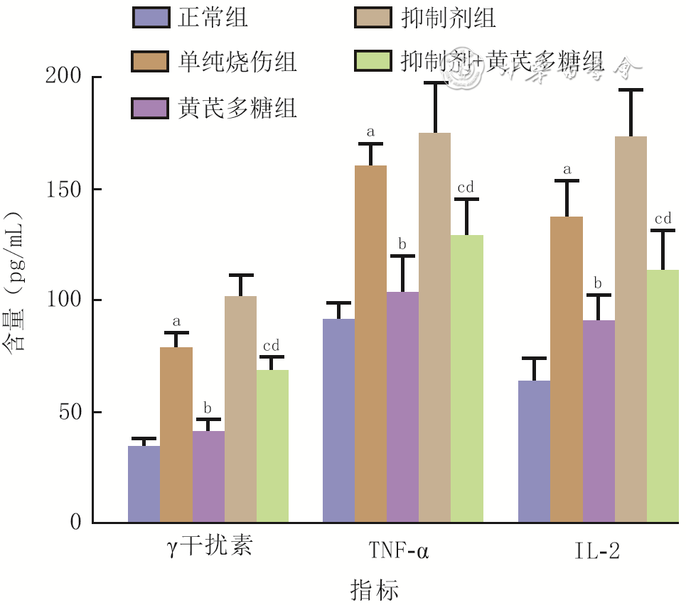
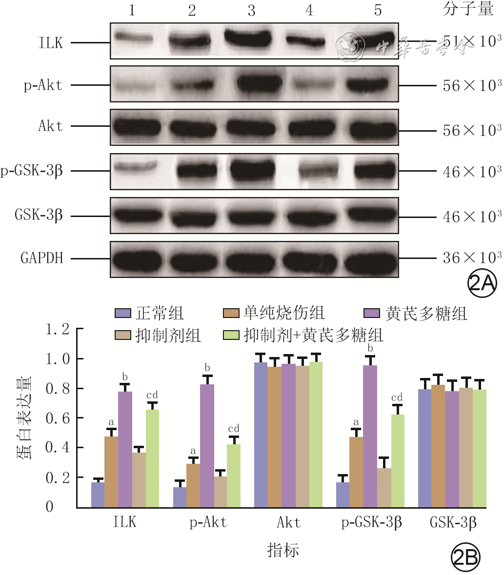
  Objective   To investigate the effects and mechanism of astragalus polysaccharide (APS) on wound healing of deep partial-thickness burns in rats.   Methods   The experimental study method was used. Fifty 7-week-old male Sprague-Dawley rats were divided into normal group, simple burn group, APS group, inhibitor group, and inhibitor+APS group according to the random number table, with 10 rats in each group. Except for normal group, rats in the other 4 groups were inflicted with a deep partial-thickness burn wound on the back. Rats in normal group and simple burn group were intraperitoneally injected with normal saline, and rats in the other three groups were injected with APS and/or integrin-linked kinase (ILK) inhibitor, respectively. After 7 days of injection, the wound healing rate of rats with burns in the four groups was calculated, and the serum content of interferon-γ, interleukin-2 (IL-2), and tumor necrosis factor α (TNF-α) in rats in 5 groups was determined by enzyme-linked immunosorbent assay (ELISA). The normal skin tissue of rats in normal group and wound tissue of rats with burns in the four groups were taken, the water content was determined and the water ratio was calculated, the content of interferon-γ, IL-2, and TNF-α was detected by ELISA, the mRNA expressions of epidermal growth factor (EGF), basic fibroblast growth factor (bFGF), and ILK were detected by real-time fluorescence quantitative reverse transcription polymerase chain reaction, and the protein expressions of ILK, protein kinase B (Akt), phosphorylated Akt (p-Akt), glycogen synthetic kinase-3β (GSK-3β), and phosphorylated GSK-3β (p-GSK-3β) were detected by Western blotting. Data were statistically analyzed with one-way analysis of variance and least significant difference test.   Results   After 7 days of injection, the wound healing rate of rats in APS group was (67±5)%, which was significantly higher than (52±4)% in simple burn group and (59±5)% in inhibitor+APS group (with all the P values <0.05). The wound healing rate of rats in inhibitor+APS group was significantly higher than (48±4)% in inhibitor group ( P<0.05). After 7 days of injection, compared with those in serum or normal skin tissue of rats in normal group, the serum content of interferon-γ, TNF-α, IL-2 and the water ratio of wound tissue of rats in simple burn group were significantly increased ( P<0.05); compared with those in APS group, the serum content of interferon-γ, TNF-α, IL-2 and the water ratio of wound tissue of rats in simple burn group and inhibitor+APS group were significantly increased ( P<0.05); compared with those in inhibitor group, the serum content of interferon-γ, TNF-α, IL-2 and the water ratio of wound tissue of rats in inhibitor+APS group were significantly decreased ( P<0.05). After 7 days of injection, compared with that in normal skin tissue of rats in normal group, the content of interferon-γ, TNF-α, and IL-2 in wound tissue of rats in simple burn group was significantly increased ( P<0.05); compared with that in APS group, the content of interferon-γ, TNF-α, and IL-2 in wound tissue of rats in simple burn group and inhibitor+APS group was significantly increased ( P<0.05); compared with that in inhibitor group, the content of interferon-γ, TNF-α, and IL-2 in wound tissue of rats in inhibitor+APS group was significantly decreased ( P<0.05). After 7 days of injection, compared with those in normal skin tissue of rats in normal group, the mRNA expressions of EGF, bFGF, ILK and protein expressions of ILK, p-Akt, p-GSK-3β in wound tissue of rats in simple burn group were significantly increased ( P<0.05); compared with those in APS group, the mRNA expressions of EGF, bFGF, ILK and protein expressions of ILK, p-Akt, p-GSK-3β in wound tissue of rats in simple burn group and inhibitor+APS group were significantly decreased ( P<0.05); compared with those in inhibitor group, the mRNA expressions of EGF, bFGF, ILK and protein expressions of ILK, p-Akt, p-GSK-3β in wound tissue of rats in inhibitor+APS group were significantly increased ( P<0.05). There were no statistically significant differences in the protein expressions of Akt and GSK-3β in normal skin tissue of rats in normal group and wound tissue of rats with burns in the four groups ( P>0.05).   Conclusions   APS can alleviate systemic and local inflammation, alleviate tissue edema, and promote the expressions of healing factors in rats with deep partial-thickness burns, thus to promote the wound healing, possibly by activating ILK/Akt/GSK-3β signaling pathway.
Reviews
Research advances on venous thromboembolism in burn patients
Shi Yiqin, Liu Lei, Li Ning, Luo Gaoxing, Li Haisheng
2023, 39(3): 264-268. doi: 10.3760/cma.j.cn501225-20220323-00083
Abstract:
Venous thromboembolism (VTE) has become a serious medical problem faced by medical personnel all over the world, due to its high incidence, high fatality, and easily missed and misdiagnosed. Patients with severe burns are at high risk for VTE due to the presence of blood hypercoagulability, central venous catheterization, repeatedly received surgical procedures, and prolonged bed rest. Identifying the risk factors of VTE in burn patients and taking targeted preventive measures are the key to reduce the incidence of VTE. However, there are no risk assessment tools or prevention guidelines for VTE in burn patients at home and abroad, and scholars from various countries are actively exploring the occurrence, influencing factors, and prevention of VTE in burn patients. This paper reviews the research progress of the occurrence situation, related risk factors, risk assessment, and prevention of VTE in burn patients in recent years, and discusses the existing problems and future research directions in this field.
Research advances on invasive fungal infections after burns
Cai Yue, Cheng Xing, Zhan Jianhua, Luo Jinhua, Liao Wenqiang
2023, 39(3): 269-274. doi: 10.3760/cma.j.cn501225-20220523-00199
Abstract:
Invasive fungal infection (IFI) is one of the serious complications in burn patients. The gradual development and application of broad-spectrum antibiotics in recent years has led to a serious dysbiosis of the flora, while the widespread prophylactic use of antifungal drugs has led to an increasing number of drug-resistant fungi. The clinical treatment of IFI is difficult and the prognosis is poor. The mortality of burn patients caused by IFI is increasing year by year. This paper reviews the epidemiologic characteristics, related risk factors, diagnostic methods, and treatment progress of IFI after burns, aiming to provide new ideas and reference for the prevention and treatment of IFI after burns.
Research advances on the application of rehabilitation exercise training in pediatric burn rehabilitation
Huo Ting, Xu Xiangyang, Xie Weiguo, Liu Shuhua
2023, 39(3): 275-279. doi: 10.3760/cma.j.cn501225-20220116-00008
Abstract:
The sequelae of pediatric burn seriously affect the physical function and quality of life of children with burns. Rehabilitation exercise training mainly based on aerobic and resistance exercise can effectively alleviate the negative effects. This article reviews the effects of rehabilitation exercise training on cardiopulmonary function, muscle function, and quality of life of children with burns, and introduces the latest rehabilitation exercise training prescription for children with burns based on type, mode, intensity, frequency, and time of exercise, so as to improve the level of rehabilitation treatment for children with burns.
Research advances on the application of music therapy in pain management of children with burns
Duan Deqing, Zhang Zhongwei, Mao Yuangui, Zhang Hongyan
2023, 39(3): 280-284. doi: 10.3760/cma.j.cn501225-20220328-00098
Abstract:
Pain after burns is a very common problem in children. Severe pain will not only hinder treatment, but also damage children's mental health if not handled in time. Therefore, pain management is very important in treating children with burns. As a safe, effective, and convenient non-drug therapy, music therapy has great advantages in relieving pain and is widely used in a variety of clinical fields. This paper focused on music therapy and its mechanism of pain relief, the current status of research on pain management of pediatric burns, the application and prospect of music therapy in pain management of pediatric burns, etc., to provide reference for clinical application.
Research advances on the prevention and treatment of burn infection in the elderly
Yang Changfa, Min Dinghong, Guo Guanghua
2023, 39(3): 285-289. doi: 10.3760/cma.j.cn501225-20220321-00078
Abstract:
Infection is a common complication after burns and the major cause of death in patients suffering severe burn injury. The infection of the elderly after burns is more serious due to their decreased immune function that is complicated with factors such as multiple chronic diseases and dysfunction of various organs. In addition, the burn infection in the elderly lacks the specific symptoms and signs, which brings great challenges to its diagnosis and treatment. To effectively prevent and control infection is very important for the treatment of elderly burn patients. Combined the clinical characteristics of burn infection in the elderly, this paper summarized the research advances of prevention and treatment for burn infection in the elderly from fluid resuscitation, wound treatment, antibiotic using, organ protection, nutritional support, and infection prevention, aiming to provide reference for clinical practice.
Research advances of stem cell-based tissue engineering repair materials in promoting the healing of chronic refractory wounds on the body surface
Peng Yu, Meng Hao, Li Pinxue, Jiang Yufeng, Fu Xiaobing
2023, 39(3): 290-295. doi: 10.3760/cma.j.cn501225-20220407-00126
Abstract:
Repairing chronic refractory wounds on the body surface is a complex medical problem involving all stages of wound healing. In recent years, stem cells (SCs) and tissue engineering (TE) have brought hope for repairing chronic refractory wounds. SCs have excellent regenerative and paracrine effects; various TE strategies have the potential to repair chronic refractory wounds on the body surface and also improve the delivery efficiency of SCs. This article reviews the pathological characteristics of chronic refractory wounds, SCs used to repair chronic refractory wounds, and SC-based TE wound repair strategies.
Research progress on the role of adipose-derived stem cell exosomes in skin scar formation
Wang Liuxin, Li Yunpeng, Wu Simo, Zhang Junrui, Kong Liang, Lu Bin, Liu Fuwei, Li Zhiye
2023, 39(3): 295-300. doi: 10.3760/cma.j.cn501225-20220308-00057
Abstract:
The adipose-derived stem cell exosomes are subcellular structures of adipose stem cells. They are nano-sized membrane vesicles that can transport various cell components and act on target cells by paracrine, and they play an important role in the exchanges of substance and information between cells. Scar healing is the commonest way of healing after skin tissue injury. Pathological scar can not only cause movement dysfunction, but also lead to deformity, which affects the appearance of patients and brings life and mental pressure to the patients. In recent years, many researches have shown that the adipose-derived stem cell exosomes contain a variety of bioactive molecules, which play an important role in reducing scar formation and scar-free wound healing, by affecting the proliferation and migration of fibroblasts and the composition of extracellular matrix. This article reviewed the recent literature on the roles and mechanisms of adipose-derived stem cell exosomes in scar formation, and prospected the future application and development of adipose-derived stem cell exosomes in scar treatment.