2023 Vol. 39, No. 8

Guidelines and Consensuses
Expert consensus on the clinical treatment of burn patients complicated with Coronavirus infection (2023 version)
2023, 39(8): 701-712. doi: 10.3760/cma.j.cn501225-20230519-00176
Abstract:
With China downgrading the management of Coronavirus infection (COVID-19) from Category A to Category B, a large number of COVID-19 patients have occurred in multiple waves across the country. Meanwhile, the long-term impact of Coronavirus on the body has gradually been noticed. However, the clinical treatment of burns complicated with COVID-19 is still a major challenge in Chinese burn centers. It is then essential to standardize the clinical treatment of such patients, improve the prognosis to the greatest extent, and provide valuable experiences for similar infectious diseases in future. Therefore, Chinese Burn Association, Burn Medicine Branch of China International Exchange and Promotion Association for Medical and Healthcare, and Editorial Committee of Chinese Journal of Burns and Wounds jointly initiated and organized multidisciplinary experts to develop this expert consensus based on the current medical evidence, clinical practice, and authoritative guidelines of other disciplines, in order to standardize the clinical treatment of burn patients complicated with COVID-19.
Expert Forums
Exploration of functional reconstruction and rehabilitation strategies for patients with destructive electric burns
Shen Yuming, Dai Qiang
2023, 39(8): 713-717. doi: 10.3760/cma.j.cn501225-20230506-00158
Abstract:
Electric burn is a kind of three-dimensional destructive damage. It is necessary to attach great importance to the functional reconstruction and rehabilitation of patients with destructive electric burns. Wound repair and limb salvage are not the end of the treatment of destructive electric burns, but functional rehabilitation and reintegration into society of patients are the goals of treatment. This paper systematically discusses the early wound repair, late functional reconstruction and rehabilitation, limb salvage and amputation, minimized damage of donor area, psychological rehabilitation, and multi-disciplinary cooperation of destructive electric burns. Only by attaching great importance to the functional reconstruction and rehabilitation, and embedding these concepts in people's brains, perfect repair and rehabilitation of destructive electric burns can be realized.
Mechanism and prevention and treatment strategy of progressive injury in high-voltage electric burns
Zhang Qingfu, Hao Jiawen
2023, 39(8): 718-723. doi: 10.3760/cma.j.cn501225-20230331-00107
Abstract:
High-voltage electric burn is one of burns that can cause severe damages to tissue and organs. Clinically, progressive injury in high-voltage electric burns is a key pathological change that affects the level of amputation and the success rate of treatment. At present, the exact definition and mechanism of progressive injury in high-voltage electric burns have not been elucidated, and the clinical treatment is mainly symptomatic treatment. Relevant research data on the mechanism and treatment of progressive injury in high-voltage electric burns are lacking. This paper analyzes and summarizes the mechanism, diagnosis, treatment, and common outcome of progressive injury in high-voltage electric burns, and provides a reference for the mechanism research, clinical diagnosis and treatment of progressive injury in high-voltage electric burns.
Original Articles·Electric Burns
Curative effects of foot microflap free transplantation in the repair of full-thickness electric burn wounds deep to tendon or even bone in fingers
Xue Jidong, Di Haiping, Liang Yan, Xing Peipeng, Guo Haina, Zhao Xiaokai, Wang Limin, Xia Chengde
2023, 39(8): 724-730. doi: 10.3760/cma.j.cn501225-20230323-00094
Abstract:
  Objective   To explore the curative effects of foot microflap free transplantation in the repair of full-thickness electric burn wounds deep to tendon or even bone in fingers.   Methods   A retrospective observational study was conducted. From July 2017 to February 2022, 20 patients with full-thickness electric burn wounds deep to tendon or even bone in fingers who met the inclusion criteria were admitted to Zhengzhou First People's Hospital, including 19 males and 1 female, aged 18 to 64 years. Among the 20 wounds, 15 wounds were located on the palm side, including 8 on the thumb, 5 on the index finger, and 2 on the middle finger; 5 wounds were located on the back, including 1 on the index finger and 4 on the middle finger. After debridement, the wound area ranged from 4.5 cm×2.0 cm to 7.0 cm×3.0 cm. According to the principle of tissue structure similarity, 10 wounds were repaired with plantar medial flaps, 5 wounds were repaired with hallux peroneal flaps, and 5 wounds were repaired with dorsalis pedis artery flaps, with flap area of 5.0 cm×2.5 cm-8.0 cm×3.5 cm. The flaps were transplanted freely and arteries and veins and/or nerves were anastomosed at the same time. The wound in the donor site was repaired with thigh medium-thick skin graft. The survival of flaps and skin grafts were observed after surgery. The appearance of flap, temperature and color of the distal end in the affected finger were observed during follow-up. At the last follow-up, the joint function and flap sensory recovery of the affected finger were evaluated with the trial standard for the evaluation of the functions of the upper limbs of the Hand Surgery Society of the Chinese Medical Association; the two-point discrimination distance of skin in the area of flaps with nerve anastomosis was measured; the satisfaction of patients with the curative effect was investigated by using the curative effect satisfaction rating scale, and the very satisfied rate was calculated; the repair effect of flap was evaluated by the comprehensive evaluation scale, and the excellent and good rate was calculated.   Results   All the flaps and skin grafts survived after surgery. During the follow-up of 10-18 months after surgery, the appearance of flap was natural and not bloated; the temperature and color of the distal end in the affected finger were basically the same as that of normal finger skin. At the last follow-up, the function recovery of the affected finger joints was as follows: 11 affected fingers were within the normal range of motion, 6 affected fingers had their total active range of motion recovered to 85% of the healthy side, and 3 affected fingers had their total active range of motion recovered to 75% of the healthy side; the flap sensory recovery was as follows: the sense of 15 flaps with nerve anastomosis all recovered to grade S3 +, and the two-point discrimination distance of skin in the flap area was 7.0-9.0 mm; the sense of 1 flap without nerve anastomosis recovered to grade S2 and the sense of 4 flaps recovered to grade S1. The satisfaction with curative effect of 20 patients was very satisfied in 16 cases and moderately satisfied in 4 cases, with the very satisfied rate of 80%; the repair result of 20 flaps was excellent in 16 cases, good in 2 cases, and fair in 2 cases, with excellent and good rate of 90%.   Conclusions   Due to the similar tissue structure of donor site and recipient site, foot microflap free transplantation in the repair of full-thickness electric burn wounds deep to tendon or even bone in fingers can achieve good appearance and function, with better functional and sensory recovery of the affected finger in the case of nerve anastomosis. Patients have high degree of satisfaction with the curative effects, which is worthy of promotion.
Treatment methods of upper limbs with destructive electric burns and its clinical efficacy
Zhang Wei, Chen Lan, Yang Fei, Zhang Weidong, Liu Feng, Xie Weiguo
2023, 39(8): 731-737. doi: 10.3760/cma.j.cn501225-20230530-00188
Abstract:
  Objective   To investigate the treatment methods of upper limbs with destructive electric burns and its clinical efficacy.   Methods   A retrospective observational study was conducted. From July 2014 to December 2020, 20 male patients with destructive electric burns in upper limbs who met the inclusion criteria were admitted to Tongren Hospital of Wuhan University & Wuhan Third Hospital, aged from 21 to 57 years, of whom 7 patients underwent emergency surgery, and a total of 20 affected limbs were treated with limb salvage. The necrotic bone was resected in 5 affected limbs, the residual hand and wrist at the distal end of left affected limb was replanted to the residual end of the right forearm in one patient in a cross heterotopic way, and short reduction and replantation after osteotomy were performed for two affected limbs with distal ulnar and radial necrosis. After thorough debridement, the area of wound proposed to be repaired by tissue flap was from 12 cm×7 cm to 58 cm×13 cm. According to the size and distribution of wound, the wounds of 2 affected limbs were repaired by transplantation of pedicled latissimus dorsi myocutaneous flap and free groin flap with vascular anastomosis. The wounds of the remaining 17 affected limbs were repaired with the transplantation of free latissimus dorsi myocutaneous flap, anterolateral thigh flaps, and paraumbilical perforator flap, with 10 affected limbs with larger wounds being jointly transplanted with the groin flap or the paraumbilical perforator flap on the other side. The total grafted tissue flap area was 20 cm×8 cm to 52 cm×20 cm. During tissue flap transplantation, according to the length of blood vessel defect in the affected limb, the distal artery of the affected limb was bridged with the distal part of flap vascular pedicle, undamaged vein on the affected side, superficial vein of abdominal wall, and great saphenous vein, etc., in 14 affected limbs, and the great saphenous vein was grafted in 3 of them with impeded distal return for recanalization of distal limb veins. The wound in the donor area was repaired by direct suture or grafting with split-thickness scalp. After the wound was basically healed, the functional rehabilitation training was started gradually, and the functional reconstruction and scar rectification surgery were started 3 months after tissue flap transplantation. The survival of tissue flaps/skin grafts, wound healing, limb salvage, and follow-up status after surgery were recorded. At the last follow-up, the function of the successfully salvaged limb was evaluated and scored by the disabilities of the arm, shoulder and hand (DASH) scoring scale.   Results   After surgery, the grafted tissue flap in the affected limb and the skin grafts transplanted on the wound at flap donor site survived, and wounds at the recipient and donor sites healed well. Two affected limbs had distal necrosis within 10 days after tissue flap transplantation, and the middle and upper forearms were amputated. The remaining 18 affected limbs were successfully salvaged (including shortened replantation and cross heterotopic replantation). During 6-48 months of follow-up, 5 affected limbs that were successfully salvaged developed aseptic dissolution of residual tendon and bone tissue 3 to 18 months after tissue flap transplantation, which gradually healed after surgical debridement combined with vacuum sealing drainage treatment. At the last follow-up, the stump of two affected limbs healed well after amputation; 18 affected limbs that were successfully salvaged all survived well, of which 8 affected limbs had good recovery of finger flexion and extension function and thumb opponensplasty and could complete daily activities independently, 9 affected limbs regained partial mobility and could complete daily activities such as dressing and eating with the assistance of the opposite upper limb or auxiliary devices, and one affected limb had no function. At the last follow-up, the functional scores of DASH scoring scale of the 18 affected limbs that were successfully salvaged ranged from 30.0 to 100.   Conclusions   Timely surgical debridement, proper treatment of the injured bone tissue, effective vascular bridging for reconstruction of the distal artery of the affected limb, and the use of blood-rich tissue flap to repair the wound, combined with early rehabilitation and functional restoration treatment, are beneficial to salvage the upper limb with destructive electric burns and improve the function of the affected limb.
Establishment and validation of a risk prediction model for disseminated intravascular coagulation patients with electrical burns
Li Quan, Ba Te, Cao Shengjun, Chen Qiang, Zhou Biao, Yan Zengqiang, Hou Zhihui, Wang Lingfeng
2023, 39(8): 738-745. doi: 10.3760/cma.j.cn501225-20230419-00132
Abstract:
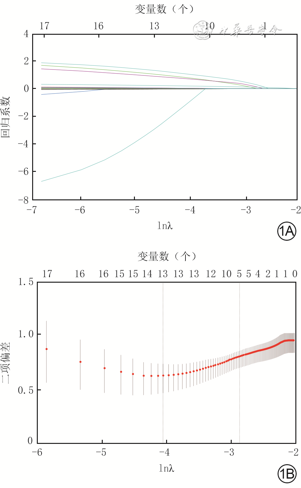
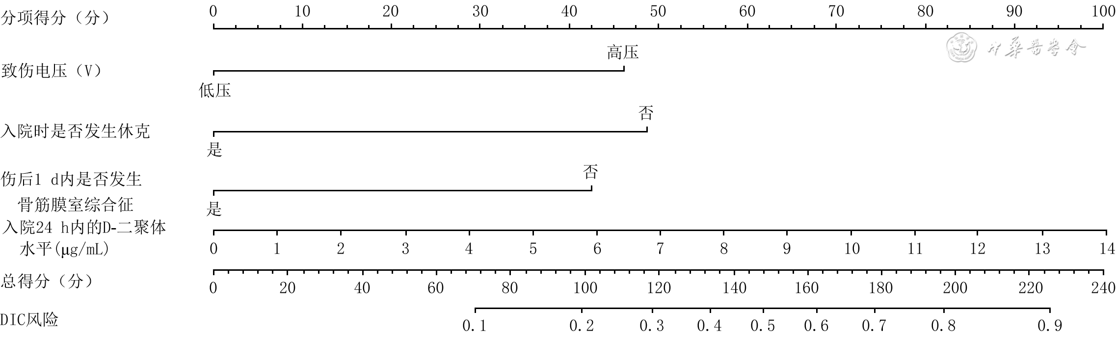
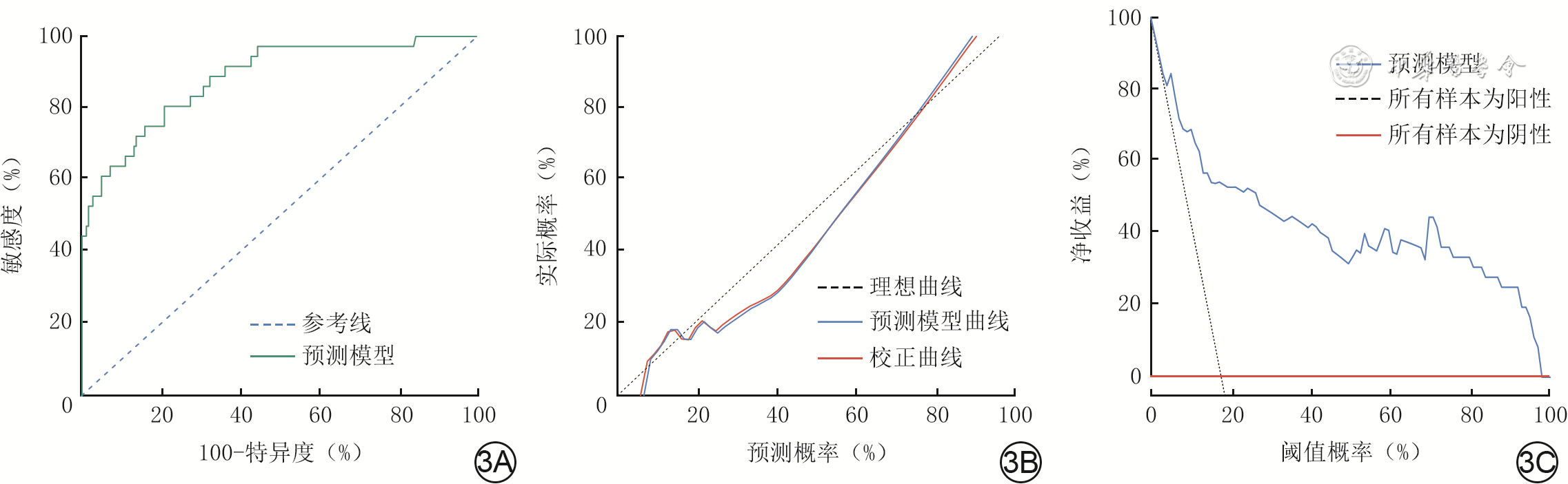
  Objective   To establish and validate a risk prediction model of disseminated intravascular coagulation (DIC) by the screening independent risk factors for the occurrence of DIC in patients with electrical burns.   Methods   The retrospective case series study was conducted. The clinical data of 218 electrical burn patients admitted to Baogang Hospital of Inner Mongolia from January 2015 to January 2023 who met the inclusion criteria were collected, including 198 males and 20 females, with the age of (38±14) years. The patients were divided into DIC group and non DIC group based on whether they were diagnosed with DIC during the treatment period. The following data of patients of two groups were collected and compared, including age, gender, total burn area, full-thickness burn area, injury voltage, whether osteofascial compartment syndrome occurred within 1 day after injury, duration of stay in burn intensive care unit, total length of hospital stay, whether combined with inhalation injury and multiple injuries, whether shock occurred upon admission, the abbreviated burn severity index score, and the acute physiology and chronic health evaluation Ⅱ score. The laboratory examination data of the patients within 24 hours after admission were also collected, including blood routine indexes: white blood cell count (WBC), hemoglobin level, platelet count (PLT), and neutrophil count; coagulation indexes: activated partial thromboplastin time (APTT), prothrombin time, thrombin time, and levels of D-dimer and fibrinogen (FIB); blood biochemistry indexes: aspartic transaminase, alanine transaminase, direct bilirubin, total bilirubin, total protein, albumin, blood glucose, creatinine, and urea nitrogen; blood gas analysis indexes: blood pH value, arterial partial pressure of oxygen, arterial partial pressure of carbon dioxide, bicarbonate, and base excess; and cardiac zymogram indexes: levels of myoglobin, troponin, lactate dehydrogenase, creatine kinase (CK), and α-hydroxybutyrate dehydrogenase. Data were statistically analyzed with chi-square test, Fisher's exact probability test, independent sample t test, and Mann-Whitney U test. For the variables with statistically significant differences in single factor analysis, the least absolute value selection and shrinkage operator (LASSO) regression was used to reduce the dimension, and the predictive factors for DIC in 218 patients with electrical burns were screened. The above-mentioned predictors were included in multivariate logistic regression analysis to find out the independent risk factors for DIC in 218 patients with electrical burns, and to draw the prediction model nomograms. The performance of the prediction model was evaluated by the receiver operating characteristic (ROC) curve and the area under the ROC curve, and the prediction model was validated by the calibration curve and clinical decision curve analysis (DCA).   Results   Compared with those in non DIC group, the total burn area, full-thickness burn area, total length of hospital stay, and the proportions of high voltage caused injury, occurrence of osteofascial compartment syndrome within 1 day after injury, combination of inhalation injury, and occurrence of shock upon admission of patients in DIC group were significantly increased/prolonged (with Z values of -2.53, -4.65, and -2.10, respectively, with χ 2 values of 11.46, 16.00, 7.98, and 18.93, respectively, P<0.05). Compared with those in non DIC group, the APTT, level of D-dimer, myoglobin, WBC, PLT, and levels of FIB, total bilirubin, and CK of patients within 24 hours after admission in DIC group were significantly prolonged/increased (with Z values of -2.02, -4.51, and -3.82, respectively, with t values of -3.84, -2.34, -2.77, -2.70, and -2.61, respectively), and the level of total protein and blood pH value were significantly reduced ( t=-2.85, Z=-2.03), P<0.05. LASSO regression analysis was carried out for the above 17 indicators with statistically significant differences. The results showed that injury voltage, the occurrence of shock upon admission, the occurrence of osteofascial compartment syndrome within 1 day after injury, and levels of D-dimer and total protein within 24 hours after admission were predictive factors for the occurrence of DIC in 218 patients with electrical burns (with regression coefficients of 0.24, 0.52, 0.35, 0.13, and -0.001, respectively). Multivariate logistic regression analysis showed that injury voltage, the occurrence of shock upon admission, the occurrence of osteofascial compartment syndrome within 1 day after injury, and D-dimer level within 24 hours after admission were independent risk factors for DIC in 218 patients with electrical burns (with odds ratios of 3.33, 4.24, 2.68, and 1.38, respectively, with 95% confidence intervals of 1.43-7.79, 1.78-10.07, 1.17-6.13, and 1.19-1.61, respectively, P<0.05). Based on the aforementioned four independent risk factors, the nomogram of prediction model for evaluating the probability of DIC in patients was drawn. The area under the ROC curve of prediction model was 0.88, and the 95% confidence interval was 0.82-0.95, indicating that the model had good predictive ability; the curve of prediction model tended to be near the ideal curve, indicating that the model had a high calibration degree; the clinical DCA of prediction model showed that the threshold probability of patients ranged from 4% to 97%, indicating that the model had good predictive ability.   Conclusions   The injury voltage, the occurrence of shock upon admission, the occurrence of osteofascial compartment syndrome within 1 day after injury, and D-dimer level within 24 hours after admission are independent risk factors for the occurrence of DIC in patients with electrical burns. The prediction model established based on the above indicators can provide early warning for the occurrence of DIC in these patients.
Original Articles
Prospective study on the influence of dobutamine on blood perfusion in free flap repair of diabetic foot wounds
Dou Xuejiao, Wang Haiying, Chen Wei, Zhou Jian, Wei Zairong
2023, 39(8): 746-752. doi: 10.3760/cma.j.cn501225-20221220-00543
Abstract:
  Objective   To investigate the influence of clinical administration of dobutamine on blood perfusion in free flap repair of diabetic foot wounds.   Methods   A prospective self‐controlled study was conducted. From January to November 2022, 20 patients with diabetic foot who met the inclusion criteria were hospitalized in the Department of Burns and Plastic Surgery of Affiliated Hospital of Zunyi Medical University, including 9 males and 11 females, aged from 44 to 75 years, with the foot wounds area ranging from 5 cm×4 cm to 20 cm×10 cm, which were repaired by free anterolateral thigh flaps. Heart rate (HR) and mean arterial pressure (MAP) were recorded before anesthesia induction, 10 minutes after vascular recanalization, when the target blood pressure (i.e., MAP being 6-10 mmHg (1 mmHg=0.133 kPa) higher than that before anesthesia induction) was reached after infusion of dobutamine, and 10 minutes after tracheal catheter removal. Additionally, indocyanine green, a contrast agent, was injected intravenously at 10 minutes after vascular recanalization and when the target blood pressure was reached after infusion of dobutamine to assess flap blood perfusion using infrared imager, and the area ratio of flaps with hyperperfusion and hypoperfusion was calculated. Other recorded variables included flap harvesting area, surgical duration, total fluid infusion amount, infusion dose and total usage of dobutamine, intraoperative adverse events, postoperative flap complications, and follow‐up outcomes. Data were statistically analyzed with paired sample t test, analysis of variance for repeated measurement, Bonferroni method, and generalized estimating equation.   Results   Compared with those before anesthesia induction, HR and MAP of patients were significantly decreased at 10 minutes after vascular recanalization ( P<0.05), while HR and MAP of patients were significantly increased when the target blood pressure was reached after infusion of dobutamine ( P<0.05). Compared with those at 10 minutes after vascular recanalization, HR and MAP of patients were significantly increased when the target blood pressure was reached after infusion of dobutamine and at 10 minutes after tracheal catheter removal ( P<0.05). Compared with those when the target blood pressure was reached after infusion of dobutamine, HR and MAP of patients were significantly decreased at 10 minutes after tracheal catheter removal ( P<0.05). The area ratio of flaps with hyperperfusion of patients was 0.63±0.11 when the target blood pressure was reached after infusion of dobutamine, which was significantly higher than 0.31±0.09 at 10 minutes after vascular recanalization ( t=-9.92, P<0.05). The area ratio of flaps with hypoperfusion of patients was 0.12±0.05 when the target blood pressure was reached after infusion of dobutamine, which was significantly lower than 0.45±0.10 at 10 minutes after vascular recanalization ( t=17.05, P<0.05). The flap harvesting area of patients was (174±35) cm², the surgical duration was (372±52) min, the total fluid infusion amount was (2 485±361) mL, the infusion dose of dobutamine was 3-13 μg·kg⁻¹·min⁻¹, and the total usage of dobutamine was 5.7 (2.1, 9.7) mg. Two patients showed a significant increase in MAP during the infusion of dobutamine compared with that at 10 minutes after vascular recanalization, but before reaching 6 mmHg higher than that before anesthesia induction, their HR had reached the maximum (over 130 beats/min). The HR gradually returned to around 90 beats/min after the infusion of dobutamine was stopped. On post operation day 2, one patient had partial necrosis at the distal part of the flap, which was repaired by transplantation of thin split-thickness skin graft from the opposite thigh. During the follow‐up of 3 to 6 months after operation, all the flaps survived well, with soft texture and well-formed shape, and no adverse cardiovascular events of patients were reported.   Conclusions   The administration of dobutamine in free flap repair of diabetic foot wounds can significantly improve the MAP of patients, expand the area of hyperperfusion, reduce the area of hypoperfusion, and enhance the flap viability, with promising short‐term follow‐up results, which is suitable for promotion in clinical applications.
Clinical effects of free perforator propeller myocutaneous flap from buttock in repairing complex wounds in the buttock with deep dead cavity
Chen Liming, Wang Gang, Liu Hong, Zhao Ruomei, Liu Xiaohui, Cao Guangtong, Yao Zongjiang, Li Zhiyong, Liu Yi
2023, 39(8): 753-757. doi: 10.3760/cma.j.cn501225-20221115-00488
Abstract:
  Objective   To investigate the feasibility and clinical effects of using free perforator propeller myocutaneous flap from buttock in repairing complex wounds in the buttock with deep dead cavity.   Methods   A retrospective observational study was conducted. From June 2020 to June 2022, 9 patients with complex wounds in the buttock with deep dead cavity who met the inclusion criteria were admitted to Lanzhou University Second Hospital, including 6 males and 3 females, aged 26 to 62 years, with original wound area ranging from 4.0 cm×3.0 cm to 8.0 cm×7.0 cm and dead cavity depth of 7 to 11 cm. All the wounds were repaired with free perforator propeller myocutaneous flap from buttock, with flap area of 6.0 cm×2.5 cm to 13.0 cm×7.0 cm and muscle flap length of 6 to 11 cm. All the wounds in the donor area were closed and sutured directly. Postoperative myocutaneous flap survival, complications, as well as donor and recipient wound healing were observed, and the shape of donor and recipient areas were followed up.   Results   Congestion occurred under the myocutaneous flap of one patient due to poor drainage on post surgery day 2, which was healed after 15 days of drainage and dressing change. The myocutaneous flaps of other patients survived successfully after surgery. The wounds in the donor and recipient areas were all well healed. During the follow-up of 3 to 10 months, the donor and recipient areas were full in shape, with little difference from the healthy side, and were able to bear pressure.   Conclusions   The free perforator propeller myocutaneous flap from buttock can repair the deep dead cavity and surface wounds at the same time. The use of this myocutaneous flap in repairing complex wounds in the buttock with deep dead cavity results in minimal damage to the donor area, allows pressure-bearing of the donor and recipient areas after surgery, and ensures a full buttock shape.
Clinical application of combination of different types of free perforator flaps in the repair of complex wounds in extremities
Li Hai, Xiao Shun'e, Deng Chengliang, Wu Bihua, Wu Xiangkui, Zhang Tianhua, Liu Zhiyuan, Wei Zairong
2023, 39(8): 758-764. doi: 10.3760/cma.j.cn501225-20220720-00300
Abstract:
  Objective   To investigate the clinical application effects of combination of different types of free perforator flaps in the repair of complex wounds in extremities.   Methods   A retrospective observational study was conducted. From January 2018 to June 2022, 11 patients with complex wounds in extremities who met the inclusion criteria was admitted to the Affiliated Hospital of Zunyi Medical University, including 8 males and 3 females, aged 28 to 55 years. The wounds in the upper extremities in 4 cases and in the lower extremities in 7 cases were repaired with different combination of free perforator flaps. After debridement, the wound area was 7.0 cm×6.0 cm-28.0 cm×12.0 cm. A combination of different types of perforator flaps were applied, including the perforator tri-leaf flap of the descending branch of the lateral femoral circumflex artery in 6 cases, the descending branch of lateral femoral circumflex artery combined with oblique branch perforating branch flap in 2 cases, the lobulated flap of the descending branch of the lateral femoral circumflex artery combined with the contralateral medial plantar artery perforator flap in 2 cases, and the bilateral perforator flap of the descending branch of lateral femoral circumflex artery combined with great toe nail flap in 1 case, with the size of a single flap ranged from 2.0 cm×2.0 cm-25.0 cm×6.0 cm. The donor site was repaired by direct suture, skin grafting, or flap transplantation. During free flap transplantation, the flap was cut and split according to the distribution of perforators, and end-to-end or end-to-side anastomosis was performed between the donor area and the recipient area. After surgery, the survival of transplanted flap in the primary recipient site, the occurrence of vascular crisis, the wound healing in the flap donor site, and the survival of transplanted skin or flap in the flap donor site were observed. During follow-up, the blood supply, appearance and texture of the transplanted flap in the primary recipient site were observed; and at the same time, the weight bearing of the plantar receiving area, the presence of sliding, ulcers, and sinus tracts of the flap, and the appearance and function of the hand were observed; the complications in the donor area were observed.   Results   After surgery, one patient's transplanted flap in the primary recipient site had vascular crisis but survived after exploration+vein graft bridging; partial necrosis occurred in one lobe of anterolateral thigh lobulated flap transplanted to the primary recipient site in one patient and recovered after dressing change+skin grafting, and the different types of perforator flap transplanted to the primary recipient site in the other 9 patients all survived. After surgery, the wound with direct suture at the donor site healed well, and the skin or flap transplanted to the donor area survived well. During 3-24 months of follow-up, the blood supply, appearance, and texture of the transplanted flap at the primary recipient site were good. In two patients, the anterolateral thigh flap combined with the medial plantar flap were used to repair plantar defects. The plantar receiving area was able to bear weight, and the texture of the flaps in the recipient area was close to the normal plantar skin, without flap sliding, ulcer, or sinus tract formation. In one patient, bilateral anterolateral thigh flap combined with great toe nail flap were used to repair hand combined with soft forearm defect, and the appearance and function of hand, especially thumb were good. Only linear scar was left in the donor site without other obvious complications.   Conclusions   The combination of different types of perforator flaps is a reliable clinical method to repair complex wounds in extremities with high safety, good efficacy, and less complications.
Effects of tibial second toe free flap bridged with blood flow and nerve in the treatment of severe flexion contracture of the proximal interphalangeal joint
Wang Kai, Jin Guangzhe, Teng Zhicheng, Ge Chengwei, Liu Zhijin, Ju Jihui, Dong Shuai, Wang Qiang, Li Yadong
2023, 39(8): 765-770. doi: 10.3760/cma.j.cn501225-20220707-00286
Abstract:
  Objective   To investigate the effects of tibial second toe free flap bridged with blood flow and nerve in the treatment of severe flexion contracture of the proximal interphalangeal joint.   Methods   A retrospective observational study was conducted. From March 2013 to October 2019, 9 patients with severe flexion contracture (type Ⅲ) of the proximal interphalangeal joint after trauma operation, conforming to the inclusion criteria, were hospitalized in Suzhou Ruihua Orthopaedic Hospital, including 5 males and 4 females, aged from 17 to 62 years. After the contracture tissue affecting the extension of the proximal interphalangeal joint was cut off, and the scar tissue was resected, the size of the volar wound near the proximal interphalangeal joint in extended position was 2.0 cm×1.0 cm-2.5 cm×1.5 cm, with the length of proper digital artery and nerve defect being 1.0-1.5 cm. A free flap of the same size as the wound was cut from the tibial side of the second toe and transplanted to repair the wound, and the defective proper digital artery and nerve was repaired by bridging with the tibial proper plantar digital artery and nerve of about 1.5 cm in length. The full-thickness skin graft was taken from the proximal tibial side of the lower leg to repair the wound at flap donor site. The wound at skin graft donor site was sutured directly. The survival of flap and skin graft was observed after operation. The patients were followed up, and at the last follow-up, the recovery of the affected finger and the second toe, including the donor and recipient areas were observed, the two-point discrimination distances of the flap repaired site and the pulp of the affected finger were observed and measured at the same time, the blood flow patency of bridged vessel of the affected finger was examined by Allen test, and the function of the proximal interphalangeal joint of the affected finger was evaluated according to Chinese Medical Association's standard for the range of motion of proximal interphalangeal joint.   Results   The flaps and skin grafts survived smoothly after operation. The follow-up after operation lasted for 5 to 22 months, with a mean of 10 months. At the last follow-up, the flap repaired site had good shape, good color and texture, with the two-point discrimination distance being 9-12 mm, and the two-point discrimination distance of the pulp of the affected finger was 6-10 mm; the Allen test results of the affected fingers were all negative (i.e., the bridged vessels had good blood flow patency), with no recurrence of flexion contracture, and the function of the proximal interphalangeal joint was evaluated as excellent; the skin graft area of the second toe was not ruptured but was a little pigmented, and the flexion and extension activities of toe were good.   Conclusions   The tibial second toe free flap bridged with blood flow and nerve has reliable therapeutic effect in the treatment of severe flexion contracture of the proximal interphalangeal joint, and the color and texture of the flap repaired area are good. Bridging to repair the severely contracted proper digital artery and nerve is beneficial to improve the blood supply of the finger body and rebuild the sensation.
Effects of advanced platelet-rich fibrin on deep partial-thickness burn wounds in nude mice
Tang Lijun, Li Xiaomei, Zhang Xiaowei, Luo Yi, Xu Gang
2023, 39(8): 771-778. doi: 10.3760/cma.j.cn501225-20220804-00334
Abstract:
  Objective   To explore the effects of advanced platelet-rich fibrin (A-PRF) on deep partial-thickness burn wounds in nude mice and its mechanism.   Methods   The experimental study method was adopted. Forty healthy volunteers in Subei People's Hospital were recruited, including 32 females and 8 males, aged 60 to 72 years. Leukocyte platelet-rich fibrin (L-PRF) and A-PRF membranes were prepared after venous blood was extracted from them. The microstructure of two kinds of platelet-rich fibrin (PRF) membranes was observed by field emission scanning electron microscope. The number of samples was 3 in the following experiments. The L-PRF and A-PRF membranes were divided into L-PRF group and A-PRF group and cultured, and then the release concentrations of platelet-derived growth factor-AB (PDGF-AB) and vascular endothelial growth factor (VEGF) in culture supernatant were determined by enzyme-linked immunosorbent assay on culture day 1, 3, 7, and 14. Mice L929 fibroblasts (Fbs) were divided into L-PRF group and A-PRF group, and cultured with L-PRF or A-PRF conditioned medium, respectively. On culture day 1, 3, and 7, the cell proliferation activity was detected by thiazole blue method. The cell migration rate was detected and calculated at 24 h after scratching by scratch test. Thirty-six male BALB/c nude mice aged 6-8 weeks were selected to make a deep partial-thickness burn wound on one hind leg, and then divided into normal saline group, L-PRF group, and A-PRF group, according to the random number table, with 12 mice in each group. The wounds of nude mice in normal saline group were only washed by normal saline, while the wounds of nude mice in L-PRF group and A-PRF group were covered with the corresponding membranes in addition. The wounds of nude mice in the 3 groups were all bandaged and fixed with dressings. On treatment day 4, 7, and 14, the wound healing was observed and the wound healing rate was calculated. Masson staining was used to observe the new collagen in wound tissue, and immunohistochemical staining was used to detect the percentage of CD31 positive cells in the wound. Data were statistically analyzed with independent sample t test, analysis of variance for repeated measurement, analysis of variance for factorial design, one-way analysis of variance, and least significant difference test.   Results   L-PRF membrane's dense network structure was composed of coarse fibrin bundles, with scattered white blood cells and platelets with complete morphology. A-PRF membrane's loose network structure was composed of fine fibrin bundles, with scattered small amount of deformed white blood cells and platelets. On culture day 1, the release concentration of PDGF-AB in PRF culture supernatant in A-PRF group was significantly higher than that in L-PRF group ( t=5.73, P<0.05), while the release concentrations of VEGF in PRF culture supernatant in the two groups were similar ( P>0.05). On culture day 3, 7, and 14, the release concentrations of PDGF-AB and VEGF in PRF culture supernatant in A-PRF group were significantly higher than those in L-PRF group (with t values of 6.93, 7.45, 5.49, 6.97, 8.97, and 13.64, respectively, P<0.05). On culture day 3, 7, and 14, the release concentrations of PDGF-AB and VEGF in PRF culture supernatant in the two groups were all significantly higher than those in the previous time points within the group ( P<0.05). On culture day 1, 3, and 7, the proliferation activity of mice Fbs in A-PRF group was 0.293±0.034, 0.582±0.054, and 0.775±0.040, respectively, which were significantly stronger than 0.117±0.013, 0.390±0.036, and 0.581±0.037 in L-PRF group (with t values of 8.38, 5.14, and 6.16, respectively, P<0.05). At 24 h after scratching, the migration rate of mice Fbs in A-PRF group was (60.9±2.2)%, which was significantly higher than (39.1±2.3)% in L-PRF group ( t=11.74, P<0.05). On treatment day 4, the wound exudates of nude mice in L-PRF group and A-PRF group were less with no obvious signs of infection, while the wounds of nude mice in normal saline group showed more exudation. On treatment day 7, the wounds of nude mice in L-PRF group and A-PRF group were dry and crusted, while there was still a small amount of exudate in the wounds of nude mice in normal saline group. On treatment day 14, the wounds of nude mice in A-PRF group tended to heal; a small portion of wounds remained in nude mice in L-PRF group; the wound of nude mice was still covered with eschar in normal saline group. On treatment day 4, 7, and 14, the wound healing rate and percentage of CD31 positive cells of nude mice in L-PRF group were all significantly higher than those in normal saline group ( P<0.05); compared with those in normal saline group and L-PRF group, the wound healing rate of nude mice in A-PRF group was significantly increased ( P<0.05), the newborn collagen was orderly and evenly distributed, with no excessive deposition, and the percentage of CD31 positive cells was significantly increased ( P<0.05).   Conclusions   The stable fibrin network structure of A-PRF can maintain the sustained release of growth factors, accelerate cell proliferation, and promote cell migration, so as to shorten the healing time and improve the healing quality of deep partial-thickness burn wounds in nude mice.
Original Article·Nursing Column
Influence of work engagement and self-efficacy of nurses on clinical practice ability in burn intensive care unit
Chen Hanxi, Liu Wenji, Liu Bing, Huang Zhifeng, Zhang Qiuping, Xiao Xiling, Lai Wen, Zheng Shaoyi
2023, 39(8): 779-786. doi: 10.3760/cma.j.cn501225-20220905-00379
Abstract:
  Objective   To analyze the influence of work engagement and self-efficacy of nurses on clinical practice ability in burn intensive care unit (BICU), and to explore its potential pathways of action.   Methods   A cross-sectional survey was conducted. From May to October 2020, a total of 30 hospitals with BICU in China were selected by stratified sampling method. Among BICU nurses who met the inclusion criteria, their clinical practice ability, work engagement, and self-efficacy were evaluated by self-evaluation scale of oriented problem-solving behavior in nursing practice (OPSN), Utrecht work engagement scale (UWES), and general self-efficacy scale (GSES), respectively. The total scale scores of each index and the average item scores were recorded. The self-designed general data questionnaire was used to investigate the nurses' gender, age, marital status, education background, working years, professional title, and the economic region of the hospital that they belonged to. The total scale scores of the above-mentioned three evaluation indexes were compared after the classification of nurses according to general data, and the data were statistically analyzed with independent sample t test or one-way analysis of variance. Pearson correlation analysis was used to analyze the correlation between the total scale scores of the three evaluation indexes. Based on the total scale scores of the above-mentioned three evaluation indexes, a structural equation model was established, the mediation analysis of the relationship among the three evaluation indexes and the pathway analysis of the structural model were conducted, and the Bootstrap method was used to verify the pathways of action.   Results   A total of 401 questionnaires were distributed, and 337 valid questionnaires were returned, with a valid return rate of 84.04%. The total scale scores of clinical practice ability, work engagement, and self-efficacy of 337 nurses were 98.2±11.7, 67.7±18.6, and 26.6±5.6, respectively, and the average item scores were 3.9±0.5, 4.5±1.2, and 2.7±0.6, respectively. Among the 337 nurses, the majority were female, aged 40 or below, married, and had a bachelor's degree with work experience of ≤10 years; both nurses with professional nurse title and nurses from the Southeast region accounted for about 50%. There were statistically significant differences in the total scale score of clinical practice ability among nurses with different ages, education backgrounds, working years, and professional titles (with F values of 3.26, 4.36, 3.12, and 2.80, respectively, P<0.05). There was statistically significant difference in the total scale score of work engagement among nurses with different working years ( F=4.50, P<0.05). There were statistically significant differences in the total scale score of self-efficacy among nurses with different ages, working years, and professional titles (with F values of 4.91, 4.50, and 2.91, respectively, P<0.05). The total scale score of nurses' work engagement was significantly positively correlated with the total scale score of clinical practice ability and the total scale score of self-efficacy (with rvalues of 0.30 and 0.51, respectively, P<0.05). The total scale score of nurses' self-efficacy was significantly positively correlated with the total scale score of clinical practice ability ( r=0.37, P<0.05). The model had good adaptability, and the intermediary model was established. Nurses' work engagement had a significantly positive effect on both self-efficacy and clinical practice ability (with βvalues of 0.54 and 0.16, respectively, P<0.05), and nurses' self-efficacy had a significantly positive effect on clinical practice ability ( β=0.29, P<0.05). Work engagement had a direct effect on self-efficacy and clinical practice ability, and self-efficacy had a direct effect on clinical practice ability and played a mediating role between work engagement and clinical practice ability. Bootstrap validation showed that self-efficacy played a significantly mediating role in the influence of work engagement on clinical practice ability (with effect size of 0.16, with 95% confidence interval of 0.08-0.24, P<0.05), accounting for half of the total effect of work engagement on clinical practice ability (with effect size of 0.32).   Conclusions   BICU nurses have an above-average level of clinical practice ability, a medium level of self-efficacy, and a high level of work engagement. Work engagement and self-efficacy are positively correlated with clinical practice ability. Work engagement can directly affect clinical practice ability or indirectly affect clinical practice ability through the mediating role of self-efficacy.
Case Report
A case with giant ulcer caused by skin diffuse large B-cell lymphoma in the axilla
Liu Feng, Zhang Wei, Xie Weiguo, Chen Lan, Li Ze
2023, 39(8): 787-789. doi: 10.3760/cma.j.cn501225-20220331-00116
Abstract:
In May 16 th, 2019, a male patient (aged 51 years) with a rare giant ulcer caused by skin diffuse large B-cell lymphoma in the axilla was admitted to Tongren Hospital of Wuhan University & Wuhan Third Hospital, and the ulcer wound was confirmed by biopsy and immunohistochemical analysis after extensive excision. Ultimately, a good prognosis was obtained by transplantation of flap and skin graft in combination with radiotherapy and chemotherapy.
Reviews
Research advances on post-burn cognitive disorders and its mechanism
Luo Yue, Liang Guangping
2023, 39(8): 790-794. doi: 10.3760/cma.j.cn501225-20221117-00492
Abstract:
Cognitive disorders after burns is an important problem that cannot be ignored in the diagnosis and treatment of burn patients. Its mechanism may be related to the activation of inflammatory response, destruction of the blood-brain barrier, hypothalamic-pituitary-adrenal axis imbalance, nerve cell changes, etc. Mechanism-specific prevention and treatment will be the best choice to reduce post-burn cognitive disorders in the future. This review introduces the research advances in epidemiology, risk factors, possible pathogenesis, diagnosis, treatment, and prevention of cognitive disorders in burn patients, in order to provide a reference for timely and accurate assessment, treatment, and prevention of cognitive disorders in burn patients.
Research advances on the role of cell senescence in chronic wound healing
Fang Shaoyihan, Liu Dewu
2023, 39(8): 795-800. doi: 10.3760/cma.j.cn501225-20220928-00424
Abstract:
Chronic wounds bring huge pressure and difficulties to patients, their families, and society due to their long-term refractory characteristics and serious poor prognosis. Recently, more and more evidences have proven that cell senescence exists in chronic wounds and affects wound healing. This article reviews the characteristics of cell senescence in chronic wounds, discusses the relationship between cell senescence and chronic wound healing, and summarizes current series of anti-senescence strategies to bring out the possibility of treating chronic wounds with cell senescence as a potential target.