


Influence and mechanism of extracellular vesicles derived from human adipose-derived mesenchymal stem cells on pyroptosis of human umbilical vein endothelial cells induced by high glucose
-
摘要:
目的 探讨人脂肪间充质干细胞(hADMSC)来源细胞外囊泡(EV),即hADMSC-EV对高糖诱导的人脐静脉内皮细胞(HUVEC)焦亡的影响及其机制,为改善糖尿病创面中血管功能提供依据。 方法 该研究为实验研究。收集2023年6—9月于南昌大学第一附属医院妇产科完成正常阴道分娩的5名25~40岁产妇的脐带,分离HUVEC并成功鉴定;取同期于该院整形外科行腹部抽脂术的6名25~35岁健康女性的脂肪组织,分离hADMSC后提取hADMSC-EV并成功鉴定。将用含物质的量浓度为33 mmol/L葡萄糖的内皮细胞培养基培养的第4代HUVEC,分为加入磷酸盐缓冲液(PBS)培养的PBS组、加入hADMSC-EV培养的EV组、加入磷脂酰肌醇3-激酶/蛋白激酶B(PI3K/Akt)信号通路抑制剂LY294002和hADMSC-EV培养的EV+LY294002组,采用蛋白质印迹法检测细胞培养48 h后PI3K/Akt信号通路相关蛋白PI3K、Akt和焦亡相关蛋白核苷酸结合寡聚化结构域样受体蛋白3(NLRP3)、胱天蛋白酶1(caspase-1)、消皮素D、白细胞介素1β(IL-1β)、IL-18的蛋白表达;采用细胞计数试剂盒-8检测细胞培养0(即刻)、12、24、36、48、60、72 h增殖水平;培养48 h后,行细胞划痕试验并计算划痕后12、24 h的细胞迁移率,行细胞Transwell试验并计算细胞24 h迁移数量,行细胞成管实验并测算细胞成管总长度与分支节点数。样本数均为3。 结果 培养48 h后,EV组细胞中PI3K和Akt的蛋白表达均明显高于PBS组(P<0.05),EV+LY294002组细胞中PI3K和Akt的蛋白表达均明显低于EV组(P<0.05)。培养48 h后,EV组细胞中NLRP3、caspase-1、消皮素D、IL-1β、IL-18的蛋白表达分别为0.54±0.08、0.96±0.11、0.525±0.061、1.216±0.039、1.317±0.023,均明显低于PBS组的2.32±0.11、1.86±0.07、1.256±0.113、2.589±0.084、2.042±0.132(P<0.05);EV+LY294002组细胞中NLRP3、caspase-1、消皮素D、IL-1β、IL-18的蛋白表达分别为1.16±0.05、1.37±0.06、0.962±0.028、1.834±0.017、1.803±0.065,均明显高于EV组(P<0.05)。培养12、24、36、48、60、72 h,EV组细胞增殖水平均明显高于PBS组(P<0.05),EV+LY294002组细胞增殖水平均明显低于EV组(P<0.05)。培养48 h后,EV组细胞划痕后12、24 h迁移率均明显高于PBS组(P<0.05),EV+LY294002组细胞划痕后12、24 h迁移率均明显低于EV组(P<0.05);EV组细胞24 h迁移数量明显多于PBS组(P<0.05),EV+LY294002组细胞24 h迁移数量明显少于EV组(P<0.05)。培养48 h后,与PBS组比较,EV组细胞成管总长度明显延长(P<0.05)且分支节点数明显增多(P<0.05);与EV组比较,EV+LY294002组细胞成管总长度明显缩短(P<0.05)且分支节点数明显减少(P<0.05)。 结论 hADMSC-EV可通过PI3K/Akt信号通路抑制高糖诱导的HUVEC焦亡相关蛋白的表达,并改善细胞增殖、迁移和血管形成能力。 Abstract:Objective To investigate the influence and mechanism of extracellular vesicles (EVs) derived from human adipose-derived mesenchymal stem cells (hADMSCs), i.e. hADMSC-EVs on pyroptosis of human umbilical vein endothelial cells (HUVECs) induced by high glucose, with the aim of providing evidence for improving vascular function in diabetic wounds. Methods This study was an experimental research. The umbilical cords from 5 women aged 25 to 40 years were collected who had normal vaginal delivery at the Department of Obstetrics and Gynecology of the First Affiliated Hospital of Nanchang University from June to September in 2023, and HUVECs were isolated and successfully identified. Adipose tissue was obtained from 6 healthy women aged 25 to 35 years who underwent abdomen liposuction at the Department of Plastic Surgery of the above-mentioned hospital in the same period. After hADMSCs were isolated, hADMSC-EVs were extracted and successfully identified. The fourth passage of HUVECs were cultured in endothelial cell medium containing glucose in a molarity of 33 mmol/L and divided into phosphate buffered solution (PBS) group cultured with PBS, EV group cultured with hADMSC-EVs, and EV+LY294002 group cultured with hADMSC-EVs and the phosphatidylinositol 3-kinase/protein kinase B (PI3K/Akt) signaling pathway inhibitor LY294002. Western blotting was used to detect the expressions of PI3K/Akt signaling pathway-related proteins PI3K and Akt, and pyroptosis-related proteins nucleotide-binding oligomerization domain-like receptor protein 3 (NLRP3), cysteinyl aspartate specific protease-1 (caspase-1), gasdermin D, interleukin-1β (IL-1β), and IL-18 of cells after 48 hours of culture. A cell counting kit-8 was used to test the proliferation levels of cells at 0 (immediately), 12, 24, 36, 48, 60, and 72 hours of culture. After 48 hours of culture, the cell scratch test was performed and the cell migration rates at 12 and 24 hours after scratching were calculated; the cell Transwell assay was conducted and the number of cells migrating in 24 hours was calculated; the cell tube formation experiment was performed, and the total length of tube formation and the number of branch nodes were measured and counted. The sample size was 3. Results After 48 hours of culture, the protein expressions of PI3K and Akt of cells in EV group were significantly higher than those in PBS group (P<0.05), and the protein expressions of PI3K and Akt of cells in EV+LY294002 group were significantly lower than those in EV group (P<0.05). After 48 hours of culture, the protein expressions of NLRP3, caspase-1, gasdermin D, IL-1β, and IL-18 of cells in EV group were 0.54±0.08, 0.96±0.11, 0.525±0.061, 1.216±0.039, and 1.317±0.023, respectively, which were significantly lower than 2.32±0.11, 1.86±0.07, 1.256±0.113, 2.589±0.084, and 2.042±0.132 in PBS group (P<0.05); the protein expressions of NLRP3, caspase-1, gasdermin D, IL-1β, and IL-18 of cells in EV+LY294002 group were 1.16±0.05, 1.37±0.06, 0.962±0.028, 1.834±0.017, and 1.803±0.065, respectively, which were significantly higher than those in EV group (P<0.05). At 12, 24, 36, 48, 60, and 72 hours of culture, the proliferation levels of cells in EV group were significantly higher than those in PBS group (P<0.05), and the proliferation levels of cells in EV+LY294002 group were significantly lower than those in EV group (P<0.05). After 48 hours of culture, the cell migration rates at 12 and 24 hours after scratching in EV group were significantly higher than those in PBS group (P<0.05), and the cell migration rates at 12 and 24 hours after scratching in EV+LY294002 group were significantly lower than those in EV group (P<0.05); the number of cells migrating in 24 hours in EV group was significantly greater than that in PBS group (P<0.05), and the number of cells migrating in 24 hours in EV+LY294002 group was significantly less than that in EV group (P<0.05). After 48 hours of culture, compared with those in PBS group, the total length of tube formation of cells in EV group was significantly prolonged (P<0.05), and the number of branch nodes was significantly increased (P<0.05); compared with those in EV group, the total length of tube formation in EV+LY294002 group was significantly shortened (P<0.05), and the number of branch nodes was significantly decreased (P<0.05). Conclusions hADMSC-EVs can inhibit the expression of pyroptosis-related proteins in HUVECs induced by high glucose through the PI3K/Akt signaling pathway and improve the proliferation, migration, and angiogenesis capabilities of HUVECs. -
参考文献
(40) [1] PatelS,SrivastavaS,SinghMR,et al.Mechanistic insight into diabetic wounds: pathogenesis, molecular targets and treatment strategies to pace wound healing[J].Biomed Pharmacother,2019,112:108615.DOI: 10.1016/j.biopha.2019.108615. [2] PopMA,AlmquistBD.Biomaterials: a potential pathway to healing chronic wounds?[J].Exp Dermatol,2017,26(9):760-763.DOI: 10.1111/exd.13290. [3] ZhouC,ZhangB,YangY,et al.Stem cell-derived exosomes: emerging therapeutic opportunities for wound healing[J].Stem Cell Res Ther,2023,14(1):107.DOI: 10.1186/s13287-023-03345-0. [4] 陈祎琦,周莹芊,魏茜,等.负载人脐带间充质干细胞来源的小细胞外囊泡的甲基丙烯酸酐化明胶水凝胶治疗小鼠全层皮肤缺损创面的效果[J].中华烧伤与创面修复杂志,2024,40(4):323-332.DOI: 10.3760/cma.j.cn501225-20231218-00248. [5] 居裔昆,方柏荣.脂肪间充质干细胞外囊泡促进创面血管生成机制的研究进展[J].中华烧伤与创面修复杂志,2023,39(1):85-90.DOI: 10.3760/cma.j.cn501225-20220322-00080. [6] LelekJ,Zuba-SurmaEK.Perspectives for future use of extracellular vesicles from umbilical cord- and adipose tissue-derived mesenchymal stem/stromal cells in regenerative therapies-synthetic review[J].Int J Mol Sci,2020,21(3):799.DOI: 10.3390/ijms21030799. [7] BergsbakenT,FinkSL,CooksonBT.Pyroptosis: host cell death and inflammation[J].Nat Rev Microbiol,2009,7(2):99-109.DOI: 10.1038/nrmicro2070. [8] MullaJ,KattiR,ScottMJ.The role of gasdermin-D-mediated pyroptosis in organ injury and its therapeutic implications[J].Organogenesis,2023,19(1):2177484.DOI: 10.1080/15476278.2023.2177484. [9] BandharamN,LockeyRF,KolliputiN.Pyroptosis inhibition in disease treatment: opportunities and challenges[J].Cell Biochem Biophys,2023,81(4):615-619.DOI: 10.1007/s12013-023-01181-w. [10] MuX,WuX,HeW,et al.Pyroptosis and inflammasomes in diabetic wound healing[J].Front Endocrinol (Lausanne),2022,13:950798.DOI: 10.3389/fendo.2022.950798. [11] Al MamunA,ShaoC,GengP,et al.The mechanism of pyroptosis and its application prospect in diabetic wound healing[J].J Inflamm Res,2024,17:1481-1501.DOI: 10.2147/JIR.S448693. [12] LiuW,YuanY,LiuD.Extracellular vesicles from adipose-derived stem cells promote diabetic wound healing via the PI3K-AKT-mTOR-HIF-1α signaling pathway[J].Tissue Eng Regen Med,2021,18(6):1035-1044.DOI: 10.1007/s13770-021-00383-8. [13] SongY,YouY,XuX,et al.Adipose-derived mesenchymal stem cell-derived exosomes biopotentiated extracellular matrix hydrogels accelerate diabetic wound healing and skin regeneration[J].Adv Sci (Weinh),2023,10(30):e2304023.DOI: 10.1002/advs.202304023. [14] SidhomK,ObiPO,SaleemA.A review of exosomal isolation methods: is size exclusion chromatography the best option?[J].Int J Mol Sci,2020,21(18):6466.DOI: 10.3390/ijms21186466. [15] HouL,ZhangX,DuH.Advances in mesenchymal stromal cells and nanomaterials for diabetic wound healing[J].Diabetes Metab Res Rev,2023,39(4):e3638.DOI: 10.1002/dmrr.3638. [16] ChangM,NguyenTT.Strategy for treatment of infected diabetic foot ulcers[J].Acc Chem Res,2021,54(5):1080-1093.DOI: 10.1021/acs.accounts.0c00864. [17] DinhT,VevesA.Microcirculation of the diabetic foot[J].Curr Pharm Des,2005,11(18):2301-2309.DOI: 10.2174/1381612054367328. [18] Ramachandra BhatL,VedanthamS,KrishnanUM,et al.Methylglyoxal-an emerging biomarker for diabetes mellitus diagnosis and its detection methods[J].Biosens Bioelectron,2019,133:107-124.DOI: 10.1016/j.bios.2019.03.010. [19] KimJH,KimKA,ShinYJ,et al.Methylglyoxal induced advanced glycation end products (AGE)/receptor for AGE (RAGE)-mediated angiogenic impairment in bone marrow-derived endothelial progenitor cells[J].J Toxicol Environ Health A,2018,81(9):266-277.DOI: 10.1080/15287394.2018.1440185. [20] KimYH,TabataY.Recruitment of mesenchymal stem cells and macrophages by dual release of stromal cell-derived factor-1 and a macrophage recruitment agent enhances wound closure[J].J Biomed Mater Res A,2016,104(4):942-956.DOI: 10.1002/jbm.a.35635. [21] IzadiR,HejaziSH,BahramikiaS.Injection of stem cells derived from allogeneic adipose tissue, a new strategy for the treatment of diabetic wounds[J].J Diabetes Complications,2023,37(7):108496.DOI: 10.1016/j.jdiacomp.2023.108496. [22] ZhangH,GuY,ZhangK,et al.Roles and mechanisms of umbilical cord mesenchymal stem cells in the treatment of diabetic foot: a review of preclinical and clinical studies[J].J Diabetes Complications,2024,38(1):108671.DOI: 10.1016/j.jdiacomp.2023.108671. [23] WeiL,XuY,ZhangL,et al.Mesenchymal stem cells promote wound healing and effects on expression of matrix metalloproteinases-8 and 9 in the wound tissue of diabetic rats[J].Stem Cells Dev,2023,32(1/2):25-31.DOI: 10.1089/scd.2021.0218. [24] UzunE,GüneyA,GönenZB,et al.Intralesional allogeneic adipose-derived stem cells application in chronic diabetic foot ulcer: phase I/2 safety study[J].Foot Ankle Surg,2021,27(6):636-642.DOI: 10.1016/j.fas.2020.08.002. [25] ShiR,JinY,CaoC,et al.Localization of human adipose-derived stem cells and their effect in repair of diabetic foot ulcers in rats[J].Stem Cell Res Ther,2016,7(1):155.DOI: 10.1186/s13287-016-0412-2. [26] CaiF,ChenW,ZhaoR,et al.The capacity of exosomes derived from adipose-derived stem cells to enhance wound healing in diabetes[J].Front Pharmacol,2023,14:1063458.DOI: 10.3389/fphar.2023.1063458. [27] KalluriR,LeBleuVS.The biology, function, and biomedical applications of exosomes[J].Science,2020,367(6478):eaau6977.DOI: 10.1126/science.aau6977. [28] TangT,ChenL,ZhangM,et al.Exosomes derived from BMSCs enhance diabetic wound healing through circ-Snhg11 delivery[J].Diabetol Metab Syndr,2024,16(1):37.DOI: 10.1186/s13098-023-01210-x. [29] TengL,MaqsoodM,ZhuM,et al.Exosomes derived from human umbilical cord mesenchymal stem cells accelerate diabetic wound healing via promoting M2 macrophage polarization, angiogenesis, and collagen deposition[J].Int J Mol Sci,2022,23(18):10421.DOI: 10.3390/ijms231810421. [30] FuS,ZhangH,LiX,et al.Exosomes derived from human amniotic mesenchymal stem cells facilitate diabetic wound healing by angiogenesis and enrich multiple lncRNAs[J].Tissue Eng Regen Med,2023,20(2):295-308.DOI: 10.1007/s13770-022-00513-w. [31] 刘文剑,刘德伍.间充质干细胞来源细胞外囊泡促进糖尿病溃疡血管生成的研究进展[J].中华烧伤与创面修复杂志,2022,38(4):393-399.DOI: 10.3760/cma.j.cn501120-20201207-00520. [32] YuP,ZhangX,LiuN,et al.Pyroptosis: mechanisms and diseases[J].Signal Transduct Target Ther,2021,6(1):128.DOI: 10.1038/s41392-021-00507-5. [33] GuC,DragaD,ZhouC,et al.miR-590-3p inhibits pyroptosis in diabetic retinopathy by targeting NLRP1 and inactivating the NOX4 signaling pathway[J].Invest Ophthalmol Vis Sci,2019,60(13):4215-4223.DOI: 10.1167/iovs.19-27825. [34] YangK,LiuJ,ZhangX,et al.H3 relaxin alleviates migration, apoptosis and pyroptosis through P2X7R-mediated nucleotide binding oligomerization domain-like receptor protein 3 inflammasome activation in retinopathy induced by hyperglycemia[J].Front Pharmacol,2020,11:603689.DOI: 10.3389/fphar.2020.603689. [35] KongH,ZhaoH,ChenT,et al.Targeted P2X7/NLRP3 signaling pathway against inflammation, apoptosis, and pyroptosis of retinal endothelial cells in diabetic retinopathy[J].Cell Death Dis,2022,13(4):336.DOI: 10.1038/s41419-022-04786-w. [36] ShorningBY,DassMS,SmalleyMJ,et al.The PI3K-AKT-mTOR pathway and prostate cancer: at the crossroads of AR, MAPK, and WNT signaling[J].Int J Mol Sci,2020,21(12):4507.DOI: 10.3390/ijms21124507. [37] GoldbraikhD,NeufeldD,Eid-MutlakY,et al.USP1 deubiquitinates Akt to inhibit PI3K-Akt-FoxO signaling in muscle during prolonged starvation[J].EMBO Rep,2020,21(4):e48791.DOI: 10.15252/embr.201948791. [38] RezaeiS,NikpanjehN,RezaeeA,et al.PI3K/Akt signaling in urological cancers: tumorigenesis function, therapeutic potential, and therapy response regulation[J].Eur J Pharmacol,2023,955:175909.DOI: 10.1016/j.ejphar.2023.175909. [39] FontanaF,GiannittiG,MarchesiS,et al.The PI3K/Akt pathway and glucose metabolism: a dangerous liaison in cancer[J].Int J Biol Sci,2024,20(8):3113-3125.DOI: 10.7150/ijbs.89942. [40] AbidMR,GuoS,MinamiT,et al.Vascular endothelial growth factor activates PI3K/Akt/forkhead signaling in endothelial cells[J].Arterioscler Thromb Vasc Biol,2004,24(2):294-300.DOI: 10.1161/01.ATV.0000110502.10593.06. -

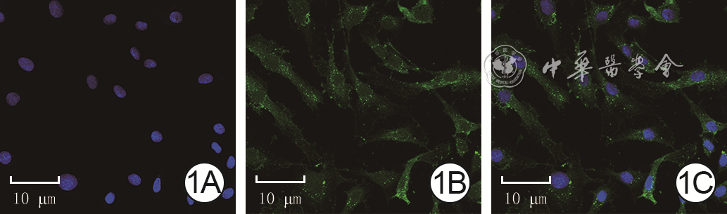
图 1 第4代人脐静脉内皮细胞形态及细胞中CD31表达情况 Alexa Fluor 488-4',6-二脒基-2-苯基吲哚×400。1A.细胞核(蓝色)染色情况;1B.细胞质中可见CD31阳性(绿色)表达;1C.1A与1B叠加图像,细胞呈燕麦状

图 2 第4代人脂肪间充质干细胞培养30 d后成脂与成骨分化情况。2A.细胞中可见大量钙结节(暗红色)形成 茜素红×100;2B.细胞中可见大小不一的脂滴(红色)形成 油红O×100

图 3 hADMSC-EV的形态与粒径及标记蛋白表达情况。3A.hADMSC-EV的形态为圆盘状,大小不等 透射电子显微镜×100 000;3B.hADMSC-EV粒径主要分布在100~150 nm;3C.蛋白质印迹法检测显示hADMSC-EV表达囊泡特异性标记蛋白CD9、CD63、Alix
注:hADMSC-EV为人脂肪间充质干细胞来源细胞外囊泡,Alix为凋亡连接基因2相互作用蛋白X;图3B为横坐标经过lg处理后生成的图

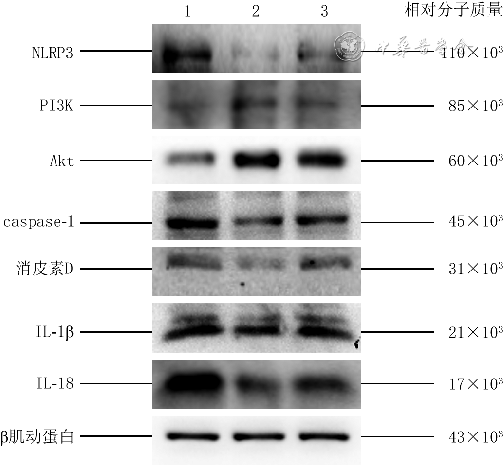
图 4 蛋白质印迹法检测的3组人脐静脉内皮细胞培养48 h后PI3K/Akt信号通路和焦亡相关蛋白的蛋白表达
注:NLRP3为核苷酸结合寡聚化结构域样受体蛋白3,PI3K为磷脂酰肌醇3-激酶,Akt为蛋白激酶B,caspase-1为胱天蛋白酶1,IL为白细胞介素;条带上方1、2、3分别指示在高糖培养条件下分别加入磷酸盐缓冲液(PBS)、人脂肪间充质干细胞来源细胞外囊泡(hADMSC-EV)、LY294002+hADMSC-EV培养细胞的PBS组、细胞外囊泡(EV)组、EV+LY294002组

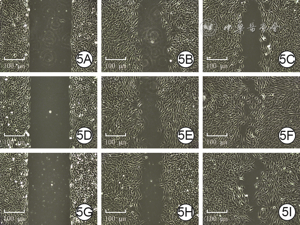
图 5 3组人脐静脉内皮细胞培养48 h后经划痕试验检测的划痕后各时间点水平迁移情况 倒置相差显微镜×50。5A、5B、5C.分别为PBS组细胞划痕后0(即刻)、12、24 h的划痕面积,逐渐缩小;5D、5E、5F.分别为EV组细胞划痕后0、12、24 h的划痕面积,图5D与图5A划痕面积相近,图5E、5F划痕面积分别明显小于图5B、5C;5G、5H、5I.分别为EV+LY294002组细胞划痕后0、12、24 h的划痕面积,图5G与图5D划痕面积相近,图5H、5I划痕面积分别明显大于图5E、5F
注:磷酸盐缓冲液(PBS)组、细胞外囊泡(EV)组、EV+LY294002组细胞于高糖培养条件下分别加入PBS、人脂肪间充质干细胞来源EV(hADMSC-EV)、LY294002+hADMSC-EV培养

图 6 3组人脐静脉内皮细胞培养48 h后经Transwell试验检测的24 h垂直迁移情况 结晶紫×200。6A.PBS组细胞迁移数量一般;6B.EV组细胞迁移数量明显多于图6A;6C.EV+LY294002组细胞迁移数量明显少于图6B
注:磷酸盐缓冲液(PBS)组、细胞外囊泡(EV)组、EV+LY294002组细胞于高糖培养条件下分别加入PBS、人脂肪间充质干细胞来源EV(hADMSC-EV)、LY294002+hADMSC-EV培养

图 7 3组人脐静脉内皮细胞培养48 h后成管情况 倒置相差显微镜×100。7A.PBS组血管较少;7B.EV组成管分支节点数明显多于图7A,成管总长度明显长于图7A;7C.EV+LY294002组成管分支节点数明显少于图7B,成管总长度明显短于图7B
注:磷酸盐缓冲液(PBS)组、细胞外囊泡(EV)组、EV+LY294002组细胞于高糖培养条件下分别加入PBS、人脂肪间充质干细胞来源EV(hADMSC-EV)、LY294002+hADMSC-EV培养
Table 1. 3组人脐静脉内皮细胞培养48 h后PI3K/Akt信号通路和焦亡相关蛋白的蛋白表达比较(
组别 样本数 PI3K Akt NLRP3 caspase-1 消皮素D IL-1β IL-18 PBS组 3 0.64±0.08 0.875±0.101 2.32±0.11 1.86±0.07 1.256±0.113 2.589±0.084 2.042±0.132 EV组 3 1.20±0.06 1.910±0.026 0.54±0.08 0.96±0.11 0.525±0.061 1.216±0.039 1.317±0.023 EV+LY294002组 3 0.05±0.04 1.676±0.043 1.16±0.05 1.37±0.06 0.962±0.028 1.834±0.017 1.803±0.065 F值 93.49 206.88 356.59 90.97 70.08 55.47 478.26 P值 <0.001 <0.001 <0.001 <0.001 <0.001 <0.001 <0.001 P1值 <0.001 <0.001 <0.001 <0.001 <0.001 <0.001 <0.001 P2值 <0.001 0.014 <0.001 0.002 0.001 0.001 <0.001 注:磷酸盐缓冲液(PBS)组、细胞外囊泡(EV)组、EV+LY294002组细胞于高糖培养条件下分别加入PBS、人脂肪间充质干细胞来源EV(hADMSC-EV)、LY294002+hADMSC-EV培养;PI3K为磷脂酰肌醇3-激酶,Akt为蛋白激酶B,NLRP3为核苷酸结合寡聚化结构域样受体蛋白3,caspase-1为胱天蛋白酶1,IL为白细胞介素;F值、P值为组间各指标总体比较所得,P1值为EV组与PBS组各指标比较所得,P2值为EV+LY294002组与EV组各指标比较所得  下载: 导出CSV
Table 2. 3组人脐静脉内皮细胞培养各时间点增殖水平比较(
组别 样本数 0 h(即刻) 12 h 24 h 36 h 48 h 60 h 72 h PBS组 3 0.086±0.021 0.137±0.019 0.218±0.026 0.325±0.024 0.431±0.036 0.547±0.032 0.647±0.039 EV组 3 0.088±0.030 0.374±0.028 0.480±0.030 0.599±0.059 0.761±0.033 0.892±0.042 1.002±0.032 EV+LY294002组 3 0.088±0.032 0.195±0.026 0.286±0.027 0.397±0.034 0.519±0.035 0.644±0.025 0.751±0.023 P1值 0.070 <0.001 <0.001 0.002 <0.001 <0.001 <0.001 P2值 0.290 0.001 0.001 0.007 <0.001 <0.001 <0.001 注:磷酸盐缓冲液(PBS)组、细胞外囊泡(EV)组、EV+LY294002组细胞于高糖培养条件下分别加入PBS、人脂肪间充质干细胞来源EV(hADMSC-EV)、LY294002+hADMSC-EV培养;处理因素主效应,F=406.06,P<0.001;时间因素主效应,F=634.43,P<0.001;两者交互作用,F=12.79,P<0.001;P1值为EV组与PBS组各时间点比较所得,P2值为EV+LY294002组与EV组各时间点比较所得
下载: 导出CSV
Table 3. 3组人脐静脉内皮细胞培养48 h后经划痕试验检测的划痕后各时间点迁移率比较(%,
组别 样本数 12 h 24 h PBS组 3 0.423±0.287 0.735±0.075 EV组 3 0.671±0.455 0.918±0.037 EV+LY294002组 3 0.532±0.024 0.820±0.019 F值 2 473.00 108.81 P值 <0.001 <0.001 P1值 0.001 0.019 P2值 0.009 0.015 注:磷酸盐缓冲液(PBS)组、细胞外囊泡(EV)组、EV+LY294002组细胞于高糖培养条件下分别加入PBS、人脂肪间充质干细胞来源EV(hADMSC-EV)、LY294002+hADMSC-EV培养;处理因素主效应,F=31.89,P<0.001;时间因素主效应,F=1 459.44,P<0.001;两者交互作用,F=11.54,P<0.001;F值、P值为组间各时间点总体比较所得,P1值为EV组与PBS组各时间点比较所得,P2值为EV+LY294002组与EV组各时间点比较所得
下载: 导出CSV
-

 下载:
下载:


计量
- 文章访问数: 1474
- HTML全文浏览量: 634
- PDF下载量: 11
- 被引次数: 0
