


Effects and mechanisms of capsaicin on full-thickness skin defects in diabetic mice
-
摘要:
目的 探讨辣椒素对糖尿病小鼠全层皮肤缺损的影响及其机制。 方法 该研究为实验研究。取36只6~8周龄雄性db/db小鼠, 在其背部制作直径6 mm圆形全层皮肤缺损创面。按照随机数字表法(分组方法下同), 将小鼠分为注射生理盐水的对照组及分别注射10、20 μmol/L辣椒素溶液的低浓度辣椒素组与高浓度辣椒素组(每组12只), 造模后即刻及第2天均于创面局部注射30 μL相应溶液。伤后4、8、12 d, 大体观察小鼠创面愈合情况并计算残余创面面积百分比。伤后12 d, 采用苏木精-伊红染色、Masson染色和免疫组织化学染色分别观测小鼠创面中炎症细胞、胶原纤维、CD31阳性表达面积的占比, 采用蛋白质印迹法检测小鼠创面中瞬时受体电位香草酸亚型1(TRPV1)的蛋白表达情况。取上海交通大学医学院附属第九人民医院整复外科于2024年10月收治的5例患者(男2例、女3例, 年龄20~45岁)正常皮肤组织, 制备人原代成纤维细胞, 取第2~5代处于对数生长期的细胞用于下述实验。取细胞, 分为对照组和高浓度辣椒素组, 分别采用不含辣椒素的完全培养基和含有20 μmol/L辣椒素的完全培养基进行培养。培养24 h后, 通过DESeq2 R包鉴定2组细胞的差异表达基因(DEG), 并对DEG进行基因本体论(GO)和京都基因和基因组百科全书(KEGG)通路富集分析。取细胞, 分为对照组、低浓度辣椒素组、高浓度辣椒素组, 分别用不含辣椒素的完全培养基和含10、20 μmol/L辣椒素的完全培养基进行培养。培养48 h后, 采用流式细胞仪检测细胞凋亡情况, 采用蛋白质印迹法检测细胞中蛋白激酶B(Akt)、磷酸化Akt(p-Akt)、哺乳动物雷帕霉素靶蛋白(mTOR)和磷酸化mTOR(p-mTOR)的蛋白表达水平并计算p-Akt与Akt、p-mTOR与mTOR的比值。伤后12 d, 采用蛋白质印迹法检测对照组和高浓度辣椒素组糖尿病小鼠创面中Akt、p-Akt、mTOR和p-mTOR的蛋白表达水平并计算p-Akt与Akt、p-mTOR与mTOR的比值。以上动物实验的样本数均为6, 细胞实验的样本数均为3。 结果 伤后4 d, 3组糖尿病小鼠创面均逐渐开始愈合, 且低浓度辣椒素组与高浓度辣椒素组小鼠残余创面面积百分比均明显低于对照组(t值分别为2.31、2.87, P < 0.05)。伤后8 d, 低浓度辣椒素组与高浓度辣椒素组小鼠残余创面面积百分比均明显低于对照组(t值分别为2.55、5.38, P < 0.05)。伤后12 d, 低浓度辣椒素组与高浓度辣椒素组小鼠残余创面面积百分比均明显低于对照组(t值分别为3.31、6.24, P < 0.05), 且高浓度辣椒素组小鼠残余创面面积百分比明显低于低浓度辣椒素组(t=3.42, P < 0.05)。伤后12 d, 高浓度辣椒素组小鼠创面中炎症细胞面积的占比为(6.2±1.8)%, 显著低于对照组的(15.5±3.0)%, t=6.45, P < 0.05;高浓度辣椒素组小鼠创面中胶原纤维面积的占比、CD31阳性表达面积的占比、TRPV1的蛋白表达量均明显高于对照组(t值分别为5.48、7.11、15.41, P < 0.05)。培养24 h后, 与对照组相比, 从高浓度辣椒素组细胞中检测到51个差异表达显著的DEG(P < 0.05), 其中31个基因显著上调, 20个基因显著下调;GO分析显示, 显著上调和显著下调的DEG主要参与了细胞外基质(ECM)聚合、细胞外结构聚合、胶原代谢过程调控、ECM成分分泌调节等生物学过程;KEGG分析显示, 显著上调和显著下调的DEG主要参与了磷脂酰肌醇3-激酶/Akt通路、肿瘤坏死因子信号通路等细胞凋亡相关通路。培养48 h后, 与对照组相比, 低浓度辣椒素组和高浓度辣椒素组的细胞凋亡率均显著下降(t值分别为6.38、9.09, P < 0.05);低浓度辣椒素组细胞中p-mTOR与mTOR的比值显著高于对照组(t=2.74, P < 0.05), 高浓度辣椒素组细胞中p-Akt与Akt、p-mTOR与mTOR的比值均分别显著高于对照组(t值分别为4.43、3.33, P < 0.05)。伤后12 d, 高浓度辣椒素组糖尿病小鼠创面中p-Akt与Akt、p-mTOR与mTOR的比值分别为0.470±0.044、0.549±0.106, 均分别明显高于对照组的0.189±0.058、0.241±0.120(t值分别为6.67、3.36, P < 0.05)。 结论 辣椒素可通过激活成纤维细胞中的Akt/mTOR信号通路, 抑制细胞凋亡, 从而促进糖尿病小鼠全层皮肤缺损创面的愈合。 Abstract:Objective To investigate the effects and mechanisms of capsaicin on full-thickness skin defects in diabetic mice. Methods This study was an experimental study. Thirty-six male db/db mice aged 6-8 weeks were taken. Circular full-thickness skin defect wounds (6 mm in diameter) were created on their backs. According to the random number table method (grouping method same below), the mice were divided into control group, low-concentration capsaicin group, and high-concentration capsaicin group injected with normal saline, 10 μmol/L capsaicin solution, and 20 μmol/L capsaicin solution, respectively (n=12). Immediately after modeling and on day 2, 30 μL of the corresponding solution was injected locally into the wounds. At 4, 8, and 12 days after injury, wound healing status was observed grossly and the percentage of residual wound area was calculated. At 12 days after injury, the proportions of inflammatory cell, collagen fiber, and CD31-positive expression areas in the wound of mice were observed and detected respectively using hematoxylin and eosin staining, Masson staining, and immunohistochemical staining, and the protein expression of transient receptor potential vanilloid type 1 (TRPV1) in the wound tissue of mice was detected using Western blotting. Human primary fibroblasts were prepared from normal skin tissue obtained from 5 patients (2 male and 3 female patients, aged 20-45 years) who were admitted to the Department of Plastic and Reconstructive Surgery of Shanghai Ninth People's Hospital of Shanghai Jiao Tong University School of Medicine in October 2024. Cells in the logarithmic growth phase (passages 2-5) were used for subsequent experiments. Cells were divided into control group and high-concentration capsaicin group, cultured in complete media without or with 20 μmol/L capsaicin, respectively. After 24 hours of culture, differentially expressed genes (DEGs) between two groups were identified using the DESeq2 R package, followed by gene ontology (GO) and Kyoto encyclopedia of genes and genomes (KEGG) pathway enrichment analysis. The cells were divided into control group, low-concentration capsaicin group, and high-concentration capsaicin group, which were cultured in complete media without capsaicin, and with 10 μmol/L capsaicin, and with 20 μmol/L capsaicin, respectively. After 48 hours of culture, cell apoptosis status was assessed using flow cytometer. The protein expression levels of protein kinase B (Akt), phosphorylated Akt (p-Akt), mammalian target of rapamycin (mTOR), and phosphorylated mTOR (p-mTOR) in cells were detected by Western blotting, and the p-Akt/Akt and p-mTOR/mTOR ratios were calculated. At 12 days after injury, the protein expression levels of Akt, p-Akt, mTOR, and p-mTOR in the wounds of diabetic mice in both control group and high-concentration capsaicin group were detected by Western blotting, and the p-Akt/Akt and p-mTOR/mTOR ratios were calculated. All animal experiments used a sample size of 6, and all cellular experiments used 3. Results At 4 days after injury, the wounds of three groups of diabetic mice began to heal gradually, and the percentages of residual wound area of mice in both low-concentration capsaicin group and high-concentration capsaicin group were significantly lower than that in control group (with t values of 2.31 and 2.87, respectively, P < 0.05). At 8 days after injury, the percentages of residual wound area of mice in both low-concentration capsaicin group and high-concentration capsaicin group were significantly lower than that in control group (with t values of 2.55 and 5.38, respectively, P < 0.05). At 12 days after injury, the percentages of residual wound area of mice in both low-concentration capsaicin group and high-concentration capsaicin group remained significantly lower than that in control group (with t values of 3.31 and 6.24, respectively, P < 0.05), with the high-concentration capsaicin group showing a significantly greater reduction compared to that in low-concentration capsaicin group (t=3.42, P < 0.05). At 12 days after injury, the proportion of inflammatory cell area in the wound of mice in high-concentration capsaicin group was (6.2±1.8)%, significantly lower than (15.5±3.0)% in control group (t=6.45, P < 0.05). The proportion of collagen fiber area, proportion of CD31-positive expression area, and protein expression of TRPV1 in the wound of mice in high-concentration capsaicin group were significantly higher compared with those in control group (with t values of 5.48, 7.11, and 15.41, respectively, P < 0.05). After 24 hours of culture, 51 DEGs with significantly differential expression were detected in high-concentration capsaicin group of cells compared with those in control group (P < 0.05), with 31 upregulated and 20 downregulated genes. GO analysis showed that the significantly upregulated and significantly downregulated DEGs mainly participated in biological processes such as extracellular matrix (ECM) polymerization, extracellular structure organization, collagen metabolic process regulation, and ECM component secretion regulation. KEGG analysis showed that the significantly upregulated and significantly downregulated DEGs mainly participated in cell apoptosis-related pathways such as the phosphatidylinositol 3-kinase/Akt pathway and tumor necrosis factor signaling pathway. After 48 hours of culture, the cell apoptosis rates in both low-concentration capsaicin group and high-concentration capsaicin group were significantly lower than that in control group (with t values of 6.38 and 9.09, respectively, P < 0.05). The p-mTOR/mTOR ratio in cells in low-concentration capsaicin group was significantly higher than that in control group (t=2.74, P < 0.05). The p-Akt/Akt and p-mTOR/mTOR ratios in cells in high-concentration capsaicin group were significantly higher than those in control group (with t values of 4.43 and 3.33, respectively, P < 0.05). At 12 days after injury, the p-Akt/Akt and p-mTOR/mTOR ratios in wounds of diabetic mice in high-concentration capsaicin group were 0.470±0.044 and 0.549±0.106, respectively, which were significantly higher than 0.189±0.058 and 0.241±0.120 in control group (with t values of 6.67 and 3.36, respectively, P < 0.05). Conclusions Capsaicin can promote the healing of full-thickness skin defect wounds in diabetic mice by activating the Akt/mTOR signaling pathway in fibroblasts, thereby inhibiting apoptosis. -
Key words:
- Diabetes mellitus /
- Fibroblasts /
- Apoptosis /
- Capsaicin /
- Wound repair /
- Eukaryotic mRNA sequencing
本文亮点(1) 初步证实使用辣椒素治疗糖尿病小鼠全层皮肤缺损创面的有效性, 并对其作用机制进行了探索和验证。(2) 基于真核mRNA测序分析, 揭示了辣椒素通过激活蛋白激酶B/哺乳动物雷帕霉素靶蛋白信号通路抑制成纤维细胞(Fb)凋亡, 改善Fb功能, 从而促进糖尿病小鼠全层皮肤缺损创面愈合。 -
参考文献
(41) [1] 夏如意, 唐棣, 杨斌. 丹参联合罗沙司他对糖尿病大鼠全层皮肤缺损创面愈合的影响及其机制[J]. 中华烧伤与创面修复杂志, 2024, 40(4): 380-388. DOI: 10.3760/cma.j.cn501225-20231020-00124. [2] Xu J, Zhang H, Ye H. Research progress on the role of fascia in skin wound healing[J/OL]. Burns Trauma, 2025, 13: tkaf002[2025-02-10]. https://pubmed.ncbi.nlm.nih.gov/40248160/ . DOI:10.1093/burnst/tkaf002 .[3] 中华医学会糖尿病学分会. 中国2型糖尿病防治指南(2020年版)[J]. 国际内分泌代谢杂志, 2021, 41(5): 482-548. DOI: 10.3760/cma.j.cn121383-20210825-08063. [4] Deng S, Tai Y, Liu C, et al. Multifunctional microneedle-mediated photothermo-gas-ion synergic therapy accelerates MRSA infacted diabetic wound healing [J]. Mater Today Bio, 2025, 32: 101903. DOI: 10.1016/j.mtbio.2025.101903. [5] Wang F, Yao J, Zuo H, et al. Diverse-origin exosomes therapeutic strategies for diabetic wound healing[J]. Int J Nanomedicine, 2025, 20: 7375-7402. DOI: 10.2147/IJN.S519379. [6] Theocharidis G, Thomas BE, Sarkar D, et al. Single cell transcriptomic landscape of diabetic foot ulcers[J]. Nat Commun, 2022, 13(1): 181. DOI: 10.1038/s41467-021-27801-8. [7] Prabhakar V, Gupta D, Kanade P, et al. Diabetes-associated depression: the serotonergic system as a novel multifunctional target[J]. Indian J Pharmacol, 2015, 47(1): 4-10. DOI: 10.4103/0253-7613.150305. [8] Halabi J, Tarshoby M. Current situation and progress of diabetic foot care in the Middle East and North Africa region[J]. Diabetes Res Clin Pract, 2025, 226: 112318. DOI: 10.1016/j.diabres.2025.112318. [9] Baltzis D, Eleftheriadou I, Veves A. Pathogenesis and treatment of impaired wound healing in diabetes mellitus: new insights[J]. Adv Ther, 2014, 31(8): 817-836. DOI: 10.1007/s12325-014-0140-x. [10] Botusan IR, Sunkari VG, Savu O, et al. Stabilization of HIF-1alpha is critical to improve wound healing in diabetic mice[J]. Proc Natl Acad Sci U S A, 2008, 105(49): 19426-19431. DOI: 10.1073/pnas.0805230105. [11] 蒋能, 汤春丽, 吴慧娴, 等. 辣椒素药理活性及其药物代谢动力学的研究进展[J]. 天然产物研究与开发, 2022, 34(9): 1597-1606. DOI: 10.16333/j.1001-6880.2022.9.017. [12] Zhang Y, Shannonhouse J, Son H, et al. Regulatory action of calcium and calcium channels in pain pathways[J]. Int J Biol Sci, 2025, 21(8): 3726-3739. DOI: 10.7150/ijbs.110504. [13] Wang J, Zhang L, Zheng K. Efficacy of capsaicin for non-allergic rhinitis: an updated systematic review and meta-analysis[J]. Clin Rev Allergy Immunol, 2024, 67(1/2/3): 40-46. DOI: 10.1007/s12016-024-09005-2. [14] O'Leary C, McGahon MK, Ashraf S, et al. Involvement of TRPV1 and TRPV4 channels in retinal angiogenesis[J]. Invest Ophthalmol Vis Sci, 2019, 60(10): 3297-3309. DOI: 10.1167/iovs.18-26344. [15] Chen Y, Lei K, Li Y, et al. Synergistic effects of NO/H2S gases on antibacterial, anti-inflammatory, and analgesic properties in oral ulcers using a gas-releasing nanoplatform[J]. Acta Biomater, 2025, 194: 288-304. DOI: 10.1016/j.actbio.2025.01.013. [16] Chen YS, Lu MJ, Huang HS, et al. Mechanosensitive transient receptor potential vanilloid type 1 channels contribute to vascular remodeling of rat fistula veins[J]. J Vasc Surg, 2010, 52(5): 1310-1320. DOI: 10.1016/j.jvs.2010.05.095. [17] Zhu Z, Jiang Y, Li Z, et al. Sensory neuron transient receptor potential vanilloid-1 channel regulates angiogenesis through CGRP in vivo[J]. Front Bioeng Biotechnol, 2024, 12: 1338504. DOI: 10.3389/fbioe.2024.1338504. [18] Kotoda Y, Hishiyama S, Shim J, et al. A novel quaternary ammonium N-propylamiodarone bromide provides long-lasting analgesia against corneal pain[J]. Drug Des Devel Ther, 2024, 18: 6199-6208. DOI: 10.2147/DDDT.S486031. [19] Chen S, Wang H, Du J, et al. Near-infrared light-activatable, analgesic nanocomposite delivery system for comprehensive therapy of diabetic wounds in rats[J]. Biomaterials, 2024, 305: 122467. DOI: 10.1016/j.biomaterials.2024.122467. [20] Li X, Yuan D, Zhang P, et al. A neuron-mast cell axis regulates skin microcirculation in diabetes[J]. Diabetes, 2024, 73(10): 1728-1741. DOI: 10.2337/db23-0862. [21] Okada Y, Sumioka T, Reinach PS, et al. Roles of epithelial and mesenchymal TRP channels in mediating inflammatory fibrosis[J]. Front Immunol, 2021, 12: 731674. DOI: 10.3389/fimmu.2021.731674. [22] Yu Q, Shen Y, Xiao F, et al. Yuhong ointment ameliorates inflammatory responses and wound healing in scalded mice[J]. J Ethnopharmacol, 2023, 306: 116118. DOI: 10.1016/j.jep.2022.116118. [23] Zhou L, Chen L, Li T, et al. Cell-free adipose tissue extracts as a novel treatment for rosacea by downregulating TRPV1[J]. Sci Rep, 2024, 14(1): 21759. DOI: 10.1038/s41598-024-72593-8. [24] Huang CJ, Pu CM, Su SY, et al. Improvement of wound healing by capsaicin through suppression of the inflammatory response and amelioration of the repair process[J]. Mol Med Rep, 2023, 28(2): 155. DOI: 10.3892/mmr.2023.13042. [25] Avishai E, Yeghiazaryan K, Golubnitschaja O. Impaired wound healing: facts and hypotheses for multi-professional considerations in predictive, preventive and personalised medicine[J]. EPMA J, 2017, 8(1): 23-33. DOI: 10.1007/s13167-017-0081-y. [26] Zhang KW, Liu SY, Jia Y, et al. Insight into the role of DPP-4 in fibrotic wound healing[J]. Biomed Pharmacother, 2022, 151: 113143. DOI: 10.1016/j.biopha.2022.113143. [27] Sarkar Z, Singh H, Iqubal MK, et al. Involvement of macromolecules in 3D printing for wound healing management: a narrative review[J]. Int J Biol Macromol, 2024, 282(Pt 3): 136991. DOI: 10.1016/j.ijbiomac.2024.136991. [28] Kaur G, Narayanan G, Garg D, et al. Biomaterials-based regenerative strategies for skin tissue wound healing[J]. ACS Appl Bio Mater, 2022, 5(5): 2069-2106. DOI: 10.1021/acsabm.2c00035. [29] Azari Z, Nazarnezhad S, Webster TJ, et al. Stem cell-mediated angiogenesis in skin tissue engineering and wound healing[J]. Wound Repair Regen, 2022, 30(4): 421-435. DOI: 10.1111/wrr.13033. [30] Wang PH, Huang BS, Horng HC, et al. Wound healing[J]. J Chin Med Assoc, 2018, 81(2): 94-101. DOI: 10.1016/j.jcma.2017.11.002. [31] Woodley DT. Distinct fibroblasts in the papillary and reticular dermis: implications for wound healing[J]. Dermatol Clin, 2017, 35(1): 95-100. DOI: 10.1016/j.det.2016.07.004. [32] Julius D. TRP channels and pain[J]. Annu Rev Cell Dev Biol, 2013, 29: 355-384. DOI: 10.1146/annurev-cellbio-101011-155833. [33] Liao M, Cao E, Julius D, et al. Structure of the TRPV1 ion channel determined by electron cryo-microscopy[J]. Nature, 2013, 504(7478): 107-112. DOI: 10.1038/nature12822. [34] Sumioka T, Okada Y, Reinach PS, et al. Impairment of corneal epithelial wound healing in a TRPV1-deficient mouse[J]. Invest Ophthalmol Vis Sci, 2014, 55(5): 3295-3302. DOI: 10.1167/iovs.13-13077. [35] Liu J, Huang S, Yu R, et al. TRPV1+ sensory nerves modulate corneal inflammation after epithelial abrasion via RAMP1 and SSTR5 signaling[J]. Mucosal Immunol, 2022, 15(5): 867-881. DOI: 10.1038/s41385-022-00533-8. [36] Maiese K. Warming up to new possibilities with the capsaicin receptor TRPV1: mTOR, AMPK, and erythropoietin[J]. Curr Neurovasc Res, 2017, 14(2): 184-189. DOI: 10.2174/1567202614666170313105337. [37] Liang Y, Chen P, Wang S, et al. SCFFBXW5-mediated degradation of AQP3 suppresses autophagic cell death through the PDPK1-AKT-MTOR axis in hepatocellular carcinoma cells[J]. Autophagy, 2024, 20(9): 1984-1999. DOI: 10.1080/15548627.2024.2353497. [38] Mossmann D, Park S, Hall MN. mTOR signalling and cellular metabolism are mutual determinants in cancer[J]. Nat Rev Cancer, 2018, 18(12): 744-757. DOI: 10.1038/s41568-018-0074-8. [39] Van Putte L, De Schrijver S, Moortgat P. The effects of advanced glycation end products (AGEs) on dermal wound healing and scar formation: a systematic review[J]. Scars Burn Heal, 2016, 2: 2059513116676828. DOI: 10.1177/2059513116676828. [40] 施彦, 易亮, 张伟强, 等. 黄芩素对糖尿病小鼠全层皮肤缺损创面愈合的影响及其机制[J]. 中华烧伤与创面修复杂志, 2024, 40(11): 1085-1094. DOI: 10.3760/cma.j.cn501225-20231104-00179. [41] 农与乐, 吕叶辉. 环状RNA在糖尿病创面愈合中的作用机制研究进展[J]. 中华烧伤与创面修复杂志, 2023, 39(5): 487-490. DOI: 10.3760/cma.j.cn501225-20220727-00317. -

图 1 3组糖尿病小鼠全层皮肤缺损创面伤后各时间点愈合情况。1A、1B、1C、1D.分别为对照组小鼠伤后0(即刻)、4、8、12 d创面, 呈逐渐愈合趋势;1E、1F、1G、1H.分别为低浓度辣椒素组小鼠伤后0、4、8、12 d创面, 图1G、1H残余创面面积分别明显小于图1C、1D;1I、1J、1K、1L.分别为高浓度辣椒素组伤后0、4、8、12 d创面, 图1J残余创面面积小于图1F, 图1K残余创面面积明显小于图1G, 图1L残余创面面积明显小于图1H
注:对照组小鼠创面注射生理盐水, 低浓度辣椒素组、高浓度辣椒素组小鼠创面分别注射10、20 μmol/L辣椒素溶液;蓝色圆形对照物的直径为6 mm
Figure 1. Healing status of full-thickness skin defect wounds in three groups of diabetic mice at various time points after injury

图 2 蛋白质印迹法检测的2组糖尿病小鼠伤后12 d全层皮肤缺损创面中TRPV1的蛋白表达情况
注:条带上方的1、2分别指采用生理盐水处理创面的对照组和采用20 μmol/L辣椒素溶液处理创面的高浓度辣椒素组;TRPV1为瞬时受体电位香草酸亚型1, GAPDH为3-磷酸甘油醛脱氢酶
Figure 2. The protein expression of TRPV1 in full-thickness skin defect wounds in two groups of diabetic mice detected by Western blotting

图 3 2组人成纤维细胞培养24 h后差异表达基因的火山图
注:差异表达基因指采用不含辣椒素完全培养基培养的对照组与采用含20 μmol/L辣椒素的完全培养基培养的高浓度辣椒素组之间表达水平存在显著差异的基因;图中红色点表示高浓度辣椒素组较对照组显著上调基因, 绿色点表示高浓度辣椒素组较对照组显著下调基因, 灰色点表示高浓度辣椒素组较对照组无显著变化基因
Figure 3. Volcano plot of differentially expressed genes between two groups of human fibroblasts after 24 hours of culture

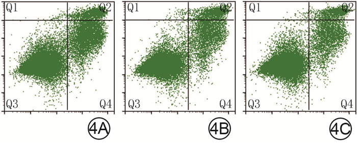
图 4 流式细胞仪检测3组人成纤维细胞培养48 h后凋亡情况。4A、4B、4C.分别为对照组、低浓度辣椒素组、高浓度辣椒素组
注:对照组、低浓度辣椒素组、高浓度辣椒素组细胞分别采用不含辣椒素的完全培养基和含10、20 μmol/L辣椒素的完全培养基进行培养;Q1区域中细胞为裸核细胞, Q2区域中细胞为坏死或晚期凋亡细胞, Q3区域中细胞为活细胞, Q4区域中细胞为早期凋亡细胞
Figure 4. The apoptosis status in three groups of human fibroblasts detected by flow cytometry after 48 hours of culture

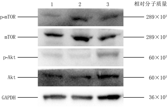
图 5 蛋白质印迹法检测的3组人成纤维细胞培养48 h后各蛋白表达情况
注:条带上方的1、2、3分别指采用不含辣椒素的完全培养基和含10、20 μmol/L辣椒素的完全培养基进行培养的对照组、低浓度辣椒素组、高浓度辣椒素组;p-mTOR为磷酸化哺乳动物雷帕霉素靶蛋白, mTOR为哺乳动物雷帕霉素靶蛋白, p-Akt为磷酸化蛋白激酶B, Akt为蛋白激酶B, GAPDH为3-磷酸甘油醛脱氢酶
Figure 5. The protein expression of various proteins in three groups of human fibroblasts detected by Western blotting after 48 hours of culture

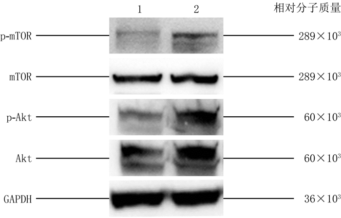
图 6 蛋白质印迹法检测的2组糖尿病小鼠伤后12 d全层皮肤缺损创面中各蛋白表达情况
注:条带上方的1、2分别指采用生理盐水处理创面的对照组和采用含20 μmol/L辣椒素溶液处理创面的高浓度辣椒素组;p-mTOR为磷酸化哺乳动物雷帕霉素靶蛋白, mTOR为哺乳动物雷帕霉素靶蛋白, p-Akt为磷酸化蛋白激酶B, Akt为蛋白激酶B, GAPDH为3-磷酸甘油醛脱氢酶
Figure 6. The protein expression of various proteins in full-thickness skin defect wounds of two groups of diabetic mice detected by Western blotting at 12 days after injury
表 1 3组糖尿病小鼠伤后各时间点残余全层皮肤缺损创面面积百分比的比较(%, x±s)
Table 1. Comparison of the percentage of residual full-thickness skin defect wound area in three groups of diabetic mice at various time points after injury
组别 样本数 4 d 8 d 12 d 对照组 6 81.8±2.2 60.0±3.4 26.6±3.8 低浓度辣椒素组 6 77.8±4.0a 55.1±3.2a 20.4±3.3a 高浓度辣椒素组 6 76.9±2.4a 49.7±3.3a 14.9±2.5ab F值 4.64 14.50 19.50 P值 0.027 < 0.001 < 0.001 注:对照组小鼠创面注射生理盐水, 低浓度辣椒素组、高浓度辣椒素组小鼠创面分别注射10、20 μmol/L辣椒素溶液;处理因素主效应, F=30.93, P < 0.001;时间因素主效应, F=2 892.00, P < 0.001;两者交互作用, F=6.14, P < 0.001;与对照组比较, a P < 0.05;与低浓度辣椒素组比较, b P < 0.05  下载: 导出CSV
-

 下载:
下载:


计量
- 文章访问数: 4776
- HTML全文浏览量: 486
- PDF下载量: 19
- 被引次数: 0
