





Expert consensus on the surgical management of pressure injuries (2026 edition)
-
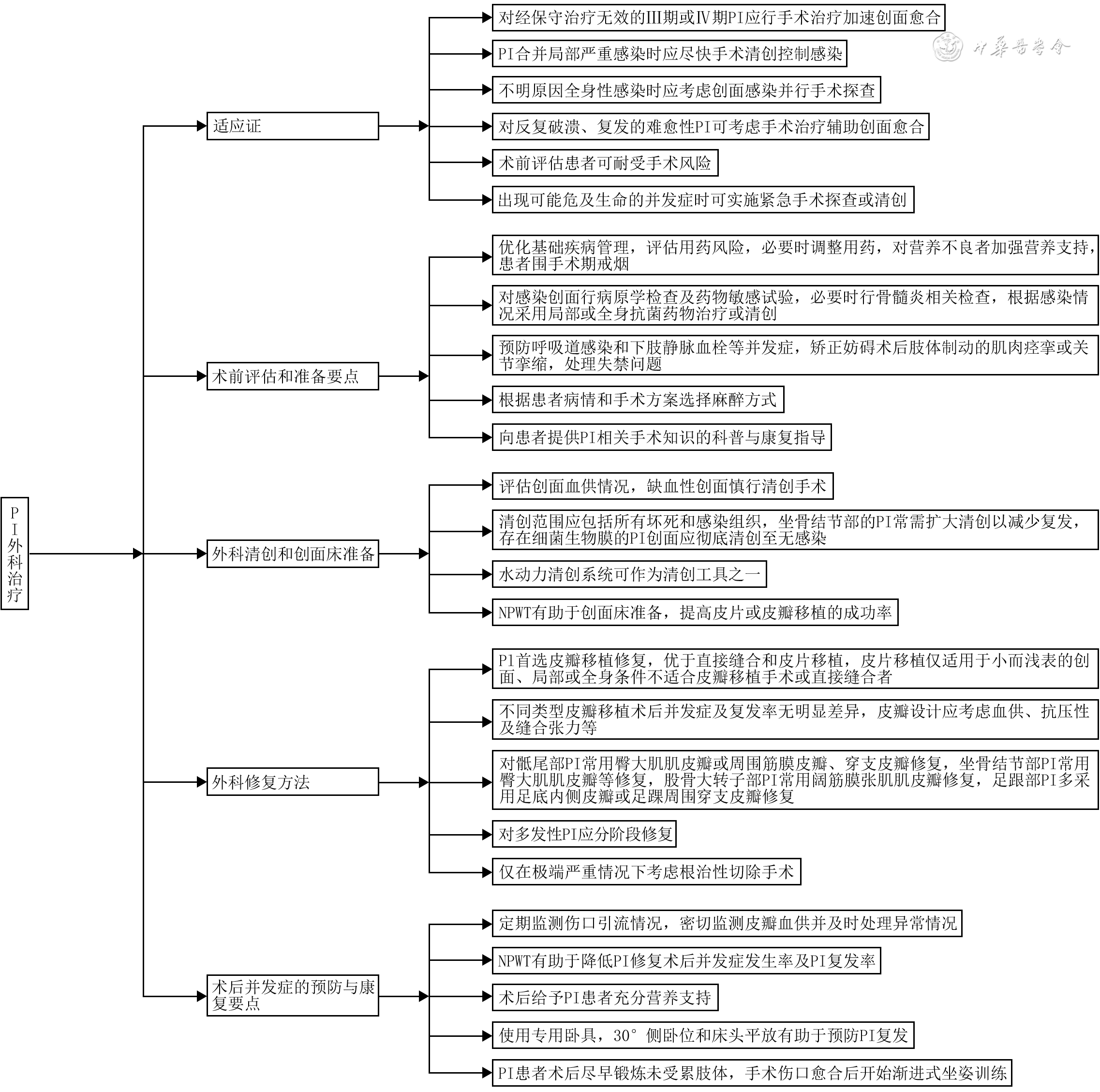
摘要: 压力性损伤是常见的慢性难愈性创面之一。随着人口老龄化加剧和慢性疾病患病率上升,压力性损伤的发生率逐年升高,外科治疗需求随之显著增长。尽管相关临床技术不断发展,但压力性损伤外科治疗的全流程仍缺乏统一的规范化路径,影响患者预后。为进一步指导临床实践,中国医师协会创面修复专业委员会组织国内多学科专家,制订了《压力性损伤外科治疗专家共识(2026版)》。该共识内容涵盖压力性损伤外科治疗的适应证、术前评估和准备要点、外科清创和创面床准备、外科修复方法及术后并发症的预防与康复要点等,旨在规范临床行为、提升治疗效果,促进各级医疗机构压力性损伤外科治疗能力的同质化提升。Abstract: Pressure injuries are common chronic hard-to-heal wounds. With population aging and the increasing prevalence of chronic diseases, the incidence of pressure injuries has gradually increased, accompanied by a growing demand for surgical management. Despite continuous advances in relevant clinical techniques, a unified standardized treatment pathway covering the entire process of surgical management of pressure injuries remains lacking, which may adversely affect patient outcomes. To further standardize clinical practice, the Wound Repair Professional Committee of the Chinese Medical Doctor Association organized a multidisciplinary panel of experts from across China to develop the Expert consensus on the surgical management of pressure injuries (2026 edition). The consensus covered key aspects of the surgical management of pressure injuries, including indications for surgical management of pressure injuries, key considerations for preoperative assessment and preparation, surgical debridement and wound-bed preparation, methods of surgical repair, and key considerations for the prevention of postoperative complications and rehabilitation. It was intended to standardize clinical practice, improve treatment outcomes, and promote consistency in the surgical management of pressure injuries across healthcare institutions at different levels.
-
Key words:
- Pressure ulcer /
- Surgical procedures, Operative /
- Debridement /
- Infections /
- Surgical flaps /
- Skin transplantation /
- Expert consensus
-
参考文献
(94) [1] EdsbergLE, BlackJM, GoldbergM, et al. Revised National Pressure Ulcer Advisory Panel pressure injury staging system: revised pressure injury staging system[J]. J Wound Ostomy Continence Nurs, 2016,43(6):585-597. DOI: 10.1097/WON.0000000000000281. [2] StansbyG, AvitalL, JonesK, et al. Prevention and management of pressure ulcers in primary and secondary care: summary of NICE guidance[J]. BMJ, 2014,348:g2592. DOI: 10.1136/bmj.g2592. [3] NugentR. Preventing and managing chronic diseases[J]. BMJ, 2019,364:l459. DOI: 10.1136/bmj.l459. [4] HajhosseiniB, LongakerMT, GurtnerGC. Pressure injury[J]. Ann Surg, 2020,271(4):671-679. DOI: 10.1097/SLA.0000000000003567. [5] LiZY, LinF, ThalibL, et al. Global prevalence and incidence of pressure injuries in hospitalised adult patients: a systematic review and meta-analysis[J]. Int J Nurs Stud, 2020, 105:103546. DOI: 10.1016/j.ijnurstu.2020.103546. [6] European Pressure Ulcer Advisory Panel, National Pressure Injury Advisory Panel, Pan Pacific Pressure Injury Alliance. Prevention and treatment of pressure ulcers/injuries: clinical practice guideline. The International Guideline[M/OL]. 3rd ed. 2019[2026-09-24]. https://www.internationalguideline.com/2019. https://www.internationalguideline.com/2019 [7] FujiwaraH, IsogaiZ, IrisawaR, et al. Wound, pressure ulcer and burn guidelines - 2: guidelines for the diagnosis and treatment of pressure ulcers, second edition[J]. J Dermatol, 2020, 47(9):929-978.DOI: 10.1111/1346-8138.14587. [8] World Health Organization. WHO handbook for guideline development[M/OL]. 2nd ed. Geneva: World Health Organization, 2014[2026-09-17]. https://www.who.int/publications/i/item/9789241548960. https://www.who.int/publications/i/item/9789241548960 [9] 蒋朱明,詹思延,贾晓巍,等. 制订/修订《临床诊疗指南》的基本方法及程序[J]. 中华医学杂志,2016,96(4):250-253.DOI: 10.3760/cma.j.issn.0376-2491.2016.04.004. [10] BrouwersMC, KhoME, BrowmanGP, et al. AGREE II: advancing guideline development, reporting and evaluation in health care[J]. J Clin Epidemiol, 2010,63(12):1308-1311. DOI: 10.1016/j.jclinepi.2010.07.001. [11] ChenY, YangK, MarušicA, et al. A reporting tool for Practice Guidelines in Health Care: the RIGHT statement[J]. Ann Intern Med, 2017, 166(2): 128-132. DOI: 10.7326/M16-1565. [12] SheaBJ, ReevesBC, WellsG, et al. AMSTAR 2: a critical appraisal tool for systematic reviews that include randomised or non-randomised studies of healthcare interventions, or both[J]. BMJ, 2017, 358: j4008. DOI: 10.1136/bmj.j4008. [13] HigginsJP, AltmanDG, GøtzschePC, et al. The Cochrane Collaboration's tool for assessing risk of bias in randomised trials[J]. BMJ, 2011, 343: d5928. DOI: 10.1136/bmj.d5928. [14] Wells GA Shea B O'Connell D et al The Newcastle–Ottawa Scale (NOS) for assessing the quality of nonrandomised studies in meta-analyses 2025-05-09 https://www.ohri.ca/programs/clinical_epidemiology/oxford.asp WellsGA, SheaB, O'ConnellD, et al. The Newcastle–Ottawa Scale (NOS) for assessing the quality of nonrandomised studies in meta-analyses[EB/OL]. 2000[2025-05-09]. https://www.ohri.ca/programs/clinical_epidemiology/oxford.asp.
[15] Müller-HeuptLK, SchiegnitzE, KayaS, et al. Diagnostic tests for titanium hypersensitivity in implant dentistry: a systematic review of the literature[J]. Int J Implant Dent, 2022, 8(1):29. DOI: 10.1186/s40729-022-00428-0. [16] Oxford Centre for Evidence-Based Medicine: levels of evidence (March 2009 2025-05-09 https://www.cebm.ox.ac.uk/resources/levels-of-evidence/oxford-centre-for-evidence-based-medicine-levels-of-evidence-march-2009 Oxford Centre for Evidence-Based Medicine: levels of evidence (March2009)[EB/OL]. 2009[2025-05-09]. https://www.cebm.ox.ac.uk/resources/levels-of-evidence/oxford-centre-for-evidence-based-medicine-levels-of-evidence-march-2009.
[17] DjedovicG, MorandiEM, MetzlerJ, et al. The posterior thigh flap for defect coverage of ischial pressure sores - a critical single-centre analysis[J]. Int Wound J, 2017, 14(6):1154-1159. DOI: 10.1111/iwj.12776. [18] LeaperD, AssadianO, EdmistonCE. Approach to chronic wound infections[J]. Br J Dermatol, 2015, 173(2):351-358. DOI: 10.1111/bjd.13677. [19] GouldLJ, AlderdenJ, AslamR, et al. WHS guidelines for the treatment of pressure ulcers-2023 update[J]. Wound Repair Regen, 2024, 32(1):6-33. DOI: 10.1111/wrr.13130. [20] MizokamiF, FurutaK, IsogaiZ. Necrotizing soft tissue infections developing from pressure ulcers[J]. J Tissue Viability, 2014, 23(1): 1-6. DOI: 10.1016/j.jtv.2013.11.001. [21] EspejoE, AndrésM, BorralloRM, et al. Bacteremia associated with pressure ulcers: a prospective cohort study[J]. Eur J Clin Microbiol Infect Dis, 2018, 37(5):969-975. DOI: 10.1007/s10096-018-3216-8. [22] GuanY, NiuH, LiuZ, et al. Sustained oxygenation accelerates diabetic wound healing by promoting epithelialization and angiogenesis and decreasing inflammation[J]. Sci Adv, 2021, 7(35):eabj0153. DOI: 10.1126/sciadv.abj0153. [23] FalangaV. Wound healing and its impairment in the diabetic foot[J]. Lancet, 2005, 366(9498): 1736-1743. DOI: 10.1016/S0140-6736(05)67700-8. [24] KeysKA, DanialiLN, WarnerKJ, et al. Multivariate predictors of failure after flap coverage of pressure ulcers[J]. Plast Reconstr Surg, 2010, 125(6):1725-1734. DOI: 10.1097/PRS.0b013e3181d51227. [25] BeitzJM. Medications and wound healing: a narrative literature review[J]. J Wound Ostomy Continence Nurs, 2026, 53(1):13-19. DOI: 10.1097/WON.0000000000001247. [26] HuangK, GuoQF. Surgical repair involving tissue flap transplantation with vascular pedicle in treating refractory pressure ulcers around hip and sacral region[J]. CURR SIGNAL TRANSD T, 2015, 10(1):36-40. DOI: 10.2174/1574362410666150414203131. [27] LjungAC, SteniusMC, BjelakS, et al. Surgery for pressure ulcers in spinal cord‐injured patients following a structured treatment programme: a 10‐year follow‐up[J]. Int Wound J, 2017, 14(2): 355-359. DOI: 10.1111/iwj.12609. [28] Di CaprioG, Serra-MestreJM, ZiccardiP, et al. Expanded flaps in surgical treatment of pressure sores: our experience for 25 years[J]. Ann Plast Surg, 2015,75(5):552-555. DOI: 10.1097/SAP.0000000000000199. [29] WenzelF, WhitakerIY. Is there a relationship between nutritional goal achievement and pressure injury risk in intensive care unit patients receiving enteral nutrition?[J]. Intensive Crit Care Nurs, 2021, 62:102926. DOI: 10.1016/j.iccn.2020.102926. [30] CeredaE, KlersyC, SerioliM, et al. A nutritional formula enriched with arginine, zinc, and antioxidants for the healing of pressure ulcers: a randomized trial[J]. Ann Intern Med, 2015, 162(3):167-174. DOI: 10.7326/M14-0696. [31] Wound/Pressure Ulcer/Burn Guidelines Drafting Committee (Pressure Ulcer Group), FujiwaraH, IrisawaR, et al. Wound, pressure ulcer, and burn guidelines (2023)–2: guidelines for the diagnosis and treatment of pressure ulcers, third edition[J]. J Dermatol, 2025, 52(9): e744-e794. DOI: 10.1111/1346-8138.17758. [32] HasegawaM, InoueY, KanekoS, et al. Wound, pressure ulcer and burn guidelines - 1: guidelines for wounds in general, second edition[J]. J Dermatol, 2020, 47(8): 807-833. DOI: 10.1111/1346-8138.15401. [33] ThiessenFE, AndradesP, BlondeelPN, et al. Flap surgery for pressure sores: should the underlying muscle be transferred or not?[J]. J Plast Reconstr Aes, 2011, 64(1): 84-90. DOI: 10.1016/j.bjps.2010.03.049. [34] HaherJN, HaherTR, DevlinVJ, et al. The release of flexion contractures as a prerequisite for the treatment of pressure sores in multiple sclerosis: a report of ten cases[J]. Ann Plast Surg, 1983, 11(3): 246-249. DOI: 10.1097/00000637-198309000-00013. [35] RekandT, HagenEM, GrønningM, et al. Spasticity following spinal cord injury[J]. Tidsskr Nor Laegeforen, 2012, 30,132(8):970-973. DOI: 10.4045/tidsskr.10.0872. [36] de la FuenteSG, LevinLS, ReynoldsJD, et al. Elective stoma construction improves outcomes in medically intractable pressure ulcers[J]. Dis Colon Rectum, 2003, 46(11): 1525-1530. DOI: 10.1007/s10350-004-6808-6. [37] GrassettiL, ScaliseA, LazzeriD, et al. Perforator flaps in late-stage pressure sore treatment: outcome analysis of 11-year-long experience with 143 patients[J]. Ann Plast Surg, 2014, 73(6):679-685. DOI: 10.1097/SAP.0b013e31828587a8. [38] O'NeillDK, RobinsB, AyelloEA, et al. Regional anaesthesia with sedation protocol to safely debride sacral pressure ulcers[J]. Int Wound J, 2012, 9(5): 525-543. DOI: 10.1111/j.1742-481X.2011.00912.x. [39] KottnerJ, CuddiganJ, CarvilleK, et al. Prevention and treatment of pressure ulcers/injuries: the protocol for the second update of the international Clinical Practice Guideline 2019[J]. J Tissue Viability, 2019, 28(2): 51-58. DOI: 10.1016/j.jtv.2019.01.001. [40] AssafN, BazziH, GhanemK, et al. Optimizing surgical outcomes through increased familial support in the perioperative period[J]. Front Anesthesiol, 2025, 4:1650491. DOI: 10.3389/fanes.2025.1650491. [41] KierneyPC, EngravLH, IsikFF, et al. Results of 268 pressure sores in 158 patients managed jointly by plastic surgery and rehabilitation medicine[J]. Plast Reconstr Surg, 1998, 102(3): 765-772. DOI: 10.1097/00006534-199809030-00022. [42] SrMills JL, ConteMS, ArmstrongDG, et al. The Society for Vascular Surgery Lower Extremity Threatened Limb Classification System: risk stratification based on wound, ischemia, and foot infection (WIfI)[J]. J Vasc Surg, 2014,59(1):220-234.e1-2. DOI: 10.1016/j.jvs.2013.08.003. [43] ErikssonE, LiuPY, SchultzGS, et al. Chronic wounds: treatment consensus[J]. Wound Repair Regen, 2022, 30(2): 156-171. DOI: 10.1111/wrr.12994. [44] AnvarB, OkonkwoH. Serial surgical debridement of common pressure injuries in the nursing home setting: outcomes and findings[J]. Wounds, 2017, 29(7): 215-221. [45] YangK, GrafA, SangerJ. Pressure ulcer reconstruction in patients with heterotopic ossification after spinal cord injury: a case series and review of literature[J]. J Plast Reconstr Aesthet Surg, 2017, 70(4): 518-528. DOI: 10.1016/j.bjps.2016.11.026. [46] MarchiM, BattagliaS, MarcheseS, et al. Surgical reconstructive procedures for treatment of ischial, sacral and trochanteric pressure ulcers[J]. G Chir, 2015, 36(3):112-116. [47] SimpsonAH, DeakinM, LathamJM. Chronic osteomyelitis. The effect of the extent of surgical resection on infection-free survival[J]. J Bone Joint Surg Br, 2001, 83(3):403-407. DOI: 10.1302/0301-620x.83b3.10727. [48] WolcottRD, RumbaughKP, JamesG, et al. Biofilm maturity studies indicate sharp debridement opens a time- dependent therapeutic window[J]. J Wound Care, 2010, 19(8): 320-328. DOI: 10.12968/jowc.2010.19.8.77709. [49] WilcoxJR, CarterMJ, CovingtonS. Frequency of debridements and time to heal: a retrospective cohort study of 312 744 wounds[J]. JAMA Dermatol, 2013, 149(9): 1050-1058. DOI: 10.1001/jamadermatol.2013.4960. [50] GranickMS, PosnettJ, JacobyM, et al. Efficacy and cost-effectiveness of a high-powered parallel waterjet for wound debridement[J]. Wound Repair Regen, 2006, 14(4): 394-397. DOI: 10.1111/j.1743-6109.2006.00136.x. [51] BowlingFL, StickingsDS, Edwards-JonesV, et al. Hydrodebridement of wounds: effectiveness in reducing wound bacterial contamination and potential for air bacterial contamination[J]. J Foot Ankle Res, 2009, 2: 13. DOI: 10.1186/1757-1146-2-13. [52] 陈礼新, 张旋, 赵遵江, 等. 老年坐骨结节4期压力性损伤分型及其修复效果的多中心研究[J]. 中华烧伤与创面修复杂志, 2025, 41(8): 749-758. DOI: 10.3760/cma.j.cn501225-20250421-00185. [53] HyldigN, Birke-SorensenH, KruseM, et al. Meta-analysis of negative-pressure wound therapy for closed surgical incisions[J]. Br J Surg, 2016, 103(5):477-486. DOI: 10.1002/bjs.10084. [54] JiS, LiuX, HuangJ, et al. Consensus on the application of negative pressure wound therapy of diabetic foot wounds[J/OL]. Burns Trauma, 2021, 9:tkab018[2025-02-13]. https://pubmed.ncbi.nlm.nih.gov/34212064/.DOI: 10.1093/burnst/tkab018. [55] HuangCY, LeeSL, ChiuWK, et al. Determinants of the success in flap reconstruction-outcome analysis of 120 flaps in 484 procedures for pressure injury[J]. Int Wound J, 2023, 20(8): 3105-3115. DOI: 10.1111/iwj.14185. [56] RicciJA, BayerLR, OrgillDP. Evidence-based medicine: the evaluation and treatment of pressure injuries[J]. Plast Reconstr Surg, 2017, 139(1): 275e-286e. DOI: 10.1097/PRS.0000000000002850. [57] CushingCA, PhillipsLG. Evidence-based medicine: pressure sores[J]. Plast Reconstr Surg, 2013, 132(6): 1720-1732. DOI: 10.1097/PRS.0b013e3182a808ba. [58] VathulyaM, PraveenAJ, BarikS, et al. A systematic review comparing outcomes of local flap options for reconstruction of pressure sores[J]. Ann Plast Surg, 2022, 88(1): 105-113. DOI: 10.1097/SAP.0000000000002941. [59] MontagE, UedaT, OkadaA, et al. Reconstruction of acquired ischiatic and perineal defects: an anatomical and clinical comparison between gluteal thigh and inferior gluteal perforator flaps[J]. Eur J Plast Surg, 2018, 41(1): 41-48. DOI: 10.1007/s00238-017-1371-2. [60] ŞibarS, FindikciogluK, GuneyK, et al. Effect of flap selection on the postoperative success of sacral pressure injuries: a retrospective analysis[J]. Wounds, 2021, 33(10): 271-276. [61] OksmanD, de AlmeidaOM, de ArrudaRG, et al. Comparative study between fasciocutaneous and myocutaneous flaps in the surgical treatment of pressure ulcers of the sacral region[J]. JPRAS Open, 2018, 16: 50-60. DOI: 10.1016/j.jpra.2017.12.001. [62] DölenUC, KoçerU. Innovation in the planning of V-Y rotation advancement flaps: a template for flap design[J]. Arch Plast Surg, 2018, 45(1): 85-88. DOI: 10.5999/aps.2017.00171. [63] KeysKA, DanialiLN, WarnerKJ, et al. Multivariate predictors of failure after flap coverage of pressure ulcers[J]. Plast Reconstr Surg, 2010, 125(6):1725-1734. DOI: 10.1097/PRS.0b013e3181d51227. [64] LemaireV, BoulangerK, HeymansO. Free flaps for pressure sore coverage[J]. Ann Plast Surg, 2008, 60(6): 631-634. DOI: 10.1097/SAP.0b013e31812c1465. [65] SarasúaJG, LópezSP, ViejoMA, et al. Treatment of pressure ulcers with autologous bone marrow nuclear cells in patients with spinal cord injury[J]. J Spinal Cord Med, 2011,34(3):301-307. DOI: 10.1179/2045772311Y.0000000010. [66] 杨力, 朱小平, 黄巧洪, 等. 臀大肌肌皮瓣修复骶尾部Ⅳ度压疮13例[J]. 中华烧伤杂志, 2014, 30(2): 146-147. DOI: 10.3760/cma.j.issn.1009-2587.2014.02.012. [67] DurgunM, BasS. Repair of sacral and ischial region defects with lateral sacral artery perforator flaps[J]. Ann Plast Surg, 2019,82(3):304-309. DOI: 10.1097/SAP.0000000000001737. [68] 邓如非,龙露瑶,陈燕微, 等. 基于窦道情况及皮肤软组织缺损范围的坐骨结节压疮临床修复策略[J]. 中华烧伤与创面修复杂志,2024,40(1):64-71. DOI: 10.3760/cma.j.cn501225-20231114-00194. [69] 周思拓,黄覛韬,曾纪章, 等. 携带知名血管穿支的新型V-Y推进皮瓣修复全身多处皮肤软组织缺损的疗效[J]. 中华烧伤杂志,2017,33(10):611-615. DOI: 10.3760/cma.j.issn.1009-2587.2017.10.005. [70] WongC, Saint-CyrM. The pedicled descending branch muscle-sparing latissimus dorsi flap for trunk and upper extremity reconstruction[J]. J Plast Reconstr Aesthet Surg, 2010,63(4):623-632. DOI: 10.1016/j.bjps.2009.01.059. [71] 王忠信,李森恺,陈树廷, 等. 阔筋膜张肌肌皮瓣的解剖研究与临床应用[J]. 中华整形外科杂志,2000,16(1):12-13,插页1. DOI: 10.3760/cma.j.issn.1009-4598.2000.01.105. [72] HanBL, ChoiHJ. Free-style dual plane recycling tensor fascia lata musculocutaneous perforator flap for reconstruction of recurrent trochanteric defects[J]. Int J Low Extrem Wounds, 2014,13(1):72-75. DOI: 10.1177/1534734613520324. [73] 赵遵江,张保德,刘勇, 等. 股直肌岛状肌皮瓣修复瘫痪患者股骨大转子压疮效果[J]. 中华烧伤杂志,2014,30(3):227-230. DOI: 10.3760/cma.j.issn.1009-2587.2014.03.011. [74] 喜雯婧,冯少清,李华, 等. 旋髂浅动脉穿支皮瓣的重新评价和手术策略[J]. 中华显微外科杂志,2018,41(4):313-318. DOI: 10.3760/cma.j.issn.1001-2036.2018.04.001. [75] TiwariP, ChewKY, TanBK, et al. Anatomical study of the medialis pedis flap and its clinical applications in Asian Singaporean patients[J]. Injury, 2021,52(10):3124-3131. DOI: 10.1016/j.injury.2021.02.012. [76] LinJ, ZhouF, SunYD, et al. Modified anterior tibial artery perforator-pedicled propeller flap for soft-tissue coverage of the ankle and heel[J]. World J Surg, 2020,44(7):2237-2242. DOI: 10.1007/s00268-020-05452-y. [77] 龙露瑶,陈燕微,邓如非, 等. 延迟腓肠神经营养血管皮瓣在糖尿病足溃疡中的应用与研究进展[J]. 中华烧伤与创面修复杂志,2024,40(3):296-300. DOI: 10.3760/cma.j.cn501225-20231102-00173. [78] 吴鹏, 彭京梁, 刘荣朋, 等. 负压创面治疗技术联合皮瓣分期修复臀股部多发Ⅳ期压疮[J]. 中国美容医学, 2020, 29(12): 87-89,101. DOI: 10.15909/j.cnki.cn61-1347/r.004115. [79] KwokAC, SimpsonAM, WillcocksonJ, et al. Complications and their associations following the surgical repair of pressure ulcers[J]. Am J Surg, 2018, 216(6): 1177-1181. DOI: 10.1016/j.amjsurg.2018.01.012. [80] BosanquetDC, WrightAM, WhiteRD, et al. A review of the surgical management of heel pressure ulcers in the 21st century[J]. Int Wound J, 2016, 13(1): 9-16. DOI: 10.1111/iwj.12416. [81] BambaR, MaddenJJ, HoffmanAN, et al. Flap reconstruction for pressure ulcers: an outcomes analysis[J]. Plast Reconstr Surg, 2017, 5(1): e1187. DOI: 10.1097/GOX.0000000000001187. [82] TadiparthiS, HartleyA, AlzweriL, et al. Improving outcomes following reconstruction of pressure sores in spinal injury patients: a multidisciplinary approach[J].J Plast Reconstr Aesthet Surg, 2016, 69(7): 994-1002. DOI: 10.1016/j.bjps.2016.02.016. [83] 中华护理学会伤口、造口、失禁护理专业委员会. 器械相关压力性损伤预防指南(2020版)[J]. 中华护理杂志, 2020, 55增刊2: S115-S121. DOI: 10.3761/j.issn.0254-1769.2020.S2.024. [84] SalgadoCJ, MoranSL, MardiniS. Flap monitoring and patient management[J]. Plast Reconstr Surg, 2009, 124(6Suppl): Se295-Se302. DOI: 10.1097/PRS.0b013e3181bcf07b. [85] KohlerLH, KöhlerH, KohlerS, et al. Hyperspectral Imaging (HSI) as a new diagnostic tool in free flap monitoring for soft tissue reconstruction: a proof of concept study[J]. BMC Surg, 2021, 21(1): 222. DOI: 10.1186/s12893-021-01232-0. [86] GroenenH, JalalzadehH, BuisDR, et al. Incisional negative pressure wound therapy for the prevention of surgical site infection: an up-to-date meta-analysis and trial sequential analysis[J]. EClinicalMedicine, 2023, 62: 102105. DOI: 10.1016/j.eclinm.2023.102105. [87] PappAA. Incisional negative pressure therapy reduces complications and costs in pressure ulcer reconstruction[J]. Int Wound J, 2019, 16(2): 394-400. DOI: 10.1111/iwj.13045. [88] Lin-YinW, Chia-MingL, LinHL, et al. Risk factors for recurrent pressure ulcers after reconstructive surgery: a retrospective study in a single medical centre[J]. Int Wound J, 2023, 20(7): 2511-2517. DOI: 10.1111/iwj.14112. [89] VecinNM, GaterDR. Pressure injuries and management after spinal cord injury[J]. J Pers Med, 2022, 12(7): 1130. DOI: 10.3390/jpm12071130. [90] PengXJ, XiongXX, LanWT. Prevention of pressure injuries in the operating room: challenges and strategies[J]. J Adv Nurs, 2025, 81(9): 6066-6067. DOI: 10.1111/jan.16410. [91] ChiangIH, WangCH, TzengYS. Surgical treatment and strategy in patients with multiple pressure sores[J]. Int Wound J, 2018, 15(6): 900-908. DOI: 10.1111/iwj.12942. [92] FinneganMJ, GazzerroL, FinneganJO, et al. Comparing the effectiveness of a specialized alternating air pressure mattress replacement system and an air-fluidized integrated bed in the management of post-operative flap patients: a randomized controlled pilot study[J]. J Tissue Viability, 2008, 17(1): 2-9. DOI: 10.1016/j.jtv.2007.09.009. [93] AwadSS, SternJD, MilneCT, et al. Surgical reconstruction of stage 3 and 4 pressure injuries: a literature review and proposed algorithm from an interprofessional working group[J]. Adv Skin Wound Care, 2023, 36(5):249-258. DOI: 10.1097/01.ASW.0000922708.95424.88. [94] HanHH, ChoiEJ, ChoiJY, et al. Efficacy of one‐stage surgical treatment and clinical features in patients with multiple pressure ulcers[J]. Int Wound J, 2016, 13Suppl 1: S7-S12. DOI: 10.1111/iwj.12537. -
Table 1. 压力性损伤的分期与评定标准
分期 评定标准 Ⅰ期 局部皮肤完好,出现压之不变白的红斑,常位于骨隆突处。与邻近组织相比,这一区域可能会出现痛感、发硬、变软、皮温升高或降低 Ⅱ期 部分皮层缺损,表现为浅表的开放性溃疡,创面呈粉红色,无腐肉;也可为完整或破裂的浆液性水疱,外观透亮或干燥,无腐肉或淤伤 Ⅲ期 仅全层皮肤缺损,可见皮下脂肪,但骨、肌腱、肌肉并未外露。可有腐肉,可伴有窦道和潜行腔隙 Ⅳ期 全层皮肤缺损,并伴有骨、肌腱或肌肉外露。在创面基底可有腐肉和焦痂覆盖,通常伴有窦道和潜行腔隙 不可分期压力性损伤 创面覆盖有腐肉和/或焦痂,除非去除足够多的腐肉和/或焦痂来暴露创面基底,否则无法判断实际深度和分期 可疑深部组织损伤 压力或剪切力导致皮下软组织受损,皮肤完整或部分缺损,局部出现持续不褪的紫色或栗色改变,或形成充血的水疱。此部位与邻近组织相比,先出现痛感、发硬、糜烂、变软、皮温升高或降低。在肤色较深的个体中,很难辨识出深层组织损伤 注:表格引自参考文献[6]  下载: 导出CSV
Table 2. 2009版牛津大学循证医学中心证据分级标准
证据等级 定义 1a级 RCT的系统评价 1b级 结果可信区间小的RCT 1c级 显示“全或无效应”的任何证据 2a级 队列研究的系统评价 2b级 单个队列研究(包括低质量的RCT,如失访率>20%) 2c级 基于患者结局的研究 3a级 病例对照研究的系统评价 3b级 单个病例对照研究 4级 病例系列报告、低质量队列研究和低质量病例对照研究 5级 专家意见(即无临床研究支持的仅依据基础研究或临床经验的推测) 注:RCT为随机对照试验
下载: 导出CSV
Table 3. 压力性损伤外科治疗推荐意见的推荐强度与定义
推荐强度 定义 强 综合现有证据、获益与风险、患者价值观与接受程度及临床实施可行性等因素,专家组认为该措施的预期获益明确超过潜在风险或负担,推荐实施 弱 综合上述因素,专家组认为该措施可能获益,但证据确定性、获益与风险平衡、患者接受程度或实施条件等仍存在一定不确定性,建议根据患者具体情况选择性实施 无特别推荐 现有证据或获益与风险平衡尚不足以支持明确推荐,或不同患者、医疗条件下选择可能存在较大差异,暂不作普遍推荐,可根据具体情况个体化选择
下载: 导出CSV
-

 下载:
下载:

图(1) / 表(3)
计量
- 文章访问数: 4
- HTML全文浏览量: 2
- PDF下载量: 1
- 被引次数: 0
