


Consensus on the Diagnosis and Treatment of Adult Necrotizing Fasciitis (2025 Edition)
-
摘要: 坏死性筋膜炎(NF)是一种罕见但极具侵袭性的软组织感染, 以筋膜和皮下组织的快速坏死为特征, 具有早期诊断困难、病情进展迅速的特点。若治疗不及时, 该疾病可迅速发展为全身性感染、脓毒症及多器官功能衰竭, 甚至导致患者死亡。目前, 我国尚未形成统一的NF诊治共识或指南, 临床诊治过程中仍面临诸多争议与挑战。为推动我国NF诊治的规范化进程, 中华医学会烧伤外科学分会、《中华烧伤与创面修复杂志》编辑委员会及中国医疗保健国际交流促进会烧伤医学分会组织相关领域专家, 依据国内外最新指南、文献及临床实践经验, 遵循循证医学原则, 经过反复讨论, 共同制订了《成人坏死性筋膜炎诊治专家共识(2025版)》, 旨在为NF的临床诊疗提供科学、规范的指导。Abstract: Necrotizing fasciitis (NF) is a rare but very aggressive soft tissue infection characterized by rapid necrosis of fascia and subcutaneous tissue, which is difficult to diagnose early and progresses rapidly. If not treated promptly, NF can quickly evolve into systemic infection, sepsis, and multiple organ failure, and even lead to the patient's death. Currently, there is not a unified consensus or guideline for the diagnosis and treatment of NF, and many controversies and challenges remain in clinical practice for its diagnosis and treatment. To promote the standardization of NF diagnosis and treatment, Chinese Burn Association, Editorial Committee of Chinese Journal of Burns and Wounds, and Burn Medicine Branch of China International Exchange and Promotion Association for Medical and Healthcare, have jointly developed the Consensus on the Diagnosis and Treatment of Adult Necrotizing Fasciitis (2025 Edition) based on the latest national and international guidelines, literature, and clinical practice experience, in accordance with the principles of evidence-based medicine, and through repeated discussion. This consensus aims to provide scientific and standardized guidance for clinical diagnosis and treatment of NF.
-
Key words:
- Fasciitis, necrotizing /
- Diagnosis /
- Treatment /
- Multidisciplinary team /
- Expert consensus
本文亮点(1) 鉴于目前对于坏死性筋膜炎的诊断和治疗尚未形成统一规范,本共识依据国内外最新指南、文献及临床实践经验,遵循循证医学原则,通过多学科团队讨论制订。(2) 针对坏死性筋膜炎相关的多个临床问题提出推荐意见,有助于规范其诊断和治疗,同时为进一步完善其诊疗策略提供依据。 -
参考文献
(118) [1] Diab J, Bannan A, Pollitt T. Necrotising fasciitis[J]. BMJ, 2020, 369: m1428. DOI: 10.1136/bmj.m1428. [2] Stevens DL, Bisno AL, Chambers HF, et al. Practice guidelines for the diagnosis and management of skin and soft tissue infections: 2014 update by the Infectious Diseases Society of America[J]. Clin Infect Dis, 2014, 59(2): e10-52. DOI: 10.1093/cid/ciu444. [3] Ajitha MB, Archana CS, Avinash K. A study on necrotizing fasciitis with the etiological factors and microbiological aspects with its prevalence[J]. Int J Surg, 2020, 4(2): 167-171. DOI: https://doi.org/ 10.33545/surgery.2020.v4.i2c.413. [4] Shivalingappa S, Manjunath KN, Waiker V, et al. Necrotising fasciitis: appearances can be deceptive[J]. World J Plast Surg, 2021, 10(1): 43-52. DOI: 10.29252/wjps.10.1.43. [5] Fais P, Viero A, Viel G, et al. Necrotizing fasciitis: case series and review of the literature on clinical and medico-legal diagnostic challenges[J]. Int J Legal Med, 2018, 132(5): 1357-1366. DOI: 10.1007/s00414-018-1838-0. [6] Guyatt GH, Oxman AD, Vist GE, et al. GRADE: an emerging consensus on rating quality of evidence and strength of recommendations[J]. BMJ, 2008, 336(7650): 924-926. DOI: 10.1136/bmj.39489.470347.AD. [7] Chen PC, Tsai SH, Wang JC, et al. An elevated glycemic gap predicts adverse outcomes in diabetic patients with necrotizing fasciitis[J]. PLoS One, 2019, 14(10): e0223126. DOI: 10.1371/journal.pone.0223126. [8] 中国医师协会肛肠医师分会临床指南工作委员会. 肛周坏死性筋膜炎临床诊治中国专家共识(2019年版)[J]. 中华胃肠外科杂志, 2019, 22(7): 689-693. DOI: 10.3760/cma.j.issn.1671-0274.2019.07.017. [9] Eggert J, Bird N, Leitze Z, et al. Diagnosis and treatment of type Ⅱ necrotizing fasciitis in a child presenting with a minor abrasion, edema, and apparent bruising[J]. Wounds, 2009, 21(3): 74-78. [10] Morgan MS. Diagnosis and management of necrotising fasciitis: a multiparametric approach[J]. J Hosp Infect, 2010, 75(4): 249-257. DOI: 10.1016/j.jhin.2010.01.028. [11] Molewa MC, Ogonowski-Bizos A, Els M, et al. The microbiological profile of necrotising fasciitis at a secondary level hospital in Gauteng[J]. S Afr JInfect Dis, 2024, 39(1): 542. DOl: 10.4102/saiid. v39i1.542. [12] Misiakos EP, Bagias G, Patapis P, et al. Current concepts in the management of necrotizing fasciitis[J]. Front Surg, 2014, 1: 36. DOI: 10.3389/fsurg.2014.00036. [13] Chen LL, Fasolka B, Treacy C. Necrotizing fasciitis: a comprehensive review[J]. Nursing, 2020, 50(9): 34-40. DOI: 10.1097/01.NURSE.0000694752.85118.62. [14] Gönüllü D, Ilgun AS, Demiray O, et al. The potential prognostic significance of the Laboratory Risk Indicator for the Necrotizing Fasciitis (LRINEC) score in necrotizing fasciitis[J]. Chirurgia (Bucur), 2019, 114(3): 376-383. DOI: 10.21614/chirurgia.114.3.376. [15] Hakkarainen TW, Kopari NM, Pham TN, et al. Necrotizing soft tissue infections: review and current concepts in treatment, systems of care, and outcomes[J]. Curr Probl Surg, 2014, 51(8): 344-362. DOI: 10.1067/j.cpsurg.2014.06.001. [16] Albadri Z, Salman K. Necrotizing fasciitis of the finger[J]. IDCases, 2019, 17: e00560. DOI: 10.1016/j.idcr.2019.e00560. [17] Wong CH, Khin LW, Heng KS, et al. The LRINEC (Laboratory Risk Indicator for Necrotizing Fasciitis) score: a tool for distinguishing necrotizing fasciitis from other soft tissue infections[J]. Crit Care Med, 2004, 32(7): 1535-1541. DOI: 10.1097/01.ccm.0000129486.35458.7d. [18] Su YC, Chen HW, Hong YC, et al. Laboratory risk indicator for necrotizing fasciitis score and the outcomes[J]. ANZ J Surg, 2008, 78(11): 968-972. DOI: 10.1111/j.1445-2197.2008.04713.x. [19] Bechar J, Sepehripour S, Hardwicke J, et al. Laboratory risk indicator for necrotising fasciitis (LRINEC) score for the assessment of early necrotising fasciitis: a systematic review of the literature[J]. Ann R Coll Surg Engl, 2017, 99(5): 341-346. DOI: 10.1308/rcsann.2017.0053. [20] El-Menyar A, Asim M, Mudali IN, et al. The laboratory risk indicator for necrotizing fasciitis (LRINEC) scoring: the diagnostic and potential prognostic role[J]. Scand J Trauma Resusc Emerg Med, 2017, 25(1): 28. DOI: 10.1186/s13049-017-0359-z. [21] Rodríguez-Roiz JM. Series of 41 cases of necrotizing fasciitis in a tertiary level hospital. Is as deadly as we think? LRINEC scale is useful today?[J]. Acta Ortop Mex, 2025, 39(1): 27-31. [22] Wu PH, Wu KH, Hsiao CT, et al. Utility of modified Laboratory Risk Indicator for Necrotizing Fasciitis (MLRINEC) score in distinguishing necrotizing from non-necrotizing soft tissue infections[J]. World J Emerg Surg, 2021, 16(1): 26. DOI: 10.1186/s13017-021-00373-0. [23] Leiblein M, Marzi I, Sander AL, et al. Necrotizing fasciitis: treatment concepts and clinical results[J]. Eur J Trauma Emerg Surg, 2018, 44(2): 279-290. DOI: 10.1007/s00068-017-0792-8. [24] Hsiao CT, Chang CP, Huang TY, et al. Prospective validation of the Laboratory Risk Indicator for Necrotizing Fasciitis (LRINEC) Score for necrotizing fasciitis of the extremities [J]. PLoS One, 2020, 15(1): e0227748. DOI: 10.1371/journal.pone.0227748. [25] Cribb BI, Wang M, Kulasegaran S, et al. The SIARI score: a novel decision support tool outperforms LRINEC score in necrotizing fasciitis[J]. World J Surg, 2019, 43(10): 2393-2400. DOI: 10.1007/s00268-019-05061-4. [26] Breidung D, Grieb G, Malsagova AT, et al. Time is fascia: laboratory and anamnestic risk indicators for necrotizing fasciitis[J]. Surg Infect (Larchmt), 2022, 23(8): 747-753. DOI: 10.1089/sur.2022.222. [27] Tso DK, Singh AK. Necrotizing fasciitis of the lower extremity: imaging pearls and pitfalls[J]. Br J Radiol, 2018, 91(1088): 20180093. DOI: 10.1259/bjr.20180093. [28] Ballard DH, Mazaheri P, Raptis CA, et al. Fournier gangrene in men and women: appearance on CT, ultrasound, and MRI and what the surgeon wants to know[J]. Can Assoc Radiol J, 2020, 71(1): 30-39. DOI: 10.1177/0846537119888396. [29] Yoon MA, Chung HW, Yeo Y, et al. Distinguishing necrotizing from non-necrotizing fasciitis: a new predictive scoring integrating MRI in the LRINEC score[J]. Eur Radiol, 2019, 29(7): 3414-3423. DOI: 10.1007/s00330-019-06103-0. [30] Castleberg E, Jenson N, Dinh VA. Diagnosis of necrotizing faciitis with bedside ultrasound: the STAFF exam[J]. West J Emerg Med, 2014, 15(1): 111-113. DOI: 10.5811/westjem.2013.8.18303. [31] Menichetti F, Giuliano S, Fortunato S. Are there any reasons to change our behavior in necrotizing fasciitis with the advent of new antibiotics?[J]. Curr Opin Infect Dis, 2017, 30(2): 172-179. DOI: 10.1097/QCO.0000000000000359. [32] Lin CN, Hsiao CT, Chang CP, et al. The relationship between fluid accumulation in ultrasonography and the diagnosis and prognosis of patients with necrotizing fasciitis[J]. Ultrasound Med Biol, 2019, 45(7): 1545-1550. DOI: 10.1016/j.ultrasmedbio.2019.02.027. [33] Kehrl T. Point-of-care ultrasound diagnosis of necrotizing fasciitis missed by computed tomography and magnetic resonance imaging[J]. J Emerg Med, 2014, 47(2): 172-175. DOI: 10.1016/j.jemermed.2013.11.087. [34] Fugitt JB, Puckett ML, Quigley MM, et al. Necrotizing fasciitis[J]. Radiographics, 2004, 24(5): 1472-1476. DOl: 10.1148/rg. 245035169. [35] Headley AJ. Necrotizing soft tissue infections: a primary care review[J]. Am Fam Physician, 2003, 68(2): 323-328. [36] Stamenkovic I, Lew PD. Early recognition of potentially fatal necrotizing fasciitis. The use of frozen-section biopsy [J]. N Engl J Med, 1984, 310(26): 1689-1693. DOI: 10.1056/NEJM198406283102601. [37] Bonne SL, Kadri SS. Evaluation and management of necrotizing soft tissue infections[J]. Infect Dis Clin North Am, 2017, 31(3): 497-511. DOI: 10.1016/j.idc.2017.05.011. [38] Huang TY, Peng KT, Hsu WH, et al. Independent predictors of mortality for aeromonas necrotizing fasciitis of limbs: an 18-year retrospective study[J]. Sci Rep, 2020, 10(1): 7716. DOI: 10.1038/s41598-020-64741-7. [39] Zhao-Fleming HH, Wilkinson JE, Larumbe E, et al. Obligate anaerobes are abundant in human necrotizing soft tissue infection samples - a metagenomics analysis[J]. APMIS, 2019, 127(8): 577-587. DOI: 10.1111/apm.12969. [40] Qu D, Qiao DF, Klintschar M, et al. High-throughput 16S rDNA sequencing assisting in the detection of bacterial pathogen candidates: a fatal case of necrotizing fasciitis in a child[J]. Int J Legal Med, 2021, 135(2): 399-407. DOI: 10.1007/s00414-020-02421-x. [41] Bellapianta JM, Ljungquist K, Tobin E, et al. Necrotizing fasciitis[J]. J Am Acad Orthop Surg, 2009, 17(3): 174-182. DOl: 10.5435/00124635-200903000-00006. doi: 10.5435/00124635-200903000-00006 [42] Hosty J, Cheema A, Magdum AA. Necrotising fasciitis: clinical judgement and early multidisciplinary collaboration is the key[J]. Age Ageing, 2022, 51(1): afab197. DOI: 10.1093/ageing/afab197. [43] Khamnuan P, Chongruksut W, Jearwattanakanok K, et al. Necrotizing fasciitis: epidemiology and clinical predictors for amputation[J]. Int J Gen Med, 2015, 8: 195-202. DOI: 10.2147/IJGM.S82999. [44] Singer M, Deutschman CS, Seymour CW, et al. The Third International Consensus Definitions for sepsis and septic shock (Sepsis-3)[J]. JAMA, 2016, 315(8): 801-810. DOI: 10.1001/jama.2016.0287. [45] Carter PS, Banwell PE. Necrotising fasciitis: a new management algorithm based on clinical classification[J]. Int Wound J, 2004, 1(3): 189-198. DOI: 10.1111/j.1742-4801.2004.00054.x. [46] Meng Z, Wang Y, Chao J, et al. Extensive necrotizing fasciitis of scrotum and abdominal wall: report of two cases and a review of the literature[J]. Front Surg, 2022, 9: 952042. DOI: 10.3389/fsurg.2022.952042. [47] Zhang N, Yu X, Zhang K, et al. A retrospective case series of Fournier's gangrene: necrotizing fasciitis in perineum and perianal region[J]. BMC Surg, 2020, 20(1): 259. DOI: 10.1186/s12893-020-00916-3. [48] Rampal S, Ganesan T, Sisubalasingam N, et al. Local trends of antibiotic prescriptions for necrotizing fasciitis patients in two tertiary care hospitals in Central Malaysia[J]. Antibiotics (Basel), 2021, 10(9): 1120. DOI: 10.3390/antibiotics10091120. [49] Hadeed GJ, Smith J, O'Keeffe T, et al. Early surgical intervention and its impact on patients presenting with necrotizing soft tissue infections: a single academic center experience[J]. J Emerg Trauma Shock, 2016, 9(1): 22-27. DOI: 10.4103/0974-2700.173868. [50] V K, Hiremath BV, V A I. Necrotising soft tissue infection-risk factors for mortality[J]. J Clin Diagn Res, 2013, 7(8): 1662-1665. DOI: 10.7860/JCDR/2013/5535.3240. [51] Wong CH, Chang HC, Pasupathy S, et al. Necrotizing fasciitis: clinical presentation, microbiology, and determinants of mortality[J]. J Bone Joint Surg Am, 2003, 85(8): 1454-1460. [52] Sartelli M, Malangoni MA, May AK, et al. World Society of Emergency Surgery (WSES) guidelines for management of skin and soft tissue infections[J]. World J Emerg Surg, 2014, 9(1): 57. DOI: 10.1186/1749-7922-9-57. [53] Osbun N, Hampson LA, Holt SK, et al. Low-volume vs high-volume centers and management of Fournier's gangrene in Washington State[J]. J Am Coll Surg, 2017, 224(3): 270-275.e1. DOI: 10.1016/j.jamcollsurg.2016.11.012. [54] Ogilvie CM, Miclau T. Necrotizing soft tissue infections of the extremities and back[J]. Clin Orthop Relat Res, 2006, 447: 179-186. DOI: 10.1097/01.blo.0000218734.46376.89. [55] Ozalay M, Ozkoc G, Akpinar S, et al. Necrotizing soft-tissue infection of a limb: clinical presentation and factors related to mortality[J]. Foot Ankle Int, 2006, 27(8): 598-605. DOI: 10.1177/107110070602700806. [56] Davies MRQ, Adendorff D, Rode H, et al. Colouring the damaged tissues on the burn wound surface[J]. Burns, 1980, 6(3): 156-159. [57] Rhodes A, Evans LE, Alhazzani W, et al. Surviving Sepsis Campaign: International Guidelines for Management of Sepsis and Septic Shock: 2016[J]. Intensive Care Med, 2017, 43(3): 304-377. DOI: 10.1007/s00134-017-4683-6. [58] Chen SJ, Chen YX, Xiao JR, et al. Negative pressure wound therapy in necrotizing fasciitis of the head and neck[J]. J Oral Maxillofac Surg, 2019, 77(1): 87-92. DOI: 10.1016/j.joms.2018.08.016. [59] Ji S, Liu X, Huang J, et al. Consensus on the application of negative pressure wound therapy of diabetic foot wounds [J/OL]. Burns Trauma, 2021, 9: tkab018[2025-03-29]. https://pubmed.ncbi.nlm.nih.gov/34212064/ . DOI:10.1093/burnst/tkab018 .[60] 李永军, 陈棉智, 张志辉, 等. 臭氧水灌洗联合负压封闭引流治疗坏死性筋膜炎的临床疗效观察[J]. 中国临床新医学, 2019, 12(9): 4. DOI: CNKI:SUN:ZYLN.0.2019-09-020 .[61] 中国老年医学学会烧创伤分会. 含银敷料在创面治疗中应用的全国专家共识(2018版)[J]. 中华烧伤杂志, 2018, 34(11): 761-765. DOI: 10.3760/cma.j.issn.1009-2587.2018.11.008. [62] 朱朝军, 张朝晖, 马静, 等. 干性与湿性愈合疗法在糖尿病足坏疽治疗中的应用[J/CD]. 中华损伤与修复杂志(电子版), 2014, 9(2): 58-60. DOI: 10.3877/cma.j.issn.1673-9450.2014.02.017 .[63] Holloway S, Ryder J. Management of a patient with postoperative necrotizing fasciitis[J]. Br J Nurs, 2002, 11(16 Suppl): S25-26, 30, 32. DOI: 10.12968/bjon.2002.11.Sup3.10552. [64] Momtaz D, Heath D, Ghali A, et al. Socioeconomic status affects amputation and mortality rates in necrotizing fasciitis patients[J]. Int Orthop, 2024, 48(10): 2505-2512. DOI: 10.1007/s00264-024-06266-6. [65] Khanna AK, Tiwary SK, Kumar P, et al. A case series describing 118 patients with lower limb necrotizing fasciitis[J]. Int J Low Extrem Wounds, 2009, 8(2): 112-116. DOI: 10.1177/1534734609334809. [66] Chang CP, Hsiao CT, Lin CN, et al. Risk factors for mortality in the late amputation of necrotizing fasciitis: a retrospective study[J]. World J Emerg Surg, 2018, 13: 45. DOI: 10.1186/s13017-018-0207-0. [67] 徐培, 张红玲, 童欢, 等. 坏死性筋膜炎的临床特征及治疗效果[J]. 中国美容整形外科杂志, 2023, 34(4): 213-216. 10.3969/j. issn. 1673-7040.2023.04.007. doi: 10.3969/j.issn.1673-7040.2023.04.007 [68] Breidung D, Billner M, Megas IF, et al. Increase in streptococcal necrotizing fasciitis during and after the coronavirus disease 2019 pandemic[J]. Surg Infect (Larchmt), 2024, 25(2): 169-174. DOI: 10.1089/sur.2023.233. [69] Siddiqui Z, Hadid S, Frishman WH. SGLT-2 inhibitors: focus on dapagliflozin[J]. Cardiol Rev, 2024. DOI: 10.1097/CRD.0000000000000694. [70] Imai T, Kato N, Kanda N, et al. Risk of urogenital bacterial infection with sodium-glucose cotransporter-2 inhibitors: a retrospective cohort study using a claims database[J]. Diabetes Ther, 2024, 15(8): 1821-1830. DOI: 10.1007/s13300-024-01613-7. [71] Mascolo A, Zinzi A, Gaio M, et al. Safety of dapagliflozin and empagliflozin in cases with diabetes mellitus or/and heart failure: a retrospective pharmacovigilance study conducted on the eudravigilance database[J]. Pharmacol Rep, 2025, 77(1): 274-286. DOI: 10.1007/s43440-024-00668-4. [72] Efem SE. The features and aetiology of Fournier's gangrene [J]. Postgrad Med J, 1994, 70(826): 568-571. DOI: 10.1136/pgmj.70.826.568. [73] Sorensen MD, Krieger JN, Rivara FP, et al. Fournier's gangrene: management and mortality predictors in a population based study[J]. J Urol, 2009, 182(6): 2742-2747. DOI: 10.1016/j.juro.2009.08.050. [74] Hernández Martínez JO, Bertrand Noriega F, Ramírez Pedraza JM, et al. Ischemic gangrene of the penis due to Fournier's gangrene following traumatic transurethral catheterization: a case report[J]. Urol Case Rep, 2024, 56: 102820. DOI: 10.1016/j.eucr.2024.102820. [75] Polistena A, Cavallaro G, D'Ermo G, et al. Fournier's gangrene: early diagnosis. How to diagnose, how to manage it[J]. Minerva Chir, 2014, 69(2): 113-119. [76] El Hajjar C, Al Hassan J, Siblini M, et al. Comprehensive management and outcomes in female patients with Fournier's gangrene: a case report[J]. SAGE Open Med Case Rep, 2024, 12: 2050313X241271829. DOI: 10.1177/2050313X241271829. [77] Eray IC, Alabaz O, Akcam AT, et al. Comparison of diverting colostomy and bowel management catheter applications in Fournier gangrene cases requiring fecal diversion[J]. Indian J Surg, 2015, 77(Suppl 2): S438-441. DOI: 10.1007/s12262-013-0868-6. [78] Spyropoulou GA, Jeng SF, Demiri E, et al. Reconstruction of perineoscrotal and vaginal defects with pedicled anterolateral thigh flap[J]. Urology, 2013, 82(2): 461-465. DOI: 10.1016/j.urology.2013.04.044. [79] Matias M, Guimarães D, Vilela M, et al. Fournier gangrene-would you KISS it?[J]. GMS Interdiscip Plast Reconstr Surg DGPW, 2023, 12: Doc12. DOI: 10.3205/iprs000182. [80] Wahl P, Guidi M, Benninger E, et al. The levels of vancomycin in the blood and the wound after the local treatment of bone and soft-tissue infection with antibiotic-loaded calcium sulphate as carrier material[J]. Bone Joint J, 2017, 99-B(11): 1537-1544. DOI: 10.1302/0301-620X.99B11.BJJ-2016-0298.R3. [81] Zhong M, Guo J, Qahar M, et al. Combination therapy of negative pressure wound therapy and antibiotic-loaded bone cement for accelerating diabetic foot ulcer healing: a prospective randomised controlled trial[J]. Int Wound J, 2024, 21(10): e70089. DOI: 10.1111/iwj.70089. [82] Wu S, Xu Y, Guo L, et al. A meta-analysis of the effectiveness of antibacterial bone cement in the treatment of diabetic foot skin wound infections[J]. Int Wound J, 2024, 21(3): e14487. DOI: 10.1111/iwj.14487. [83] Chen H, Yao L, Zhou Y, et al. Evaluation of antibiotic-loaded bone cement in treatment of infected diabetic foot: systematic review and meta-analysis[J]. Diabetes Metab Res Rev, 2024, 40(8): e70002. DOI: 10.1002/dmrr.70002. [84] Guo H, Xue Z, Mei S, et al. Clinical efficacy of antibiotic-loaded bone cement and negative pressure wound therapy in multidrug-resistant organisms diabetic foot ulcers: a retrospective analysis[J]. Front Cell Infect Microbiol, 2024, 14: 1521199. DOI: 10.3389/fcimb.2024.1521199. [85] Du Y, Yu Y, Xu S, et al. Antibiotic bone cement combined with vacuum sealing drainage effectively repairs sacrococcygeal pressure ulcer[J]. Am J Transl Res, 2024, 16(8): 4042-4051. DOI: 10.62347/RYCD5610. [86] Li L, Zhang G, Sun Y. Antibiotic bone cement accelerates diabetic foot wound healing-Elucidating the role of ROCK1 protein expression[J]. Int Wound J, 2024, 21(6): e14945. DOI: 10.1111/iwj.14945. [87] 郭晓峰, 金柱成, 邓鑫鑫, 等. 抗生素骨水泥联合负压封闭引流治疗糖尿病并发坏死性筋膜炎的临床效果[J]. 中华烧伤与创面修复杂志, 2023, 39(12): 1158-1162. DOI: 10.3760/cma.j.cn501225-20231030-00151. [88] Hersant B, SidAhmed-Mezi M, Bosc R, et al. Autologous platelet-rich plasma/thrombin gel combined with split-thickness skin graft to manage postinfectious skin defects: a randomized controlled study[J]. Adv Skin Wound Care, 2017, 30(11): 502-508. DOI: 10.1097/01.ASW.0000524399.74460.87. [89] Çetinkaya RA, Yenilmez E, Petrone P, et al. Platelet-rich plasma as an additional therapeutic option for infected wounds with multi-drug resistant bacteria: in vitro antibacterial activity study[J]. Eur J Trauma Emerg Surg, 2019, 45(3): 555-565. DOI: 10.1007/s00068-018-0957-0. [90] Chen X, Chen H, Zhang G. Management of wounds with exposed bone structures using an artificial dermis and skin grafting technique[J]. Clin Plast Surg, 2012, 39(1): 69-75. DOI: 10.1016/j.cps.2011.09.011. [91] Hulsen J, Diederich R, Neumeister MW, et al. Integra® dermal regenerative template application on exposed tendon[J]. Hand (N Y), 2014, 9(4): 539-542. DOI: 10.1007/s11552-014-9630-1. [92] 《双层人工真皮临床应用专家共识(2019版)》编写组. 双层人工真皮临床应用专家共识(2019版)[J]. 中华烧伤杂志, 2019, 35(10): 705-711. DOI: 10.3760/cma.j.issn.1009-2587.2019.10.001. [93] 陈秀梅, 宋西成. 颈部坏死性筋膜炎7例并文献复习[J]. 山东大学耳鼻喉眼学报, 2016, 30(3): 65-67, 72. [94] Whitesides L, Cotto-Cumba C, Myers RA. Cervical necrotizing fasciitis of odontogenic origin: a case report and review of 12 cases[J]. J Oral Maxillofac Surg, 2000, 58(2): 144-151; discussion 152. DOI: 10.1016/s0278-2391(00)90327-6. [95] Umeda M, Minamikawa T, Komatsubara H, et al. Necrotizing fasciitis caused by dental infection: a retrospective analysis of 9 cases and a review of the literature[J]. Oral Surg Oral Med Oral Pathol Oral Radiol Endod, 2003, 95(3): 283-290. DOI: 10.1067/moe.2003.85. [96] Mastronikolis NS, Stathas T, Naxakis SS, et al. Necrotizing fasciitis of the head and neck: report of 5 cases and review of the literature[J]. Eur Rev Med Pharmacol Sci, 2010, 14(2): 123-134. [97] Kadri SS, Swihart BJ, Bonne SL, et al. Impact of intravenous immunoglobulin on survival in necrotizing fasciitis with vasopressor-dependent shock: a propensity score-matched analysis from 130 US hospitals[J]. Clin Infect Dis, 2017, 64(7): 877-885. DOI: 10.1093/cid/ciw871. [98] Hofmaenner DA, Wendel Garcia PD, Blum MR, et al. The importance of intravenous immunoglobulin treatment in critically ill patients with necrotizing soft tissue infection: a retrospective cohort study[J]. BMC Infect Dis, 2022, 22(1): 168. DOI: 10.1186/s12879-022-07135-6. [99] Sato S, Ito M, Sakai T, et al. Successful treatment of streptococcal toxic shock syndrome with both diffuse peritonitis and necrotizing fasciitis[J]. Case Rep Surg, 2018, 2018: 8260968. DOI: 10.1155/2018/8260968. [100] Eckmann C, Heizmann W, Bodmann KF, et al. Tigecycline in the treatment of patients with necrotizing skin and soft tissue infections due to multiresistant bacteria[J]. Surg Infect (Larchmt), 2015, 16(5): 618-625. DOI: 10.1089/sur.2014.089. [101] Petitpas F, Blancal JP, Mateo J, et al. Factors associated with the mediastinal spread of cervical necrotizing fasciitis[J]. Ann Thorac Surg, 2012, 93(1): 234-238. DOI: 10.1016/j.athoracsur.2011.09.012. [102] Ciavola L, Sogni F, Mucci B, et al. Analgosedation in pediatric emergency care: a comprehensive scoping review [J]. Pharmaceuticals (Basel), 2024, 17 (11): 1506. DOI: 10.3390/ph17111506. [103] Mugabo EN, Kulimushi YM, Pollach G, et al. Clonidine and dexmedetomidine for controlled hypotension during functional endoscopic sinus surgery: a comparative study [J]. BMC Anesthesiol, 2024, 24(1): 425. DOI: 10.1186/s12871-024-02809-x. [104] 急诊创伤疼痛管理共识专家组. 急诊创伤疼痛管理专家共识[J]. 中华急诊医学杂志, 2022, 31(4): 436-441. DOI: 10.3760/cma.j.issn.1671-0282.2022.04.003. [105] Hull RD, Gersh MH. The current landscape of treatment options for venous thromboembolism: a focus on novel oral anticoagulants[J]. Curr Med Res Opin, 2015, 31(2): 197-210. DOI: 10.1185/03007995.2014.975786. [106] 中华医学会外科学分会血管外科学组. 深静脉血栓形成的诊断和治疗指南(第三版)[J/CD]. 中国血管外科杂志(电子版), 2017, 9(4): 250-257. DOI: CNKI:SUN:XGWK.0.2017-04-005 .[107] AlMarshad FA, Shah Mardan Q, Mahabbat NA, et al. Skin preservation in the debridement of necrotizing fasciitis: a demonstrative case report[J]. Plast Reconstr Surg Glob Open, 2022, 10(4): e4227. DOI: 10.1097/GOX.0000000000004227. [108] Memar MY, Yekani M, Alizadeh N, et al. Hyperbaric oxygen therapy: antimicrobial mechanisms and clinical application for infections[J]. Biomed Pharmacother, 2019, 109: 440-447. DOI: 10.1016/j.biopha.2018.10.142. [109] Mladenov A, Diehl K, Müller O, et al. Outcome of necrotizing fasciitis and Fournier's gangrene with and without hyperbaric oxygen therapy: a retrospective analysis over 10 years[J]. World J Emerg Surg, 2022, 17(1): 43. DOI: 10.1186/s13017-022-00448-6. [110] 李小林. 高压氧治疗肛周坏死性筋膜炎(附12例)[J]. 中国中西医结合外科杂志, 2013, 19(1): 88-89. DOI: 10.3969/j.issn.1007-6948.2013.01.036. [111] Hung MC, Chou CL, Cheng LC, et al. The role of hyperbaric oxygen therapy in treating extensive Fournier's gangrene [J]. Urol Sci, 2016, 27(3): 148-153. DOI: 10.1016/j.urols.2015.06.294. [112] Oguz H, Yilmaz MS. Diagnosis and management of necrotizing fasciitis of the head and neck[J]. Curr Infect Dis Rep, 2012, 14(2): 161-165. DOI: 10.1007/s11908-012-0240-1. [113] Escobar SJ, Slade JB, Hunt TK, et al. Adjuvant hyperbaric oxygen therapy (HBO2) for treatment of necrotizing fasciitis reduces mortality and amputation rate[J]. Undersea Hyperb Med, 2005, 32(6): 437-443. [114] 郑珺珺. 奥氮平联合抗抑郁和抗焦虑剂对伴疼痛躯体症状的焦虑、抑郁患者的疗效[J]. 实用临床医学, 2017, 18(12): 11-14, 45. DOI: 10.13764/j.cnki.lcsy.2017.12.004. [115] Sudenis T, Hall K, Cartotto R. Enteral nutrition: what the dietitian prescribes is not what the burn patient gets![J] J Burn Care Res, 2015, 36(2): 297-305. [116] 中华医学会肠外肠内营养学分会. 中国成人患者肠外肠内营养临床应用指南(2023版)[J]. 中华医学杂志, 2023, 103(13): 946-974. DOI: 10.3760/cma.j.cn112137-20221116-02407. [117] Majeski JA, Alexander JW. Early diagnosis, nutritional support, and immediate extensive debridement improve survival in necrotizing fasciitis[J]. Am J Surg, 1983, 145(6): 784-787. DOI: 10.1016/0002-9610(83)90140-x. [118] Graves C, Saffle J, Morris S, et al. Caloric requirements in patients with necrotizing fasciitis[J]. Burns, 2005, 31(1): 55-59. DOI: 10.1016/j.burns.2004.07.008. -

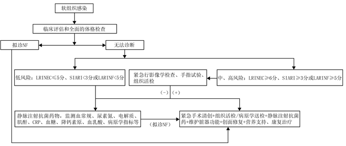
图 1 成人NF诊治简化流程图
注:NF指坏死性筋膜炎, LRINEC评分指坏死性筋膜炎实验室风险指标评分, SIARI指Site other than the lower limb, Immunosuppression, Age, Renal impairment and Inflammatory markers, LARINF指laboratory and anamnestic risk indicator for necrotizing fasciitis, CRP为C反应蛋白;“(-)”“(+)”分别表示阴性、阳性检查结果
Figure 1. Simplified flowchart for the diagnosis and management of adult NF
表 1 基于GRADE系统的坏死性筋膜炎推荐意见的推荐强度及证据等级描述
Table 1. The description of recommendation strength and evidence level of the GRADE system-based recommendations for necrotizing fasciitis
项目与分类 描述 推荐强度 强 强烈推荐采用 弱 结合临床审慎采用 证据等级 高 证据来源于基于创伤、坏死性筋膜炎人群良好设计的RCT, 或来源于包含至少1项高质量RCT的系统评价/荟萃分析、权威指南 中 证据来源于质量降低的RCT, 或来源于设计合理的非RCT、队列研究、病例对照研究、多中心的注册研究, 或来源于非创伤、坏死性筋膜炎人群的高质量RCT 低 证据来源于观察性研究, 或来源于非创伤、坏死性筋膜炎人群的非RCT、队列研究、病例对照研究等 极低 证据来源于个案报道、临床经验总结, 或来源于非创伤、坏死性筋膜炎人群的观察性研究 注:GRADE指推荐意见分级的评估、制订和评价, RCT指随机对照试验  下载: 导出CSV
表 2 坏死性筋膜炎实验室风险指标评分(分)
Table 2. Laboratory risk indicator score of necrotizing fasciitis
指标 C反应蛋白 白细胞计数 血红蛋白 血清钠 血肌酐 血糖 < 150 ≥150 < 15 15~25 > 25 > 135 110~135 < 110 ≥135 < 135 ≤141 > 141 ≤10 > 10 得分 0 4 0 1 2 0 1 2 0 2 0 2 0 1 注:该表内容来自参考文献[17];C反应蛋白、白细胞计数、血红蛋白、血清钠、血肌酐、血糖的单位分别为mg/L、×109/L、g/L、mmol/L、μmol/L、mmol/L;患者的总得分≤5分为低风险, 发生坏死性筋膜炎的概率 < 50%;总得分6或7分为中度风险, 发生坏死性筋膜炎的概率为50%~75%;总得分≥8分为高风险, 发生坏死性筋膜炎的概率 > 75%
下载: 导出CSV
表 3 坏死性筋膜炎的SIARI评分(分)
Table 3. SIARI score of necrotizing fasciitis
指标 分值 感染部位为非下肢 3 免疫缺陷史(获得性免疫缺陷综合征、化学治疗或使用激素) 3 年龄≤60岁 2 血肌酐 > 141 μmol/L 1 白细胞计数 > 25×109/L 1 C反应蛋白≥150 mg/L 1 注:SIARI指Site other than the lower limb, Immunosuppression, Age, Renal impairment and Inflammatory markers;该表内容来自参考文献[25]
下载: 导出CSV
表 4 坏死性筋膜炎的LARINF评分(分)
Table 4. LARINF score of necrotizing fasciitis
指标 血红蛋白 降钙素原 C反应蛋白 心肝肾功能不全 免疫抑制 肥胖 > 135 g/L 110~135 g/L < 110 g/L < 1 ng/mL ≥1 ng/mL < 10 mg/dL ≥10 mg/dL 否 是 否 是 否 是 得分 0 1 2 0 3 0 1 0 2 0 2 0 1 注:LARINF指laboratory and anamnestic risk indicator for necrotizing fasciitis;该表内容来自参考文献[26];1 dL=10 mL
下载: 导出CSV
-
 周灵 网刊视频.mp4

-

 下载:
下载:


计量
- 文章访问数: 2740
- HTML全文浏览量: 1270
- PDF下载量: 395
- 被引次数: 0
