


Indicative effect of indocyanine green-near-infrared fluorescence imaging in the thickness of necrotic dermal tissue in porcine burn wounds
-
摘要:
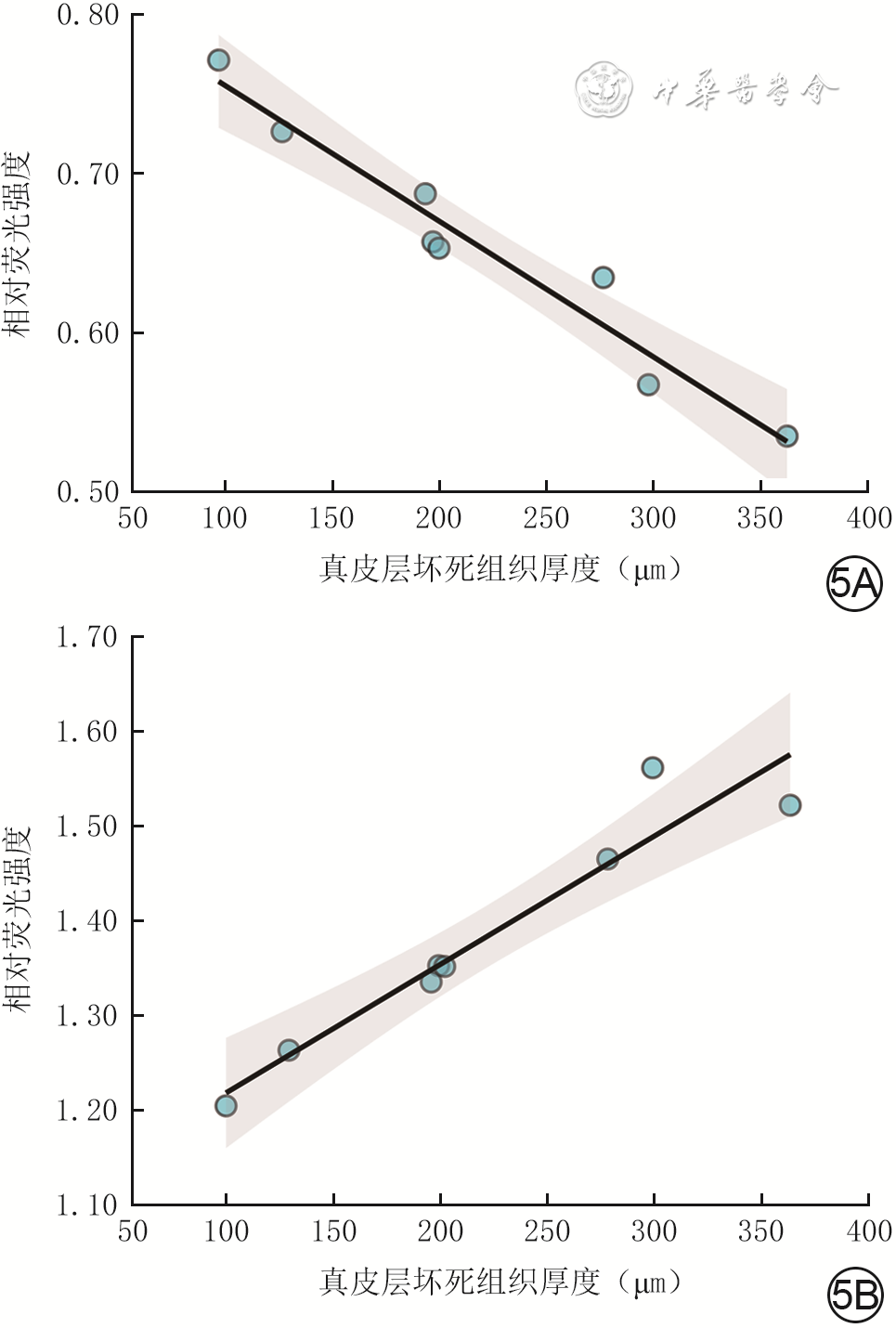
目的 探讨吲哚菁绿近红外荧光显影(ICG-NIFI)对猪烧伤创面真皮层坏死组织厚度的指示效果。 方法 该研究为医工交叉应用基础研究。选择1头3个月龄雄性巴马香猪,将烫伤仪(致伤温度75 ℃)与猪背部皮肤分别接触3、7、11 s造成直径2 cm的圆形烧伤创面,每个致伤时长2个创面。采用ICG-NIFI技术检测推注吲哚菁绿后(下称给药后)700 s内创面和创周正常皮肤感兴趣区的荧光强度,观察归一化处理的荧光强度的变化趋势并确定创面和创周正常皮肤感兴趣区的荧光强度达到峰值的时间(下称达峰时间)。另外选择2头3个月龄雄性巴马香猪,将烫伤仪(致伤温度75 ℃)与两侧胸壁皮肤分别接触5、7、9、11、13、15、17、19 s造成直径2 cm的圆形烧伤创面,每个致伤时长共4个创面。采用激光散斑对比成像技术检测创面感兴趣区的血流强度;采用ICG-NIFI技术检测并计算创面和创周正常皮肤感兴趣区的达峰时间时创面感兴趣区的相对荧光强度。取创面全层皮肤组织,行苏木精-伊红染色,测量真皮层坏死组织厚度。分析创面感兴趣区的血流强度及创面和创周皮肤达峰时间时创面感兴趣区的相对荧光强度与创面真皮层坏死组织厚度的相关性。 结果 创周正常皮肤感兴趣区的荧光强度呈先快速升高、后缓慢下降的趋势,达峰时间约为给药后60 s;创面感兴趣区的荧光强度呈先缓慢上升、后持续平稳的趋势,达峰时间约为给药后600 s。致伤时长5、7、9、11、13、15、17、19 s造成的创面真皮层坏死组织厚度分别为(101±8)、(130±6)、(201±19)、(197±30)、(204±21)、(280±39)、(302±35)和(366±27)μm,创面感兴趣区的血流强度与创面真皮层坏死组织厚度的相关性不显著(P>0.05)。给药后60 s,创面感兴趣区的相对荧光强度与创面真皮层坏死组织厚度具有显著的负相关性(R²=0.97,P<0.05);给药后600 s,创面感兴趣区的相对荧光强度与创面真皮层坏死组织厚度具有显著的正相关性(R²=0.96,P<0.05)。 结论 ICG-NIFI检测的猪烧伤创面感兴趣区在给药后60和600 s时的相对荧光强度均与创面真皮层坏死组织厚度具有显著的相关性,说明ICG-NIFI对猪烧伤创面真皮层坏死组织厚度具有显著灵敏的指示作用。 Abstract:Objective To explore the indicative effects of indocyanine green-near-infrared fluorescence imaging (ICG-NIFI) in the thickness of necrotic dermal tissue in porcine burn wounds. Methods This study was a medical-engineering interdisciplinary basic research. One 3-month-old male Bama miniature pig was selected, and circular burn wounds with a diameter of 2 cm were created by applying a scalding instrument with inflicting temperature of 75 ℃ to the skin on the dorsal side of the pig for 3, 7, and 11 seconds, respectively, with two wounds for each duration of injury. The fluorescence intensity of the regions of interest in the wounds and the surrounding normal skin was detected by ICG-NIFI technology within 700 seconds after injecting indocyanine green (hereinafter referred to as after administration). The trend of the normalized fluorescence intensity was observed, and the time when the fluorescence intensity of the regions of interest in the wounds and the surrounding normal skin reached the peak (hereinafter referred to as the peak time) was determined. Additional two 3-month-old male Bama miniature pigs were selected, and circular burn wounds with a diameter of 2 cm were created on the skin of bilateral thoracic walls by applying a scalding instrument with inflicting temperature of 75 ℃ for 5, 7, 9, 11, 13, 15, 17, and 19 seconds, respectively, with 4 wounds for each duration of injury. The blood flow intensity of the regions of interest in the wounds was detected by laser speckle contrast imaging technology; the relative fluorescence intensity of the regions of interest in the wounds was detected by ICG-NIFI technology at the peak time of the wounds and the surrounding normal skin. The full-thickness skin tissue of the wounds was taken for hematoxylin-eosin staining, and the thickness of necrotic dermal tissue was measured. The correlation between the blood flow intensity of the regions of interest in the wounds and the relative fluorescence intensity at the peak time of the regions of interest in the wounds and the surrounding normal skin and the thickness of necrotic dermal tissue in the wounds was analyzed. Results The fluorescence intensity of the regions of interest in the surrounding normal skin showed a rapid increase followed by a slow decrease, with the peak time of approximately 60 seconds after administration. The fluorescence intensity of the regions of interest in the wounds showed a slow increase followed by a sustained stable trend, with the peak time of approximately 600 seconds after administration. The thickness of necrotic dermal tissue in the wounds caused by durations of injury of 5, 7, 9, 11, 13, 15, 17, and 19 seconds was (101±8), (130±6), (201±19), (197±30), (204±21), (280±39), (302±35), and (366±27) μm, respectively. The correlation between the blood flow intensity of the regions of interest in the wounds and the thickness of necrotic dermal tissue in the wounds was not significant (P>0.05). The relative fluorescence intensity of the regions of interest in the wounds at 60 seconds after administration was significantly negatively correlated with the thickness of necrotic dermal tissue in the wounds (R²=0.97, P<0.05), and the relative fluorescence intensity of the regions of interest in the wounds at 600 seconds after administration was significantly positively correlated with the thickness of necrotic dermal tissue in the wounds (R²=0.96, P<0.05). Conclusions The relative fluorescence intensity of the regions of interest in porcine burn wounds detected by ICG-NIFI at 60 and 600 seconds after administration was significantly correlated with the thickness of necrotic dermal tissue in the wounds, indicating that ICG-NIFI has a significantly sensitive indicative effects for the thickness of necrotic dermal tissue in porcine burn wounds. -
Key words:
- Burns /
- Dermis /
- Laser speckle contrast imaging /
- Laser-Doppler flowmetry /
- Fluorescence imaging /
- Indocyanine green
-
参考文献
(41) [1] KarimAS,ShaumK,GibsonALF.Indeterminate-depth burn injury-exploring the uncertainty[J].J Surg Res,2020,245:183-197.DOI: 10.1016/j.jss.2019.07.063. [2] 刘一嘉,吴鹏,安纲,等.烧伤创面深度诊断技术的研究进展[J].中华烧伤与创面修复杂志,2022,38(5):481-485.DOI: 10.3760/cma.j.cn501120-20210518-00195. [3] 粘永健,陈志强,薛冬冬,等. 烧伤深度诊断技术研究进展[J]. 中华烧伤杂志,2016,32(11):698-701. DOI: 10.3760/cma.j.issn.1009-2587.2016.11.014. [4] JaspersMEH,van HaasterechtL,van ZuijlenPPM,et al.A systematic review on the quality of measurement techniques for the assessment of burn wound depth or healing potential[J].Burns,2019,45(2):261-281.DOI: 10.1016/j.burns.2018.05.015. [5] SchulzT,MarotzJ,SeiderS,et al.Burn depth assessment using hyperspectral imaging in a prospective single center study[J].Burns,2022,48(5):1112-1119.DOI: 10.1016/j.burns.2021.09.010. [6] DijkstraA,GuvenG,van BaarME,et al.Laser speckle contrast imaging, an alternative to laser doppler imaging in clinical practice of burn wound care derivation of a color code[J].Burns,2023,49(8):1907-1915.DOI: 10.1016/j.burns.2023.04.009. [7] ŁawnickaA, NowakowskiJ, MurawaD, et al. Uso de verde de indocianina en cirugía reconstructiva y quemaduras[J]. Rev Cir, 2022, 74(4):426-431. DOI: 10.35687/s2452-454920220041515. [8] LiuA,OchoaM,ReedMS,et al.Indocyanine green and protoporphyrin IX fluorescence imaging of inflammation, hypoxia, and necrosis of burns[J/OL].Burns Trauma,2025,13:tkaf021[2025-07-31]. https://pubmed.ncbi.nlm.nih.gov/40400790/. DOI: 10.1093/burnst/tkaf021. [9] YinM,LiY,LuoY,et al.A novel method for objectively, rapidly and accurately evaluating burn depth via near infrared spectroscopy[J/OL].Burns Trauma,2021,9:tkab014[2025-07-31]. https://pubmed.ncbi.nlm.nih.gov/34258302/. DOI: 10.1093/burnst/tkab014. [10] 黄通村,黄品同.吲哚菁绿在乳腺癌肿瘤坏死模型中的光学成像研究[J].实用肿瘤杂志,2022,37(6):508-513.DOI: 10.13267/j.cnki.syzlzz.2022.086. [11] FangC,WangK,ZengC,et al.Illuminating necrosis: from mechanistic exploration to preclinical application using fluorescence molecular imaging with indocyanine green[J].Sci Rep,2016,6:21013.DOI: 10.1038/srep21013. [12] HaradaT,KurodaT,TsutsumiH,et al.Heat shock zone of the burn wound[J].Burns,1996,22(7):578-579.DOI: 10.1016/0305-4179(96)88886-3. [13] VaupelP,KallinowskiF,OkunieffP.Blood flow, oxygen and nutrient supply, and metabolic microenvironment of human tumors: a review[J].Cancer Res,1989,49(23):6449-6465. [14] 蒋梅君,褚志刚,谢琼慧,等.激光散斑血流成像在预测烧伤患者创面愈合时间中的应用[J].中华烧伤杂志,2016,32(12):721-724.DOI: 10.3760/cma.j.issn.1009-2587.2016.12.004. [15] ZhengKJ,MiddelkoopE,StoopM,et al.Validity of laser speckle contrast imaging for the prediction of burn wound healing potential[J].Burns,2022,48(2):319-327.DOI: 10.1016/j.burns.2021.04.028. [16] 赖子森,吕嘉晖,刘广文,等.超声引导下经皮经肝门静脉穿刺注射吲哚菁绿在腹腔镜解剖性肝切除术中的应用价值[J].中华消化外科杂志,2024,23(12):1544-1549.DOI: 10.3760/cma.j.cn115610-20241120-00500. [17] 王宏光,王之浩.腹腔镜解剖性肝段切除术中吲哚菁绿荧光染色方法选择和术中超声应用策略[J].中华消化外科杂志,2024,23(2):228-235.DOI: 10.3760/cma.j.cn115610-20231203-00231. [18] McUmberH,DabekRJ,BojovicB,et al.Burn depth analysis using indocyanine green fluorescence: a review[J].J Burn Care Res,2019,40(4):513-516.DOI: 10.1093/jbcr/irz054. [19] JanSN,KhanFA,BashirMM,et al.Comparison of laser Doppler imaging (LDI) and clinical assessment in differentiating between superficial and deep partial thickness burn wounds[J].Burns,2018,44(2):405-413.DOI: 10.1016/j.burns.2017.08.020. [20] JiangZ,WuJ,QiuY,et al.Perfusion analysis using high-definition indocyanine green angiography in burn comb model[J].J Burn Care Res,2024,45(2):373-383.DOI: 10.1093/jbcr/irad156. [21] ÖnerÇ,IrmakF,EkenG,et al.The effect of stromal vascular fraction in an experimental frostbite injury model[J].Burns,2023,49(1):149-161.DOI: 10.1016/j.burns.2022.02.011. [22] WongkietkachornA,SurakunpraphaP,JenwitheesukK,et al.An inconvenient truth of clinical assessment and indocyanine green angiography precise marking for indeterminate burn excision[J].Plast Reconstr Surg Glob Open,2021,9(3):e3497.DOI: 10.1097/GOX.0000000000003497. [23] ChaudhryMA,MercerJB,de WeerdL.In vivo perforasome perfusion in hemi-DIEP flaps evaluated with indocyanine-green fluorescence angiography and infrared thermography[J].Plast Reconstr Surg Glob Open,2021,9(5):e3560.DOI: 10.1097/GOX.0000000000003560. [24] LauritzenE,BredgaardR,Laustsen-KielCM,et al.Indocyanine green angiography in oncoplastic breast surgery, a prospective study[J].J Plast Reconstr Aesthet Surg,2023,85:276-286.DOI: 10.1016/j.bjps.2023.07.022. [25] 窦雪娇,王海英,陈伟,等.多巴酚丁胺对糖尿病足创面游离皮瓣修复术中血流灌注影响的前瞻性研究[J].中华烧伤与创面修复杂志,2023,39(8):746-752.DOI: 10.3760/cma.j.cn501225-20221220-00543. [26] 谢婷珺,刘元波,韩婷璐,等. 吲哚菁绿血管造影技术辅助肱动脉穿支螺旋桨皮瓣修复躯干和上肢软组织缺损[J]. 中国修复重建外科杂志,2021,35(2):200-205. DOI: 10.7507/1002-1892.202008094. [27] 王石,董帅,曹阳,等.高选择性动脉吲哚菁绿造影在游离股前外侧皮瓣设计中的应用[J].中华烧伤与创面修复杂志,2024,40(10):948-954.DOI: 10.3760/cma.j.cn501225-20240513-00174. [28] ThomasB,FalknerF,DidzunO,et al.Optimizing ALT flap harvest: the role of combined preoperative duplex ultrasound and intraoperative ICG angiography for perforator selection[J].Life (Basel),2025,15(4):620.DOI: 10.3390/life15040620. [29] 胡雅楠,谢婷珺,刘元波,等. 吲哚菁绿血管造影辅助下设计切取扩张皮瓣整复瘢痕的临床效果[J]. 中华烧伤与创面修复杂志,2025,41(4):341-347. DOI: 10.3760/cma.j.cn501225-20250108-00013. [30] ZajacJC,LiuA,UselmannAJ,et al.Lighting the way for necrosis excision through indocyanine green fluorescence-guided surgery[J].J Am Coll Surg,2022,235(5):743-755.DOI: 10.1097/XCS.0000000000000329. [31] ShahzadF,FabbriN.Real-time indocyanine green imaging to aid in closure of radiated wounds[J].J Plast Reconstr Aesthet Surg,2024,89:51-52.DOI: 10.1016/j.bjps.2023.12.002. [32] AbdelrahmanH,El-MenyarA,PeraltaR,et al.Application of indocyanine green in surgery: a review of current evidence and implementation in trauma patients[J].World J Gastrointest Surg,2023,15(5):757-775.DOI: 10.4240/wjgs.v15.i5.757. [33] BreukingEA,de FraitureEJ,KrijghDD,et al.Current applications of indocyanine green fluorescence angiography in trauma patients and its potential impact: a systematic review[J].BMJ Open,2025,15(5):e099755.DOI: 10.1136/bmjopen-2025-099755. [34] PereiraN,OñateV,RoaR.A comprehensive approach to posttraumatic lymphedema surgical treatment[J].Arch Plast Surg,2023,50(4):422-431.DOI: 10.1055/s-0043-1768645. [35] YuanZ, WuJ, XiaoY, et al. A photo-therapeutic nanocomposite with bio-responsive oxygen self-supplying combats biofilm infections and inflammation from drug-resistant bacteria[J]. Adv Funct Mater, 2023,33(37):2302908. DOI: 10.1002/adfm.202302908. [36] ZöttermanJ,TesselaarE,ElawaS,et al.Correlation between indocyanine green fluorescence angiography and laser speckle contrast imaging in a flap model[J].Plast Reconstr Surg Glob Open,2023,11(9):e5187.DOI: 10.1097/GOX.0000000000005187. [37] DesmettreT,DevoisselleJM,MordonS.Fluorescence properties and metabolic features of indocyanine green (ICG) as related to angiography[J].Surv Ophthalmol,2000,45(1):15-27.DOI: 10.1016/s0039-6257(00)00123-5. [38] FarrakhovaD,MaklyginaY,RomanishkinI,et al.Fluorescence imaging analysis of distribution of indocyanine green in molecular and nanoform in tumor model[J].Photodiagnosis Photodyn Ther,2022,37:102636.DOI: 10.1016/j.pdpdt.2021.102636. [39] CassinottiE,Al-TaherM,AntoniouSA,et al.European Association for Endoscopic Surgery (EAES) consensus on indocyanine green (ICG) fluorescence-guided surgery[J].Surg Endosc,2023,37(3):1629-1648.DOI: 10.1007/s00464-023-09928-5. [40] SimionL,IonescuS,ChitoranE,et al.Indocyanine green (ICG) and colorectal surgery: a literature review on qualitative and quantitative methods of usage[J].Medicina (Kaunas),2023,59(9):1530.DOI: 10.3390/medicina59091530. [41] GarcíaHA,JunakMI,DonahueB,et al.Indocyanine green angiography processing and analysis pipeline for the assessment of indeterminate burn wounds[J].J Biomed Opt,2025,30(6):065002.DOI: 10.1117/1.JBO.30.6.065002. -

图 1 不同致伤时长造成的猪烧伤创面及用吲哚菁绿近红外荧光显影技术检测的创面和创周正常皮肤感兴趣区。1A.左侧2个创面的致伤时长为11 s,中间2个创面的致伤时长为7 s,右侧2个创面的致伤时长为3 s;1B.共16个感兴趣区,其中1~4、5~8、9~12分别表示致伤时长为11、7、3 s造成的创面的感兴趣区,13~16表示创周正常皮肤感兴趣区

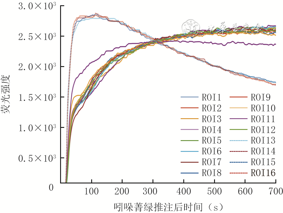
图 2 猪烧伤创面和创周正常皮肤感兴趣区的荧光强度-时间曲线
注:ROI1~4、5~8、9~12分别表示致伤时长为11、7、3 s造成的创面的感兴趣区,ROI13~16表示创周正常皮肤感兴趣区;采用吲哚菁绿近红外荧光显影技术测量荧光强度,所有荧光强度数据进行了归一化处理

图 3 不同致伤时长造成的猪烧伤创面真皮层坏死组织厚度 苏木精-伊红×20。3A、3B、3C、3D、3E、3F、3G、3H.分别为致伤时长5、7、9、11、13、15、17、19 s造成的创面
注:图中黑色线段和数据表示真皮层坏死组织的厚度

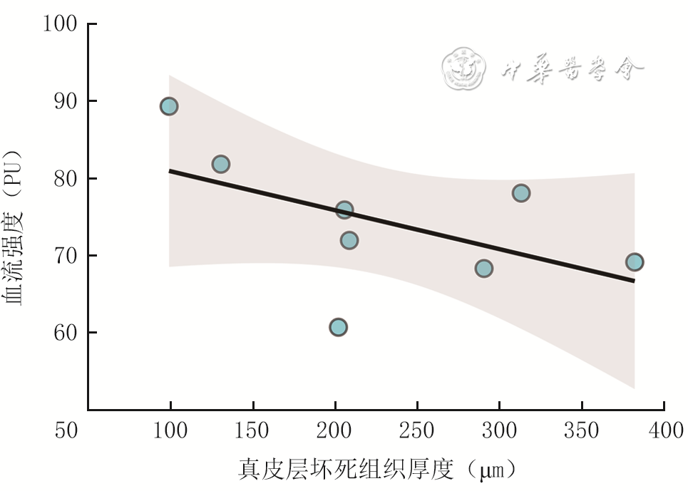
图 4 不同致伤时长造成的猪烧伤创面感兴趣区的血流强度与创面真皮层坏死组织厚度的相关性(总样本数为8×4)
注:烧伤创面致伤时长分别为5、7、9、11、13、15、17、19 s;采用激光散斑对比成像技术检测创面感兴趣区的血流强度;图中灰色为95%置信区间;创面感兴趣区的血流强度与创面真皮层坏死组织厚度无显著相关性(R2=0.29,P=0.170)
-
 董帅 11月03日.mp4

-

 下载:
下载:


计量
- 文章访问数: 1437
- HTML全文浏览量: 279
- PDF下载量: 15
- 被引次数: 0
