Abstract:
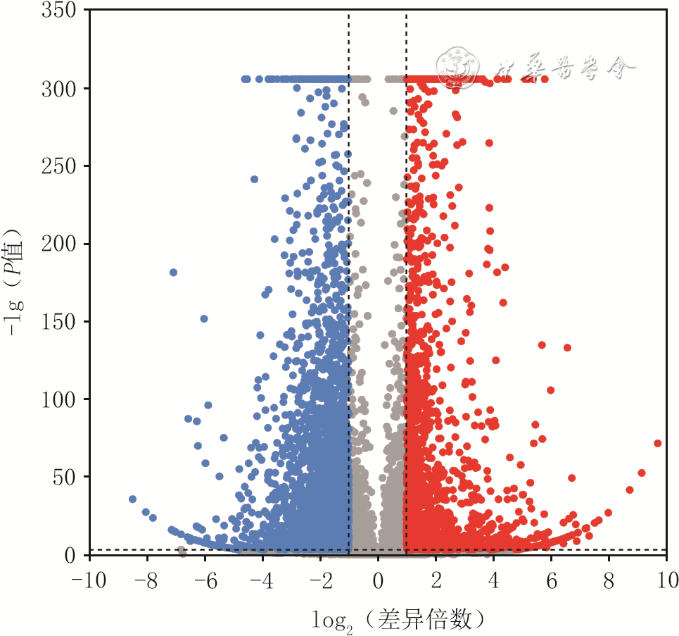
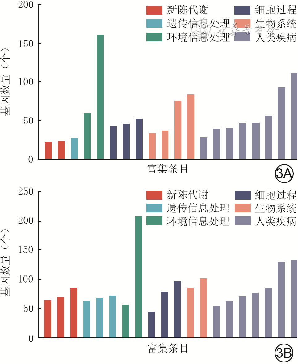
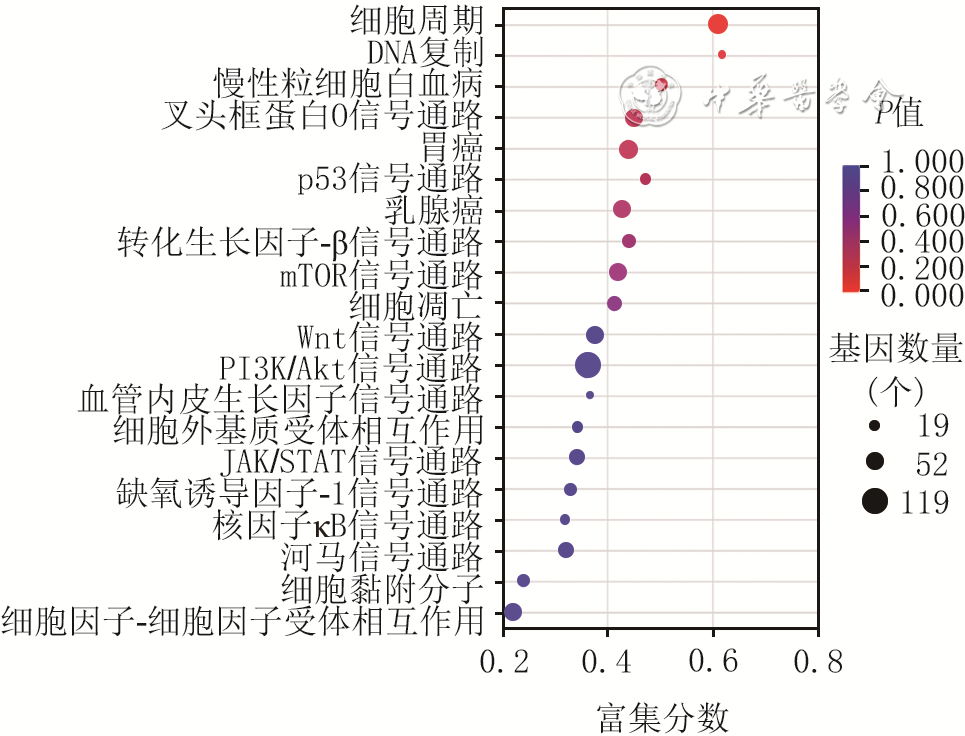
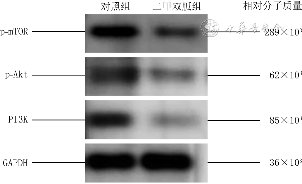
Objective To investigate the influence and mechanisms of metformin on the proliferation and apoptosis of human keloid fibroblasts (Fbs). Methods This study was an experimental research. The keloid tissue was collected from 7 keloid patients (2 males and 5 females, aged 20-65 years, with a disease course of more than 1 year) who underwent keloid excision surgery at the Department of Plastic, Cosmetic and Maxillofacial Surgery of the First Affiliated Hospital of Xi'an Jiaotong University from September 2020 to September 2023. The primary Fbs were isolated and cultured, and cells from passages 3 to 6 were used for experiments. The cells were divided into control group and metformin group, and were cultured in complete medium. The medium for metformin group was supplemented with metformin at a final molarity of 60 mmol/L. The cell counting kit-8 was used to assess the proliferation activity of cells in two groups after 12 and 24 hours of culture, and the proliferation inhibition rate of cells in metformin group after 12 and 24 hours of culture was calculated, with a sample size of 6. The apoptosis detection kit was used to detect the apoptotic distribution of cells in control group after 0 hour (immediately) of culture and in metformin group after 12 and 24 hours of culture, with a sample size of 3. The cell cycle detection kit was used to detect the cycle distribution of cells in two groups after 12 and 24 hours of culture, with a sample size of 3. The eukaryotic mRNA sequencing was performed on suitable number of cells of two groups after 24 hours of culture, and the Kyoto encyclopedia of genes and genomes functional annotation analysis and functional enrichment analysis were performed after screening for differentially expressed genes (DEGs) with significantly differential expression between two groups. Western blotting was conducted to detect the protein expressions of phosphatidylinositol 3-kinase (PI3K), phosphorylated protein kinase B (p-Akt), and phosphorylated mammalian target of rapamycin (p-mTOR) in the PI3K/protein kinase B (Akt)/mammalian target of rapamycin (mTOR) signaling pathway of cells in two groups after 24 hours of culture, with a sample size of 3. Results After 12 and 24 hours of culture, the proliferation activity of cells in metformin group was significantly lower than that in control group (with t values of 4.70 and 24.02, respectively, P<0.05); the proliferation activity of cells in metformin group after 24 hours of culture was significantly lower than that after 12 hours of culture within the group (t=4.73, P<0.05). Compared with that after 12 hours of culture within the group, the proliferation inhibition rate of cells in metformin group was significantly increased after 24 hours of culture (t=5.29, P<0.05). Compared with that in control group after 0 hour of culture, the proportion of early apoptotic cells in metformin group was significantly increased (with t values of 6.62 and 4.58, respectively, P<0.05), and the proportion of early and late apoptotic cells was significantly increased after 12 and 24 hours of culture (with t values of 4.84 and 3.75, respectively, P<0.05). After 24 hours of culture, the proportion of late apoptotic cells in metformin group was significantly higher than that after 12 hours of culture within the group (t=4.55, P<0.05). After 12 hours of culture, the proportion of S-phase cells in metformin group was significantly lower than that in control group (t=5.90, P<0.05). After 24 hours of culture, compared with that in control group, the proportion of G0/G1-phase cells in metformin group was significantly increased (t=5.36, P<0.05), while the proportion of G2/M-phase cells was significantly decreased (t=17.63, P<0.05). The proportion of S-phase cells in metformin group after 24 hours of culture was significantly higher than that after 12 hours of culture within the group (t=7.60, P<0.05). After 24 hours of culture, 4 814 DEGs with significantly differential expression were detected in the cells of metformin group compared with control group. The significantly upregulated and downregulated DEGs were mainly involved in biological functions related to signal transduction, cell growth and death, transport and catabolism, the endocrine system, the immune system, and cancer. The pathways that were significantly enriched with DEGs with significantly differential expression included the cell cycle and DNA replication, with the highest number of genes in the PI3K/Akt signaling pathway. After 24 hours of culture, the protein expressions of PI3K, p-Akt, and p-mTOR of cells in metformin group were 0.190±0.017, 0.170±0.017, and 0.247±0.005, respectively, which were significantly lower than 0.440±0.026, 0.300±0.060, and 0.547±0.025 in control group (with t values of 13.69, 3.61, and 20.12, respectively, P values all <0.05). Conclusions Metformin can significantly inhibit the proliferation of human keloid Fbs through the PI3K/Akt/mTOR signaling pathway and effectively induce its apoptotic process, thereby exerting antifibrotic effects.
Wu ML,Wang R,Zheng XN,et al.Influence and mechanisms of metformin on the proliferation and apoptosis of human keloid fibroblasts[J].Chin J Burns Wounds,2025,41(4):355-363.DOI: 10.3760/cma.j.cn501225-20241216-00489.


 Abstract
 PDF
PDF