2021 Vol. 37, No. 11

Expert Forum
Application of functional materials to promote cutaneous wound healing
Luo Gaoxing, Liu Menglong
2021, 37(11): 1005-1010. doi: 10.3760/cma.j.cn501120-20210930-00340
Abstract:
Cutaneous wound is one of the common clinical diseases. Functional materials can provide targeted wound protection and promote wound healing through the structural adjustment and functional integration. Currently, functional materials have been widely used in the field of wound repair, becoming one of the important tools for clinical wound treatment. This paper summarizes the application of functional materials of following categories including hemostasis, antibacterial, anti-inflammation, vascularization, and regulation of wound microenvironment in wound repair.
Original Articles•Wound Repair and Functional materials
Preliminary investigation on the wound healing effect of three-dimensional bioprinting ink containing human adipose-derived protein complexes
Zhang Chao, Li Zhao, Song Wei, Yao Bin, Enhejirigala, Zhang Mengde, Liang Liting, Jiang Yufeng, Fu Xiaobing, Huang Sha
2021, 37(11): 1011-1023. doi: 10.3760/cma.j.cn501120-20210813-00282
Abstract:
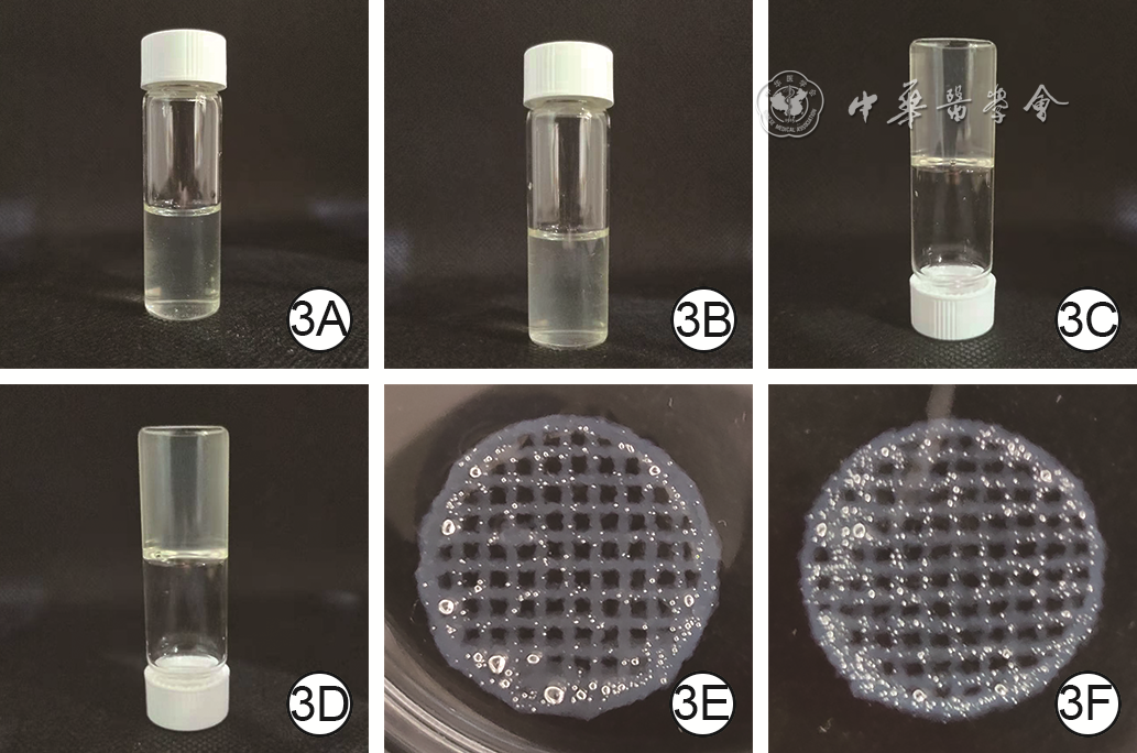
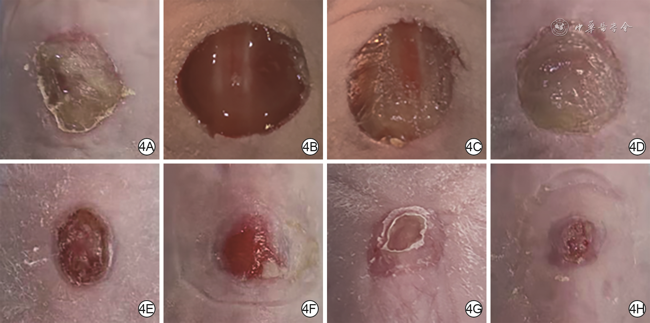
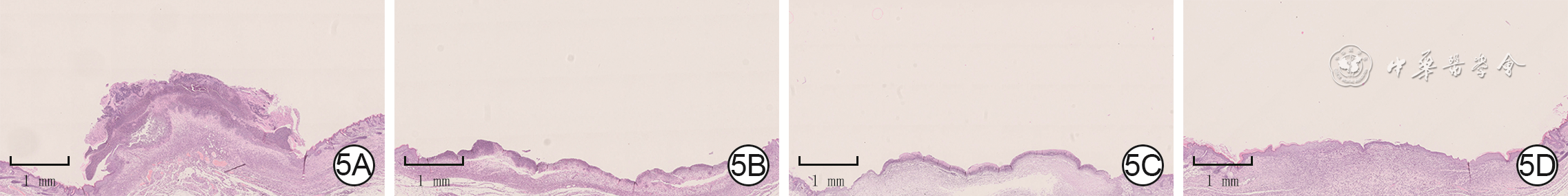
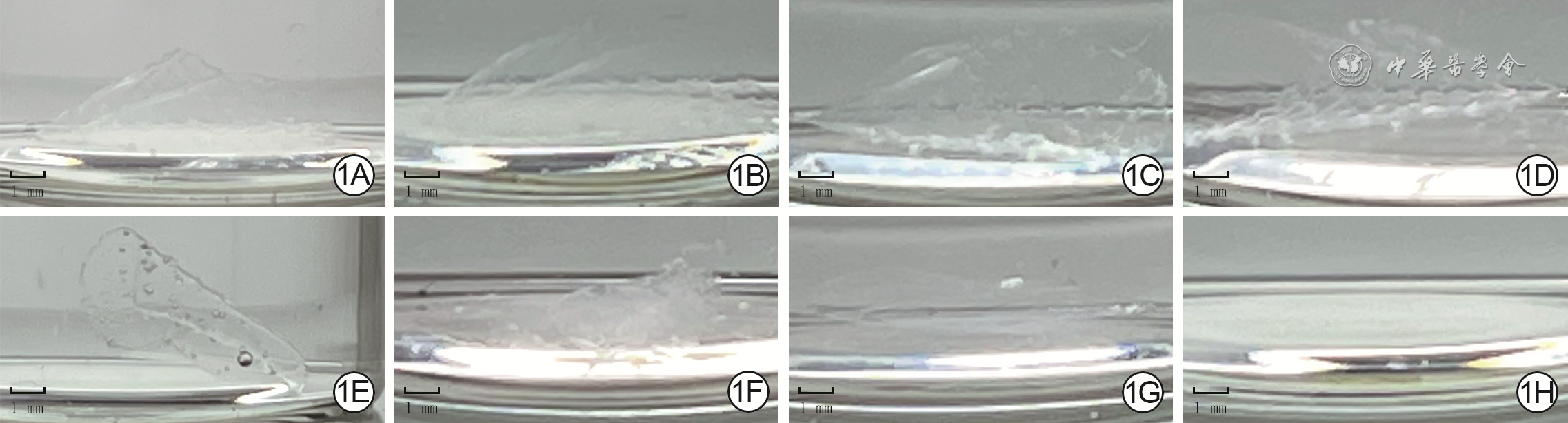
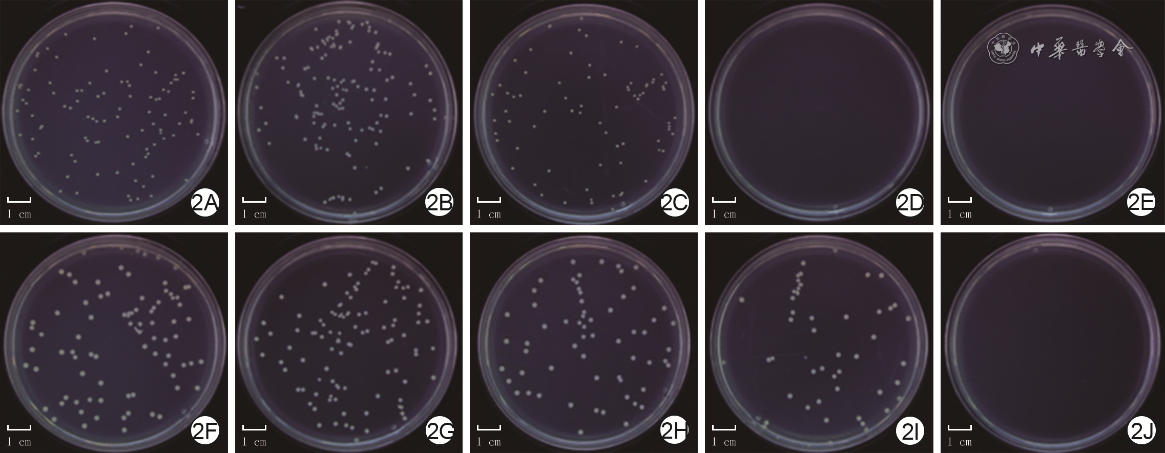
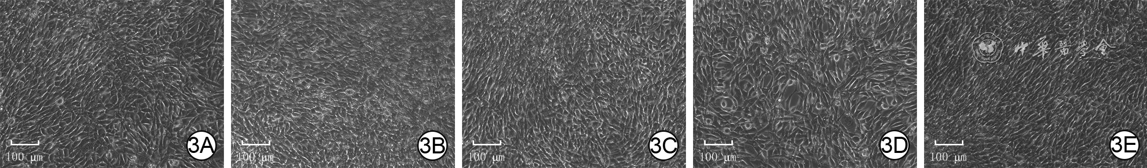
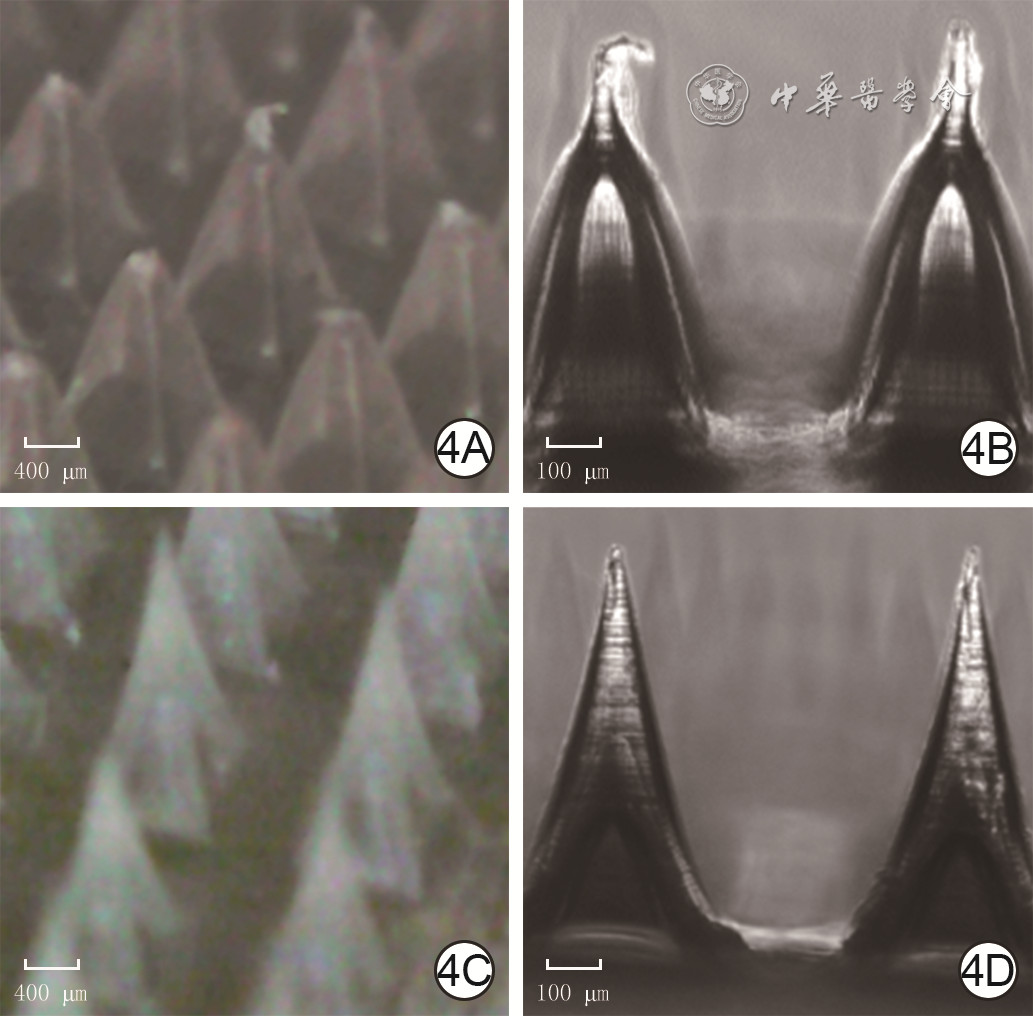
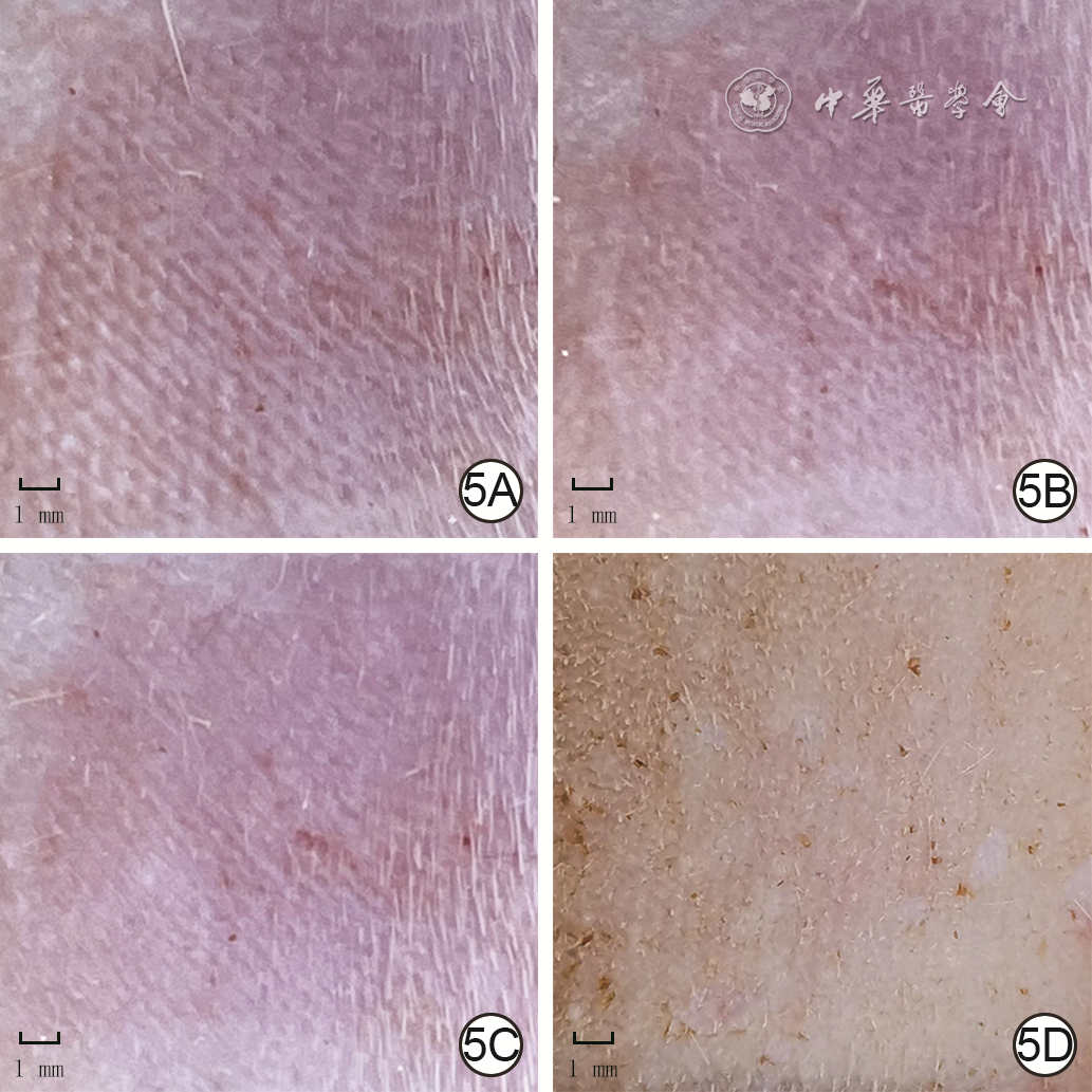
    Objective   To investigate the effects of human adipose-derived protein complex (ADPC) on the proliferation and migration ability of human skin fibroblasts (HSFs) and human umbilical vein endothelial cells (HUVECs), and the repairing effects of ADPC-containing three-dimensional (3D) bioprinting ink (Bioink) in full-thickness skin defect wounds of nude mice.    Methods   The experimental research method was used. Discarded subcutaneous adipose tissue from 3 female patients with chronic wounds (aged 29-34 years) admitted to PLA General Hospital for abdominal flap transfer from October 2020 to March 2021 and discarded liposuction adipose tissue from 3 healthy female (aged 24-36 years) for abdominal liposuction during the same period were collected to prepare normal ADPC (nADPC) and liposuction-derived ADPC (lADPC), respectively. The protein concentration of the two kinds of ADPC was measured by bicinchoninic acid method, and the extraction efficiency of them was calculated. The sample numbers were 3. HSFs and HUVECs in logarithmic growth phase were taken for the subsequent experiments. HSFs and HUVECs were divided into phosphate buffered saline (PBS) control group, 4 μg/mL nADPC group, 20 μg/mL nADPC group, 100 μg/mL nADPC group, and 200 μg/mL nADPC group according to the random number table (the same grouping method below), with 5 wells in each group. Cells in PBS control group were cultured with PBS, and the cells in the 4 remaining groups were cultured with the corresponding final mass concentration of nADPC. After 24 h of conventional culture, the cell proliferation viability was detected by cell counting kit 8 method. HSFs and HUVECs were taken and divided into PBS control group, nADPC alone group, lADPC alone group, tumor necrosis factor-α (TNF-α) alone group, TNF-α+nADPC group, and TNF-α+lADPC group. Cells in PBS control group and TNF-α alone group were added with PBS. nADPC or lADPC was added to the cells in nADPC alone group, lADPC alone group, TNF-α+nADPC group, and TNF-α+lADPC group with a final mass concentration of 100 μg/mL, respectively. TNF-α with a final mass concentration of 20 ng/mL was added to the cells in TNF-α alone group, TNF-α+nADPC group, and TNF-α+lADPC group. The cell migration rate was calculated after the scratch test at 24 h after scratching (n=3), and the cell proliferation viability was detected after 24 h of culture as above (n=5). Gelatin-alginate composite Bioink (Bioink AG) was taken. Bioink AG containing 100 μg/mL lADPC (lADPC-Bioink AG) was prepared. The morphology of the two at room temperature and after condensation was observed. The morphology after 3D bioprinting and cross-linking was observed. The low-temperature gel formation time was recorded when detecting rheological properties using rheometer (n=3). Twenty BALB/c-NU female nude mice of 8-10 weeks old were taken to establish the full-thickness skin defect wounds on the back, and then they were divided into routine dressing change group, lADPC alone group, Bioink AG alone group, and lADPC-Bioink AG group, with 5 nude mice in each group. The wounds of nude mice in routine dressing change group were covered with hydrocolloid dressings and performed with routine dressing changes only, while the wounds of nude mice in the remaining 3 groups were treated with lADPC, Bioink AG, and lADPC-Bioink AG accordingly in addition. General observation was performed from treatment day (TD) 0, and the wound healing rate was calculated on TD 2, 6, and 10. On TD 10, histopathological observation of wounds was performed with hematoxylin eosin staining. Data were statistically analyzed with independent samples t test, one-way analysis of variance, analysis of variance for repeated measurement, Student-Newman-Keuls q test, and least significant difference t test.    Results   The protein concentration and extraction efficiency of lADPC were respectively (1.306±0.011) mg/mL and (11.1±1.5)%, which were significantly lower than (2.039±0.042) mg/mL and (22.2±2.0)% of nADPC (t=23.83, 6.38, P<0.05 or P<0.01). After 24 h of culture, compared with those in PBS control group, the proliferation viabilities of HSFs (q=6.943, 6.375, P<0.01) and HUVECs (q=6.301, 6.496, P<0.01) were significantly decreased in 100 μg/mL nADPC group and 200 μg/mL nADPC group; compared with those in 100 μg/mL nADPC group, the proliferation viabilities of HSFs and HUVECs in 200 μg/mL nADPC group did not change significantly (P>0.05). At 24 h after scratching, compared with those in PBS control group, the HSF and HUVEC migration rates were significantly lower in nADPC alone group, lADPC alone group, and TNF-α alone group (q=5.642, 6.645, 11.480, 4.772, 6.298, 10.420, P<0.05 or P<0.01); compared with those in nADPC alone group, there were no significant changes in the HSF and HUVEC migration rates in lADPC alone group (P>0.05); compared with those in TNF-α alone group, there were no significant changes in the HSF migration rates in TNF-α+nADPC group or TNF-α+lADPC group (P>0.05), the HUVEC migration rates were significantly higher in TNF-α+nADPC group and TNF-α+lADPC group (q=8.585, 7.253, P<0.01); compared with those in TNF-α+nADPC group, there were no significant changes in the HSF and HUVEC migration rates in TNF-α+lADPC group (P>0.05). After 24 h of culture, compared with those in PBS control group, the HSF and HUVEC proliferation viabilities were significantly lower in nADPC alone group, lADPC alone group, and TNF-α alone group (q=5.803, 5.371, 9.136, 11.580, 9.493, 13.510, P<0.05 or P<0.01); compared with those in nADPC alone group, the HSF and HUVEC proliferation viabilities in lADPC alone group did not change significantly (P>0.05); compared with those in TNF-α alone group, the HSF (q=14.990, 10.850, P<0.01) and HUVEC (q=7.066, 8.942, P<0.01) proliferation viabilities were significantly higher in TNF-α+nADPC group and TNF-α+lADPC group; compared with those in TNF-α+nADPC group, the HSF and HUVEC proliferation viabilities in TNF-α+lADPC group did not change significantly (P>0.05). At room temperature and in the condensed state, lADPC-Bioink AG had a more slightly turbid appearance than Bioink AG. lADPC-Bioink AG had a similar morphology to Bioink AG after 3D bioprinting and cross-linking. At 10 ℃, the coagulation time of lADPC-Bioink AG was (76.6±0.4) s, which was significantly slower than (74.4±0.6) s of Bioink AG (t=4.64, P<0.01). On TD 2, the nude mice in routine dressing change group had more wound exudation, while the nude mice in the remaining 3 groups had no significant exudation. On TD 8, the nude mice in lADPC-Bioink AG group had the smallest residual wound area and obvious epithelial coverage. On TD 2, the wound healing rate of nude mice in lADPC-Bioink AG group was significantly higher than that in lADPC alone group (t=3.59, P<0.05) and similar to the rates in routine dressing change group and Bioink AG alone group (P>0.05). On TD 6, the wound healing rate of nude mice in lADPC-Bioink AG group was significantly higher than the rates in routine dressing change group, lADPC alone group, and Bioink AG alone group (t=18.70, 15.70, 3.15, P<0.05 or P<0.01). On TD 10, the wound healing rate of nude mice in lADPC-Bioink AG group was significantly higher than the rates in routine dressing change group and lADPC alone group (t=12.51, 4.84, P<0.01) but similar to that in Bioink AG alone group (P>0.05). On TD 10, the wounds of nude mice in lADPC-Bioink AG group had moderate vascularization of the traumatic tissue, adequate epithelialization, and the best healing effect.    Conclusions   Liposuction-related operations have little effect on the characterization of ADPC protein concentration and extraction efficiency. LADPC and nADPC have the same biological effects, which can inhibit the proliferation and migration ability of HSFs and HUVECs in non-inflammatory environment and improve the proliferation viabilities of HSFs and HUVECs in inflammatory environment, while improving the migration ability of HUVECs. Adding lADPC to Bioink AG does not significantly affect the physical properties or printing performance of Bioink AG, but can enhance the wound repair effect of full-thickness skin defect wounds in nude mice.
Effects of reactive oxygen species-responsive antibacterial microneedles on the full-thickness skin defect wounds with bacterial colonization in diabetic mice
Zhang Qingrong, Yang Xiao, Li Zheng, Jia Jiezhi, Luo Gaoxing, Yu Yunlong, Zhang Yi
2021, 37(11): 1024-1035. doi: 10.3760/cma.j.cn501120-20210831-00299
Abstract:
    Objective   To study the effects of reactive oxygen species (ROS)-responsive antibacterial microneedles (MNs) on the full-thickness skin defect wounds with bacterial colonization in diabetic mice.    Methods   Experimental research methods were adopted. The ROS-responsive crosslinker N1-(4-boronobenzyl)-N3-(4-boronophenyl)-N1, N1, N3, N3-tetramethylpropane-1,3-diaminium (TSPBA) was first synthesized, and then the polyvinyl alcohol (PVA)-TSPBA MNs, PVA-ε-polylysine (ε-PL)-TSPBA MNs, PVA-TSPBA-sodium hyaluronate (SH) MNs, and PVA-ε-PL-TSPBA-SH MNs were prepared by mixing corresponding ingredients, respectively. The PVA-TSPBA MNs were placed in pure phosphate buffer solution (PBS) and PBS containing hydrogen peroxide, respectively. The degradation of MNs immersed for 0 (immediately), 3, 7, and 10 days was observed to indicate their ROS responsiveness. The standard strains of Staphylococcus aureus (S. aureus) and Escherichia coli (E. coli) cultured in Luria-Bertani medium containing hydrogen peroxide were divided according to the random number table (the same grouping method below) into blank control group (without any treatment, the same below) and 0 g/L ε-PL group, 1.0 g/L ε-PL group, 5.0 g/L ε-PL group, and 10.0 g/L ε-PL group with which PVA-ε-PL-TSPBA MNs containing the corresponding concentration of ε-PL were co-cultured, respectively. Bacterial growth was observed after 24 h of culture, and the relative survival rate of bacteria was calculated (n=3). The mouse fibroblast cell line 3T3 cells at logarithmic growth stage (the same growth stage below) were divided into blank control group and 0 g/L ε-PL group, 1.0 g /L ε-PL group, 5.0 g /L ε-PL group, and 10.0 g /L ε-PL group in which cells were cultured in medium with the extract from PVA-ε-PL-TSPBA MNs containing the corresponding concentration of ε-PL, respectively. Cell growth was observed after 24 h of culture by optical microscopy, and the relative survival rate of cells was detected and calculated by cell counting kit 8 (CCK-8) assay to indicate the cytotoxicity (n=6). Both PVA-TSPBA MNs and PVA-TSPBA-SH MNs were taken, the morphology of the two kinds of MNs was observed by optical microscopy, and the mechanical properties of the two kinds of MNs were tested by microcomputer controlled electronic universal testing machine (denoted as critical force, n=6). Six male BALB/c mice aged 6-8 weeks (the same gender and age below) were divided into PVA-TSPBA group and PVA-TSPBA-SH group, with 3 mice in each group. After pressing the skin on the back of mice vertically with the corresponding MNs for 1 minute, the skin condition was observed at 0, 10, and 20 min after pressing. Another batch of 3T3 cells were divided into blank control group, 0 g/L ε-PL group and simple 5.0 g/L ε-PL group which were cultured with the extract of PVA-ε-PL-TSPBA MNs containing the corresponding concentration of ε-PL, and 5.0 g/L ε-PL+SH group which were cultured with the extract of PVA-ε-PL-TSPBA-SH MNs with 5.0 g/L ε-PL. The CCK-8 assay was performed to detect and calculate the relative survival rate of cells cultured for 24, 48, and 72 h to indicate the cell proliferation activity (n=6). Eighteen BALB/c mice were induced into diabetic mice model by high-sugar and high-fat diet combined with streptozotocin injection and then divided into sterile dressing group, 0 g/L ε-PL+SH group, and 5.0 g/L ε-PL+SH group, with 6 mice in each group. A full-thickness skin defect wound was made on the back of each mouse, and S. aureus solution was added to make a full-thickness skin defect wound with bacterial colonization model for diabetic mouse. The wounds of mice in 0 g/L ε-PL+SH group and 5.0 g/L ε-PL+SH group were covered with PVA-ε-PL-TSPBA-SH MNs with the corresponding concentration of ε-PL, and the wounds of mice in the 3 groups were all covered with sterile surgical dressings. The wound healing was observed on post injury day (PID) 0, 3, 7, and 12, and the wound healing rate on PID 3, 7, and 12 was calculated. On PID 12, the skin tissue of the wound and the wound margin were stained with hematoxylin and eosin to observe the growth of new epithelium and the infiltration of inflammatory cells. Data were statistically analyzed with one-way analysis of variance, analysis of variance for repeated measurement, Mann-Whitney U test, and Bonferroni test.    Results   With the extension of the immersion time, the PVA-TSPBA MNs in PBS containing hydrogen peroxide gradually dissolved and completely degraded after 10 days of immersion. The PVA-TSPBA MNs in pure PBS only swelled but did not dissolve. After 24 h of culture, there was no growth of S. aureus in 5.0 g/L ε-PL group or 10.0 g/L ε-PL group, and there was no growth of E. coli in 10.0 g/L ε-PL group. The relative survival rate of S. aureus was significantly lower in 1.0 g/L ε-PL group, 5.0 g/L ε-PL group, and 10.0 g/L ε-PL group than in blank control group (P<0.05 or P<0.01). The relative survival rate ofE. coli was significantly lower in 5.0 g/L ε-PL group and 10.0 g/L ε-PL group than in blank control group (P<0.01). After 24 h of culture, the cells in blank control group, 0 g/L ε-PL group, 1.0 g/L ε-PL group, 5.0 g/L ε-PL group, and 10.0 g/L ε-PL group all grew well, and the relative survival rate of cells was similar among the groups (P>0.05). The needle bodies of PVA-TSPBA MNs and PVA-TSPBA-SH MNs were both quadrangular pyramid-shaped and neatly arranged, and the needle bodies of PVA-TSPBA-SH MNs was more three-dimensional and more angular. The critical force of PVA-TSPBA-SH MNs was significantly higher than that of PVA-TSPBA MNs (Z=3.317, P<0.01). The MNs in PVA-TSPBA+SH group penetrated the skin of mice at 0 min after pressing, and the pinholes partially disappeared after 10 min and completely disappeared after 20 min, while the MNs in PVA-TSPBA group failed to penetrate the skin of mice. After 24, 48, and 72 h of culture, the proliferation activity of the cells in 5.0 g/L ε-PL+SH group was significantly higher than that of blank control group (P<0.05 or P<0.01). In sterile dressing group, the wounds of mice healed slowly and exuded more. The wound healing speed of mice in 0 g/L ε-PL+SH group was similar to that of sterile dressing group in the early stage but was faster than that of sterile dressing group in the later stage, with moderate exudation. The wound healing of mice in 5.0 g/L ε-PL+SH group was faster than that in the other two groups, with less exudation. The wound healing rates of mice in 5.0 g/L ε-PL+SH group were (40.6±4.2)%, (64.3±4.1)%, and (95.8±2.4)% on PID 3, 7, and 12, which were significantly higher than (20.4±2.7)%, (38.9±2.2)%, and (59.1±6.2)% in sterile dressing group and (21.6±2.6)%, (44.0±1.7)%, and (82.2±5.3)% in 0 g/L ε-PL+SH group (P<0.01). The wound healing rates of mice in 0 g/L ε-PL+SH group on PID 7 and 12 were significantly higher than those in sterile dressing group (P<0.05 or P<0.01). On PID 12, the wounds of mice in 5.0 g/L ε-PL+SH group were almost completely epithelialized with less inflammatory cell infiltration, the wounds of mice in 0 g/L ε-PL+SH group were partially epithelialized with a large number of inflammatory cell infiltration, and no obvious epithelialization but a large number of inflammatory cell infiltration was found in the wounds of mice in sterile dressing group.    Conclusions   The composite MNs prepared by TSPBA, PVA, ε-PL, and SH can successfully penetrate mouse skin and slowly respond to ROS in the wound to resolve and release antibacterial substances, inhibit bacterial colonization, and promote the repair of full-thickness skin defect wounds with bacterial colonization in diabetic mice.
Preparation and roles of sliver-loaded viscous hydrogel in healing of full-thickness skin defect wounds with bacterial colonization in mice
Dong Yunqing, Li Linlin, Zhu Xuanru, Feng Longbao, Jia Keyao, Guo Rui, Cheng Biao
2021, 37(11): 1036-1047. doi: 10.3760/cma.j.cn501120-20210906-00304
Abstract:
  Objective  To prepare the modified hyaluronic acid viscous hydrogel loaded with sliver particles and to explore the roles and mechanism of the hydrogel in healing of full-thickness skin defect wounds with bacterial colonization in mice.  Methods  The experimental research method was adopted. Dopamine modified hyaluronic acid (HA-DA) and phenylboric acid modified hyaluronic acid (HA-PBA) were prepared, and their characteristic peaks were detected by Fourier-transform infrared spectroscopy. Different mass of acrylamides was added to HA-DA and HA-PBA to prepare the viscous hydrogel with mass fraction of acrylamide in 10%, 15%, and 20%. The gelation of the viscous hydrogel with mass fraction of acrylamide in 20% was observed in the state of tilt and inversion at 37 ℃, and the storage modulus and loss modulus of the above 3 kinds of viscous hydrogels were detected by rotational rheometer. The sliver-loaded viscous hydrogel was prepared by adding nano silver ions to the viscous hydrogel with mass fraction of acrylamide in 20%. The concentration of silver ions released by sliver-loaded viscous hydrogel was measured by inductively coupled plasma mass spectrometer, and the cumulative release rate of silver ion was calculated (n=5). The mouse fibroblasts L929 were divided into phosphate buffered saline (PBS) group, viscous hydrogel group, and sliver-loaded viscous hydrogel group, which were dealt correspondingly, and the cell survival was detected by cell counting kit 8 method after 1, 2, and 3 d of culture (n=5). Twenty-four male C57BL/6 mice aged 6-8 weeks were selected, and forty-eight full-thickness skin defect wounds were inflicted and inoculated with the mixture of Escherichia coli and Staphylococcus aureus in the back of the mice, with two wounds in each mouse. The wounds were divided into normal saline group, viscous hydrogel group, and sliver-loaded viscous hydrogel group, which were dealt correspondingly, with 16 wounds in each group, and two wounds in each mouse were divided into different groups. On post injury day (PID) 3, 7, 10, and 14, the wound healing was observed and the wound healing rate was calculated. On PID 3, the colony forming units of Escherichia coli and Staphylococcus aureus in wounds were observed and counted. On PID 14, the epithelized epidermal thickness and the optical density of collagen fiber in wounds were observed and analyzed after hematoxylin eosin staining and Masson staining, respectively. On PID 3, 7, and 10, the expressions of tumor necrosis factor α (TNF-α), transforming growth factor β1 (TGF-β1), and vascular endothelial growth factor (VEGF) were detected by immunohistochemistry. The number of wounds in each index detecting at each time point was four. Data were statistically analyzed with analysis of variance for factorial design, one-way analysis of variance, and Bonferroni correction.  Results  The characteristic peaks of HA-PBA were detected at the wave numbers of 1 369 and 1 425 cm-1, indicating that phenylboric acid had been successfully grafted on hyaluronic acid, and the characteristic peaks of HA-DA were detected at the wave numbers of 1 516 and 1 431 cm-1, indicating that dopamine had been successfully grafted on hyaluronic acid. The viscous hydrogel with mass fraction of acrylamide in 20% maintained the stable and no-flow condition of gelation in the state of tilt and inversion at 37 ℃. The storage modulus and loss modulus of the viscous hydrogel increased with the increase of acrylamide content, the storage modulus and loss modulus of the 3 kinds of viscous hydrogels had no obvious changes with the increase of the oscillation frequency or time, and the storage modulus of the 3 kinds of acrylamide hydrogels were greater than the loss modulus. The release of silver ion in the sliver-loaded viscous hydrogel lasted for 7 days, and the cumulative release rate of silver ion was up to 65%. After 1, 2, and 3 d of culture, the cell survival rates in sliver-loaded viscous hydrogel group were significantly lower than those in PBS group and viscous hydrogel group (P<0.05 or P<0.01), while after 1 d of culture, the cell survival rate in viscous hydrogel group was significantly lower than that in PBS group (P<0.01). With extension of time after injury, the wounds of mice in the 3 groups shrank gradually. On PID 3, 7, 10, and 14, the wound healing rates in sliver-loaded viscous hydrogel group were (53.0±3.6)%, (75.3±6.9)%, (93.3±1.2)%, and (96.7±0.8)%, which were significantly higher than (21.8±6.4)%, (53.9±8.2)%, (72.0±7.8)%, and (92.5±0.4)% in normal saline group (P<0.01). On PID 3 and 14, the wound healing rates in sliver-loaded viscous hydrogel group were significantly higher than (43.5±2.4)% and (94.1±1.5)% in viscous hydrogel group (P<0.05). On PID 3 and 10, the wound healing rates in viscous hydrogel group were significantly higher than those in normal saline group (P<0.01). On PID 3, the colony forming units of two bacteria in wound of sliver-loaded viscous hydrogel group were significantly less than those in normal saline group and viscous hydrogel group (P<0.01), while the colony forming units of two bacteria in wound of viscous hydrogel group were significantly less than those in normal saline group (P<0.05). On PID 14, the wounds were basically epithelialized and the epidermis was thicker, with collagen protein content being increased significantly and more orderly arranged collagen in sliver-loaded viscous hydrogel group compared with those in the other 2 groups. On PID 14, the epidermal thickness in wounds of sliver-loaded viscous hydrogel group was significantly increased compared with that in the other two groups (P<0.05), and the optical density of collagen fiber was significantly increased compared with those in normal saline group (P<0.05). On PID 3, the expressions of TGF-β1 and VEGF in wounds of sliver-loaded viscous hydrogel group were significantly higher than those in normal saline group (P<0.05 or P<0.01), while the expression of VEGF in wounds of viscous hydrogel group was significantly higher than that in normal saline group (P<0.01). On PID 7, the expression of TGF-β1 in wounds of sliver-loaded viscous hydrogel group was significantly higher than that in the other 2 groups (P<0.01), and the expression of VEGF was significantly higher than that in normal saline group (P<0.01). On PID 10, the expression of TNF-α in wounds of sliver-loaded viscous hydrogel group was significantly lower than that in normal saline group (P<0.05), the expressions of TGF-β1 and VEGF in wounds of sliver-loaded viscous hydrogel group were significantly higher than those in normal saline group (P<0.05 or P<0.01), and the expression of VEGF in wounds of sliver-loaded viscous hydrogel group was significantly higher than that in viscous hydrogel group (P<0.05).  Conclusions  The sliver-loaded viscous hydrogel prepared in this study has good stability and elasticity, which can continuously release silver ions and help to accelerate the healing of full-thickness defect wounds with bacterial colonization in mice. Besides, the sliver-loaded viscous hydrogel has low biological toxicity and can promote re-epithelialization, collagen deposition as well as angiogenesis of wounds, which may be related to the infiltration and regression of inflammatory cells.
Original Articles
Clinical effects of bridge-type continuous negative pressure suction in postoperative fixation of upper limb soft tissue defect wound repaired with pedicled abdominal flap
Tong Lin, Hu Xiaolong, Zhang Wanfu, Han Fei, Ji Peng, Hu Dahai, Guan Hao
2021, 37(11): 1048-1053. doi: 10.3760/cma.j.cn501120-20210204-00050
Abstract:
  Objective  To observe the clinical effects of bridge-type continuous negative pressure suction in postoperative fixation of upper limb soft tissue defect wound repaired with pedicled abdominal flap.  Methods  The retrospective observational study was conducted. From April 2018 to October 2020, ninety-five patients who met the inclusion criteria were admitted to the First Affiliated Hospital of Air Force Medical University, including 55 males and 40 females, aged 5-78 years, with a defect wound area of 82 (9, 216) cm2. All patients underwent abdominal flap repair for soft tissue defects of hand and forearm. According to the different fixation methods adopted in the operation area, the patients were divided into negative pressure group (n=48) and plaster group (n=47). Wounds of the injury sites of patients in the 2 groups were repaired by flap transplantation after debridement. The negative pressure suction device was placed after dry gauze dressing to form a "bridge" to fix the affected upper limb and chest and abdomen in negative pressure group. Patients in plaster group were treated with conventional dry gauze matting and plaster fixation. On post surgery day (PSD) 1, 3, 5, 7, and 14, flap blood circulation and pain intensity of patients in the 2 groups were calculated by self-made blood flow scoring scale and Changhai Pain Ruler, respectively. On PSD 5, the common complications in operative area and surrounding skin were observed and their incidences were calculated. On PSD 7, satisfaction degree of patients was scored. During follow-up of one month after pedicle division, the appearance and functional recovery of the affected limb were observed. Data were statistically analyzed with analysis of variance for repeated measurement, independent samples t est, Cochran & Cox approximate t test, chi-square test, and Wilcoxon rank-sum test.  Results  On PSD 1, 3, 5, 7, and 14, the flap blood circulation scores of patients in negative pressure group did not change significantly, while that of patients in plaster group showed a time-dependent decrease, and the flap blood circulation scores of patients in negative pressure group were significantly higher than those in plaster group (t=2.259, 2.552, 2.784, 2.821, 3.003, P<0.05 or P<0.01). There were no significant changes in the pain intensity scores of patients in negative pressure group, while those of patients in plaster group increased in a time-dependent manner, and the pain intensity scores of patients in negative pressure group were significantly lower than those in plaster group (t=-4.818, -4.944, -5.011, -5.976, -6.721, P<0.05). On PSD 5, the incidences of common complications in operative area and surrounding skin of patients in negative pressure group were significantly lower than those in plaster group (χ2=6.773, 5.269, P<0.05 or P<0.01). On PSD 7, the satisfaction score of patients in negative pressure group was 14.7±1.1, which was significantly higher than 7.4±1.8 in plaster group (t=23.934, P<0.01). During follow-up of one month after pedicle division, the appearance and function of the affected limb of patients in the 2 groups recovered well.  Conclusions  After repairing the upper limb soft tissue defect wound with pedicled abdominal flap, the bridge-type continuous negative-pressure suction fixation can effectively immobilize the affected limbs, chest and abdomen, reduce the incidence of common complications in the operative area and surrounding skin, relieve the pain of immobilization of patients, improve the blood circulation of flap and patient's satisfaction. Thus, it is an effective, portable, comfortable, and easy-to-operate method.
Clinical effects of negative pressure wound therapy in treating the poor healing of incisions after different abdominal operations
Wang Xuexin, Xiang Yang, Meng Yao, Ma Bing, Hu Xiaoyan, Tang Hongtai, Ben Daofeng, Xiao Shichu
2021, 37(11): 1054-1060. doi: 10.3760/cma.j.cn501120-20210518-00194
Abstract:
  Objective  To investigate the clinical effects of negative pressure wound therapy (NPWT) in treating the poor healing of incisions after different abdominal operations.  Methods  The retrospective observational study was conducted. From June 2019 to December 2020, 42 patients with poor healing of incisions after abdominal surgery were admitted to Center of Burns and Trauma of the First Affiliated Hospital of Naval Medical University, including 29 males and 13 females, aged 23-81 years. The disease course of poor healing of abdominal incision was 3-60 d. The preoperative examination of patients was completed after admission, and NPWT was used after debridement. According to the dehiscence level of incision, the negative pressure value of -10.64 to -6.65 kPa was set. The incisions were sutured in the second stage when the incisions had good blood circulation. The cause of abdominal surgery, the dehiscence level and the cause of poor healing of abdominal incision were investigated, and the final healing of abdominal incision and the occurrence of complication were observed.  Results  The causes of abdominal operations in this group of patients who ocurred poor healing of abdominal incisions were ranked according to the composition ratio, with the top 4 causes being colon cancer (9 cases, accounting for 21.4%), bile duct disease (8 cases, accounting for 19.0%), liver cancer (5 cases, accounting for 11.9%), and appendicitis (4 cases, accounting for 9.5%). There were 25 cases (59.5%) with dehiscence of abdominal incision in the deep fascia layer, and the other 17 cases (40.5%) with dehiscence of abdominal incision in the superficial fascia layer. The causes of poor healing of abdominal incision were ranked according to the composition ratio, with the top 3 causes being infection (24 cases, accounting for 57.1%), fat liquefaction (11 cases, accounting for 26.2%), and suture reaction (5 cases, accounting for 11.9%). The blood circulation in 40 patients was improved after being treated with NPWT, and the incisions were sutured in the second stage. The incisions healed well when the suture lines were removed in the second to third week. Intestinal fistula and bile leakage developed during the NPWT treatment, respectively in the other 2 patients, in which negative pressure equipment was removed subsequently, and the incisions healed after adequate drainage and conventional dressing changes.  Conclusions  NPWT is effective in treating poor healing of abdominal incision after different abdominal surgeries. The clinicians need to comprehensively assess the patient's condition to determine when and how to use NPWT to avoid the occurrence of intestinal fistula, bile leakage, and other complications.
Effects and mechanisms of allogeneic epidermal stem cells on the survival of allogeneic full-thickness skin grafts in nude mice with full-thickness skin defect wounds
Huang Shaobin, Hu Zhicheng, Zhang Yi, Tang Bing, Wang Peng, Xu Hailin, Wang Zhiyong, Dong Yunxian, Cheng Pu, Rong Yanchao, Wu Jun, Zhu Jiayuan
2021, 37(11): 1061-1069. doi: 10.3760/cma.j.cn501120-20200704-00339
Abstract:
  Objective  To investigate the effects and mechanisms of allogeneic epidermal stem cells (ESCs) on the survival of allogeneic full-thickness skin grafts in nude mice with full-thickness skin defect wounds.  Methods  Experimental research methods were applied. Primary ESCs that appeared paving stone-like after being cultured for 7 d were obtained by enzymatic digestion method from one 4-week-old male BALB/c-NU nude mouse (the same strain, age, and sex below). The cells of third passage were identified by flow cytometry to positively express ESC marker CD44 and negatively express CD45, meanwhile, the positive expression of ESC markers of p63 and integrin 6α, and negative expression of CD71 were identified by immunofluorescence method. The ESCs of third passage in the logarithmic growth phase were used for the following experiments. Twenty-six nude mice were equally divided into phosphate buffered saline (PBS) group and ESCs group according to the random number table. A full-thickness skin defect wound was made on the back of each nude mouse, and then the wounds of the two groups were sprayed with equal volumes of PBS and ESCs, respectively. The wounds were transplanted with full-thickness skin grafts cut from the backs of 4 other nude mice. Each ten nude mice from the two groups were selected, the wound healing and skin survival on post surgery day (PSD) 0 (immediately), 3, 7, 14, and 21 were observed, and the survival ratio and shrinkage rate of skin grafts on PSD 3, 7, 14, and 21 were calculated (the number of sample was the number of surviving skin grafts at each time point); the blood perfusion in the skin grafts on PSD 3, 7, and 14 was detected by the laser speckle blood flow imager, and the blood flow ratio of nude mice skin grafts in ESCs group to PBS group at each time point was calculated (the number of sample was the pair number of surviving skin grafts in group pairing at each time point). The skin graft tissue of each 3 nude mice remained in the two groups were collected on PSD 7, and the mRNA expressions and protein expressions of tumor necrosis factor α (TNF-α), interleukin 8 (IL-8), IL-10, type Ⅰ collagen, type Ⅲ collagen, and matrix metalloproteinase 9 (MMP-9) in the tissue were detected by real-time fluorescent quantitative reverse transcription polymerase chain reaction and Western blotting, respectively. Data were statistically analyzed with Log-rank test, analysis of variance for repeated measurement, one-way analysis of variance, independent sample t test, and Bonferroni correction.  Results  Taking the condition on PSD 0 as a reference, the wounds of nude mice in the two groups healed gradually on PSD 3, 7, 14, and 21, and the shrinkage of skin grafts was gradually obvious. Among them, the shrinkage healing of wound of nude mice in PBS group was more significant than that in ESCs group. On PSD 3, the skin graft of 1 nude mouse failed in ESCs group, while the skin graft of 3 nude mice failed in PBS group. On PSD 7, the skin graft of another nude mouse failed in PBS group. The survival ratio of skin grafts of nude mice in the two groups was similar on PSD 3, 7, 14, and 21 (P>0.05). On PSD 3, 7, 14, and 21, the shrinkage rates of skin grafts of nude mice in ESCs group were (9.2±0.4)%, (19.7±1.2)%, (53.6±3.5)%, and (62.2±5.1)%, respectively, which was significantly lower than (11.0±0.9)%, (47.8±2.8)%, (86.1±7.1)%, and (89.7±9.0)% in PBS group (t=5.719, 26.650, 11.940, 7.617, P<0.01). On PSD 3, 7, and 14, blood perfusion signals were observed in the skin grafts of nude mice in the two groups. The average blood perfusion ratios of the skin grafts of nude mice in ESCs group to PBS group were greater than 1, and there was no statistically significant difference in the overall comparison of 3 time points (P>0.05). On PSD 7, compared with those of PBS group, the mRNA and protein expressions of TNF-α, IL-8, type Ⅰ collagen, and type Ⅲ collagen in the skin graft tissue of nude mice in ESCs group were significantly reduced, while the mRNA and protein expressions of IL-10 and MMP-9 in the skin graft tissue of nude mice in ESCs group were significantly increased (in mRNA comparison, t=2.823, 2.934, 2.845, 2.860, 3.877, 2.916, P<0.05).  Conclusions  Allogeneic ESCs can reduce the shrinkage of allogeneic full-thickness skin grafts transplanted on full-thickness skin defect wounds in nude mice, promote the formation of new blood vessels between the skin graft and the wound, reduce inflammation and collagen protein expression, and promote the expression of MMP-9, thus improving the survival quality of skin grafts.
Original Article·Nursing Column
Application of a standard communication system-based continuous home remote visit mode in the management of visits to severely burned patients in the post-pandemic era of coronavirus disease 2019
Chen Hualing, Li Maojun, Luo Yimei, Li Ning
2021, 37(11): 1070-1077. doi: 10.3760/cma.j.cn501120-20210315-00088
Abstract:
  Objective  To observe the application effects of a standard communication system-based continuous home remote visit mode in the management of visits to severely burned patients in the post-pandemic era of coronavirus disease 2019 (hereinafter referred to as post-pandemic era).  Methods  A retrospective cohort study was conducted. A total of 69 severely burned patients who met the inclusion criteria and were admitted to the First Affiliated Hospital of Army Medical University (the Third Military Medical University) from February to December, 2020 and their 69 family members were recruited into this study. According to the difference of visit model, 33 severely burned patients who were admitted into burn intensive care unit (BICU) from February to June, 2020 and received completely restricted visits according to the requirements for epidemic prevention and control and their corresponding 33 family members were included into conventional visit group; 36 severely burned patients who were admitted into BICU from July to December, 2020 and received the standard communication system-based continuous home remote visits and their 36 corresponding family members were recruited into remote visit group. The family members in conventional visit group could only communicate with the primary nurses by telephone, while the patients and family members in remote visit group could communicate with the primary nurses, responsible doctors, rehabilitation therapists, and nutrition therapists through WeChat video call. The survey results of general information questionnaire and Hamilton Anxiety Scale (HAMA) scores of two groups of patients and their family members at BICU admission of patients, HAMA scores of the two groups of patients and their family members in the second week of BICU admission and at discharge from BICU of patients, the scores of the Visiting Effect Evaluation Scale of the two groups of patients and their family members and the scores of Chinese version of Critical Care Family Satisfaction Survey (C-CCFSS) of the two groups of family members at discharge from BICU, and the scores of Satisfaction Survey and Evaluation Scale During Hospitalization of patients within the first week after the discharge of patients from BICU were compared. Data were statistically analyzed with chi-square test, Fisher's exact probability test, and Mann-Whitney U test.  Results  In remote visit group, there were 29 males and 7 females in patients, aged 48 (34, 53) years, and 15 males and 21 females in family members, aged 45 (30, 48) years. In conventional visit group, there were 24 males and 9 females in patients, aged 49 (38, 54) years, and 9 males and 24 females in family members, aged 44 (35, 48) years. At BICU admission of patients, the HAMA scores of the two groups of patients and family members were both similar (P>0.05). The total HAMA scores of patients and family members in remote visit group were significantly lower than those in conventional visit group in the second week when the patients were admitted to BICU (Z=-3.195, -3.018, P<0.01) and discharged from BICU (Z=-2.118, -2.380, P<0.05). At discharge from BICU of patients, the scores of each dimension in Visiting Effect Evaluation Scale of the patients in the two groups were similar (P>0.05); while scores of 3 dimensions including patient safety information, diagnosis and treatment quality information, and psychological support information in Visiting Effect Evaluation Scale of family members in remote visit group were significantly higher than those in conventional visit group (Z=-2.372, -2.209, -2.174, P<0.05), and only the scores of visit perception of family members were close to those in conventional visit group (P>0.05). At discharge from BICU of patients, the C-CCFSS scores of the family members in remote visit group was 99 (98, 108), which were significantly higher than 98 (97, 100) in conventional visit group (Z=-2.545, P<0.05). Within the first week after the discharge of patients from BICU, the scores of Satisfaction Survey and Evaluation Scale During Hospitalization of patients in remote visit group were 99 (98, 100), which were significantly higher than 96 (94, 98) in conventional visit group (Z=-5.213, P<0.01).  Conclusions  In the management of visits to severely burned patients in the post-pandemic era, application of the standard communication system-based continuous home remote visit mode can improve the visit effect and satisfaction, and effectively alleviate the anxiety of patients and their family members.
Application effects of failure mode and effect analysis on the limb posture positioning nursing of extremely severe burn patients
Lu Ying, Zhou Qin, Wang Lina, He Ting, Zhao Haiyang, Cao Xiaoqin
2021, 37(11): 1078-1084. doi: 10.3760/cma.j.cn501120-20210412-00126
Abstract:
    Objective   To explore the application effects of risk assessment method of failure mode and effect analysis (FMEA) on the limb posture positioning nursing of extremely severe burn patients.    Methods   A retrospective observational study was conducted. According to the different limb posture positioning methods, 30 extremely severe burn patients who met the inclusion criteria and underwent routine limb posture positioning in the First Affiliated Hospital of Air Force Medical University from January 2018 to June 2019 were included into routine limb positioning group (19 males and 11 females, aged (40±10) years), and 30 extremely severe burn patients who met the inclusion criteria and underwent limb posture positioning with FMEA risk assessment from July 2019 to December 2020 in the department were included into FMEA limb positioning group (20 males and 10 females, aged (38±10) years). Patients in routine limb positioning group received only routine limb posture positioning by rehabilitation therapists with bare hand every day from the time when their limb wounds healed until they were discharged from hospital. Patients in FMEA limb positioning group received FMEA risk assessment by physicians, rehabilitation therapists, and nurses within 24 hours after admission to analyze the potential failure modes of limb posture positioning, and target-directed limb posture positioning measures were adopted until they were discharged. The risk priority numbers (RPNs) of six major failure modes of patients in FMEA limb positioning group before and after intervention were compared. The range of motion (ROM) of shoulder abduction, elbow extension, wrist dorsiflexion, ankle plantarflexion, total action motion of hand, and modified Barthel index scores of the patients in two groups before and after intervention were also assessed. Data were statistically analyzed with independent sample t test, chi-square test, and paired sample t test.    Results   The RPNs of 6 main potential failure modes of patients in FMEA limb positioning group i.e. untimely interference of limb posture positioning, not strong awareness of limb posture positioning of nurses, inconsistent of evaluation standards of limb posture positioning, nurses' lacking knowledge about limb posture positioning, nurses' lacking active participation, unsatisfying effects of patients' limb posture positioning were respectively (146±31), (140±22), (125±34), (136±23), (110±28), and (110±5) points after intervention, which were significantly lower than (578±64), (543±57), (419±89), (269±64), (240±41), and (222±48) points before intervention (t=18.441, 23.681, 10.035, 5.362, 9.438, 7.171, P<0.01). After intervention, the ROMs of shoulder abduction, elbow extension, wrist dorsiflexion, and ankle plantarflexion of patients in FMEA limb positioning group were significantly better than those in routine limb positioning group (t=-4.250, 11.400, -15.928, 10.963, -7.470, P<0.01); the ROMs of shoulder abduction, elbow extension, wrist dorsiflexion, and ankle plantarflexion of patients in FMEA limb positioning group and routine limb positioning group were significantly better than those before intervention (t=-35.573, 33.670, -31.090, 32.902, -19.647, -14.952, 11.411, -33.462, -12.818, -13.672, P<0.01). After intervention, the Barthel index score of patients in FMEA limb positioning group (78±9) was significantly higher than 57±9 in routine limb positioning group (t=-9.055, P<0.01), and the Barthel index scores of patients in FMEA limb positioning group and routine limb positioning group were significantly higher than those before intervention (35±5 and 34±4, t=-22.964, -12.329, P<0.01).    Conclusions   In the limb posture positioning nursing of extremely severe burn patients, risk assessment method of FMEA can effectively avoid the high risk factors in the limb posture positioning of patients, thus maintain the effects of limb posture positioning and improve the ROM of patients, as well as increase the daily living ability of patients in prognosis.
Wound Repair
A prospective randomized controlled study of the application effect of hydrogel dressings on deep partial-thickness burn wounds after dermabrasion and tangential excision
Shang Niansheng, Cui Binghuan, Wang Cheng, Gao Hua, Xu Bin, Zhao Ran, Huo Ran
2021, 37(11): 1085-1089. doi: 10.3760/cma.j.cn501120-20210419-00133
Abstract:
  Objective  To investigate the clinical effect of applying hydrogel dressings in deep partial-thickness burn wounds after dermabrasion and tangential excision.  Methods  A prospective randomized controlled study was conducted. From November 2015 to August 2019, 168 patients with deep partial-thickness burns hospitalized in Zibo Sixth People's Hospital met the inclusion criteria. According to the random number table, the patients were divided into hydrogel dressing+vaseline gauze group (84 cases, 67 males and 17 females) and vaseline gauze only group (84 cases, 65 males and 19 females) who were aged (31±16) and (35±17) years, respectively, and were given corresponding treatment after dermabrasion and tangential excision. The dressings were changed every 3-5 days. Autologous skin grafting was performed with split-thickness or medium-thickness skin grafts taken from trunk or thigh if the wounds failed to heal over 21 days or the wounds were less than 21 days but located in the joints, which might affect the functional activities at later stage. After operation, the general condition of the wounds was observed continuously and dynamically till the wounds were healed. The degree of dressing adhesion was evaluated during the first 4 dressing changes after operation, and the degree of pain was evaluated using Numerical Rating Scale (NRS). The wound healing rate on post operation day (POD) 3, 6, and 15 was calculated. The positive proportion of bacterial culture of wound exudates/cleanout fluid on admission and at dressing change on POD 3, 6, and 15 was calculated. The number of dressing changes were recorded, the rate of skin grafting operation was calculated, and the time of complete wound healing was recorded. Vancouver Scar Scale (VSS) was used in 6 months of follow-up to evaluate the hyperplasia of scar. Data were statistically analyzed with analysis of variance for repeated measurement, independent sample t test, Mann-Whitney U test, chi-square test or Fisher's exact probability test, and Bonferroni correction.  Results  During the observation period after operation, the wound was moist with less exudates and the wound healed much faster in patients of hydrogel dressing+vaseline gauze group, with the inner dressing being easier to remove with mild pain, while the wounds showed more exudates and slower healing in patients of vaseline gauze only group with the obvious adhesions of inner dressing, stronger pain, and bleeding in the wounds. Compared with those in vaseline gauze only group, the degree of dressing adhesion and pain NRS score of patients in hydrogel dressing+vaseline gauze group decreased significantly during the first 4 dressing changes after operation (χ2=52.625, Z=-10.854, P<0.01), the wound healing rate increased significantly at dressing change on POD 3, 6, and 15 (t=10.347, 41.150, 167.627, P<0.01), the positive proportion of wound exudates/cleanout fluid bacterial culture did not change significantly on admission or at dressing change on POD 3 (P>0.05) but decreased significantly at dressing change on POD 6 and 15 (χ2=15.616, 15.226, P<0.01), the dressing change times was significantly reduced (t=-11.986, P<0.01), and the rate of skin grafting operation was significantly decreased (χ2=35.850, P<0.01). The complete wound healing time of patients in hydrogel dressing+vaseline gauze group was (17.6±2.8) d, significantly shorter than (27.1±3.0) d in vaseline gauze only group (t=-21.288, P<0.01). During the follow-up of 6 months, the VSS score of scar hyperplasia of patients in hydrogel dressing+vaseline gauze group was significantly lower than that in vaseline gauze only group (Z=-11.287, P<0.01).  Conclusions  Compared with the use of vaseline gauze only, the application of vaseline gauze coated with hydrogel dressing in deep partial-thickness burn wounds after dermabrasion and tangential excision is more effective in moisturizing, significantly reducing the degree of dressing adhesion and patients' pain during dressing change, increasing wound healing rate, decreasing wound infection rate and the rate of skin grafting operation, and reducing the number of dressing changes. It shortens the time for complete wound healing, and effectively relieves scar hyperplasia.
Review•Wound Repair and Functional materials
Research advances on multifunctional hydrogel dressings for treatment of diabetic chronic wounds
Cao Wangbei, Gao Changyou
2021, 37(11): 1090-1098. doi: 10.3760/cma.j.cn501120-20210715-00249
Abstract:

Diabetes can lead to a variety of complications, such as chronic wound (diabetic foot), which is one of the important causes of death for patients with diabetes. Unfavorable factors such as high blood glucose, high level of oxidative stress and inflammation, and susceptibility to infection lead to difficult healing and even worsening of diabetic chronic wounds. Due to the advantages of high water content, good biocompatibility, and tunable physicochemical properties, the hydrogels have become hot-spot materials in wound dressing research. Compared with the traditional dressings such as gauze, the hydrogel dressings can provide a moist environment that is beneficial for wound healing. By loading of bioactive components and modulation of compositions and structures of hydrogels, the hydrogel dressings can be endowed with excellent tissue adhesion, antibacterial ability, anti-oxidation, and inflammation regulation effect, etc., and thus show great prospects in wound dressing applications. Based on the characteristics of hydrogel materials and microenvironment of diabetic chronic wound, this review summarized the research advances on new multifunctional hydrogel dressings for the treatment of diabetic chronic wounds in recent years, and discussed the drawbacks of current hydrogel dressings with prospects proposed.

Reviews
Research advances on skin wounds suturing techniques and their clinical application
Jia Shanshan, Wang Xiaochuan, Jiao Ya, Jiang Duyin, Zhao Jie
2021, 37(11): 1099-1104. doi: 10.3760/cma.j.cn501120-20200701-00334
Abstract:
Stitching skin wounds is one of the essential skills of a surgeon. Whether it is a traumatic wound or a surgical incision, choosing the most appropriate closure technique according to its characteristics is an important factor for good healing. Various skin wounds suturing techniques have been created and improved over the years, which have advantages of simple operation, precise alignment, reducing tension of the wound edges, and reducing scar formation, etc. Although these techniques provide more options for wound suture, they also put forward requirements for the judgment and operation ability of the operators. This article summarizes the advantages and disadvantages of the different skin wounds suturing techniques and their clinical application.