2023 Vol. 39, No. 6

Expert Forums
Opinion on the photoelectric therapy of scars
Zhang Yixin, Li Jie
2023, 39(6): 501-506. doi: 10.3760/cma.j.cn501225-20220821-00353
Abstract:
Scars are problems that inevitably develop after deep dermal injury in the skin. Selecting appropriate photoelectric therapy for scars at different stages is an important part of scar management, which can shorten the acute inflammatory phase, accelerate scar maturation and regression, improve scar appearance and function, and reduce associated discomfort. Based on our team's practical experience and the current literature, this paper provides targeted photoelectric management protocols in the stages of wound healing, early scarring, hypertrophic scarring, and contracture scarring, with the aim of providing a reference for the development of standardized photoelectric therapy protocols for scars.
Elite Comments
Clinical experience of free superficial temporal fascia flap/anterolateral femoral fascial flap combined with skin grafting in repairing deep tissue defects of special parts of extremities
Yang Xuekang, Zhang Dongliang, He Ting, Zhang Yue, Xu Zhigang, Wei Jingtao, Li Jun, Liu Mengdong
2023, 39(6): 507-511. doi: 10.3760/cma.j.cn501225-20220915-00407
Abstract:
The deep tissue defects of extremities are prone to cause the exposure of tendons, blood vessels, nerves, and bones, which are commonly repaired with free flaps in clinical practice. However, for special parts such as fingers, toes, posterior ankles, anterior tibias, and dorsum of feet, the appearances are usually bulky after being repaired with free flaps and need lipectomy operations, which bring great physiological, psychological, and economic burden to patients. As the fascia flap is soft and thin with reliable blood supply and strong anti-infection ability, the free fascia flap combined with skin grafting offers some advantages in repairing the above-mentioned wounds. However, its clinical application is severely limited due to the complexity of surgical operation and the difficulty in observing blood supply after operation. In recent years, our team has carried out a lot of work and accumulated rich experience in repairing deep tissue defects of special parts of extremities with free superficial temporal fascia flap/anterolateral femoral fascial flap combined with skin grafting. From the clinical perspective, this paper mainly introduces the anatomy and harvesting method of free superficial temporal fascia flap/anterolateral femoral fascial flap, as well as the advantages, difficulties, and precautions of clinical application, for reference of peers.
Original Articles·Diagnosis and Treatment of Scar
Prospective randomized controlled trial on 2 940 nm fractional erbium laser combined with fractional micro-plasma radiofrequency in the treatment of facial atrophic acne scars
Guo Qige, Wang Shen, Yan Min, Dong Jiying, Yao Min
2023, 39(6): 512-517. doi: 10.3760/cma.j.cn501225-20230116-00018
Abstract:
  Objective   To compare the efficacy and safety of 2 940 nm fractional erbium laser combined with fractional micro-plasma radiofrequency (FMR) therapy and 2 940 nm fractional erbium laser in the treatment of atrophic acne scars.   Methods   A prospective randomized controlled research was conducted. A total of 100 atrophic acne scar patients (38 males and 62 females, aged 18-37 years) who were treated in the Scar Laser Clinic of the Department of Plastic and Reconstructive Surgery of Shanghai Ninth People's Hospital of Shanghai Jiao Tong University School of Medicine from March 2018 to March 2021 and conformed to the inclusion criteria were recruited. The patients were randomly divided into erbium laser+FMR group and erbium laser alone group, with 50 cases in each group. The facial acne scars of patients in erbium laser alone group were treated with 2 940 nm fractional erbium laser, while the facial acne scars of patients in erbium laser+FMR group were treated with erbium laser as above, besides, the scars of U and M types were treated with FMR, once every 3 months for a total of 3 times. Before the first treatment and 3 months after each treatment, the Echelle D'Assessment Clinique des Cicatrices D'Acne (ECCA) was used to score the scar. The occurrence of adverse reaction during the treatment process was observed and recorded, and the incidence was calculated. Three months after the last treatment, the 5-level classification method was used to evaluate the satisfaction of patients with the treatment effect, and the satisfaction rate was calculated. Data were statistically analyzed with independent sample t test and chi-square test.   Results   A total of 89 patients completed the study, including 46 patients in erbium laser+FMR group (19 males and 27 females, aged (26±5) years) and 43 patients in erbium laser alone group (15 males and 28 females, aged (27±6) years). The ECCA scores before the first treatment and 3 months after the first treatment of patients were similar between the two groups ( P>0.05). The ECCA scores of patients in erbium laser+FMR group at 3 months after the second and third treatment were 72±23 and 61±18, respectively, which were significantly lower than 85±26 and 76±25 in erbium laser alone group (with t values of -2.45 and -3.26, respectively, P<0.05). During the treatment process, the incidence of adverse reaction of patients in erbium laser+FMR group and erbium laser alone group were 23.91% (11/46) and 16.28% (7/43), respectively, and there was no statistically significant difference between the two groups ( P>0.05). The satisfaction rate of patients in erbium laser+FMR group was 78.26% (36/46) at 3 months after the last treatment, which was significantly higher than 53.49% (23/43) in erbium laser alone group ( χ 2=6.10, P<0.05).   Conclusions   The 2 940 nm fractional erbium laser combined with FMR is superior to 2 940 nm fractional erbium laser alone in the treatment of facial atrophic acne scars, achieving significantly higher efficacy without significantly increasing the incidence of adverse reaction, and patients are more satisfied with the efficacy. It can be used as a recommended therapy in clinical practice.
Pan-cancer analysis of ubiquitin-specific protease 7 and its expression changes in the carcinogenesis of scar ulcer
Zhang Siyu, Ruan Jingjing, Jin Dongmei, Chen Nuo, Xie Weiguo, Ruan Qiongfang
2023, 39(6): 518-526. doi: 10.3760/cma.j.cn501225-20230421-00137
Abstract:
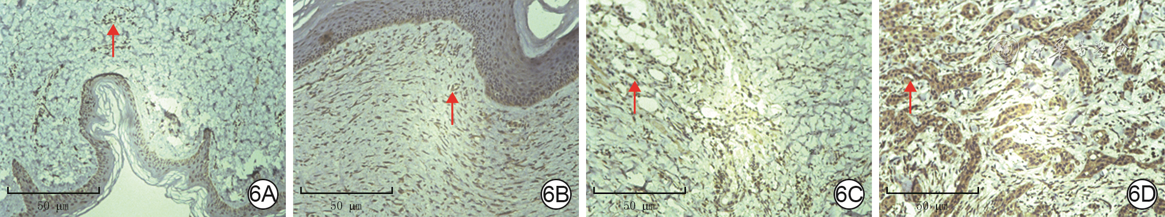
  Objective   To explore the biological role and clinical significance of ubiquitin-specific protease 7 (USP7) in the carcinogenesis of scar ulcer.   Methods   A retrospective observational study combined with bioinformatics analysis was used. The RNA expression profile data of USP7 in tumor and/or its corresponding paracancular normal tissue were obtained from The Cancer Genome Atlas (TCGA) database and the Gene Expression Omnibus database, and the RNA sequencing data were transformed by log 2. The variations of USP7 gene were analyzed by cBioPortal database. The USP7 mRNA expression in tumor and adjacent normal tissue in TCGA database were obtained by using the "Gene_DE" module in TIMER 2.0 database. The survival rates of patients with high and low USP7 expression in cutaneous melanoma (SKCM), cervical squamous cell carcinoma (CESC), lung squamous cell carcinoma (LUSC), and head and neck squamous cell carcinoma (HNSC) were analyzed using the Gene Expression Profile Interactive Analysis 2 (GEPIA2) database, and the Kaplan-Meier survival curves were drawn. Sangerbox database was used to analyze the correlation of USP7 expression in pan-cancer with microsatellite instability (MSI) or tumor mutation burden (TMB) pan-cancer. Through the "correlation analysis" module in the GEPIA2 database, the correlation of USP7 expression in pan-cancer with the expression levels of five DNA mismatch repair genes ( MLH1, MSH2, MSH6, PMS2, and EPCAM) and three essential DNA methyltransferases (DNMT)--DNMT1, DNMT3A, and DNMT3B were evaluated. The USP7 expression in CESC, HNSC, LUSC, and SKCM and its correlation with infiltration of immune cells (B cells, CD4 + T cells, CD8 + T cells, neutrophils, macrophages, and dendritic cells) were analyzed by the "Immune-Gene" module in TIMER 2.0 database. The "Similar Genes Detection" module of GEPIA2 database was used to obtain the top 100 protein sets with similar expression patterns to USP7. Intersection analysis was performed between the aforementioned protein sets and the top 50 protein sets that were directly physically bound to USP7 obtained by using the STRING database. Kyoto Encyclopedia of Genes and Genomes (KEGG) and Gene Ontology (GO) enrichment analysis were performed for the two protein sets mentioned above using the DAVID database. The samples of normal skin, hypertrophic scar, scar ulcer, and scar carcinoma with corresponding clinicopathologic features were collected from the Department of Pathology of Tongren Hospital of Wuhan University & Wuhan Third Hospital from October 2018 to October 2022, and the USP7 expression in tissue was detected by immunohistochemical method, with the number of samples of 6. Data were statistically analyzed with Log-rank test, one-way analysis of variance, and Bonferroni test.   Results   In pan-cancer, the main gene variations of USP7 were mutation and amplification, and the top 3 tumors with the highest variation frequency (>6%) were bladder urothelial carcinoma, SKCM, and endometrial carcinoma. The main mutation of USP7 gene in pan-cancer was missense mutation. In SKCM with the highest mutation frequency, the main type of mutation was missense mutation in USP7_ICP0_bdg domain. USP7 mRNA expression in breast invasive carcinoma, bile duct carcinoma, colon carcinoma, esophageal carcinoma, HNSC, renal chromophobe cell carcinoma, hepatocellular carcinoma, lung adenocarcinoma, LUSC, prostate carcinoma, and gastric carcinoma was significantly higher than that in corresponding paracancer normal tissue ( P<0.05). USP7 mRNA expression in glioblastoma multiforme, renal clear cell carcinoma, renal papillary cell carcinoma, and thyroid carcinoma was significantly lower than that in corresponding paracancular normal tissue ( P<0.05). In addition, USP7 mRNA expression in SKCM metastases was much higher than that in primary tumor tissue ( P<0.05). Survival curves showed no significant difference in survival rate between patients with high USP7 expression and patients with low USP7 expression in CESC, HNSC, LUSC, and SKCM (Log-rank P>0.05, with hazard ratios of 1.00, 0.99, 1.00, and 1.30, respectively). USP7 expression in colon cancer, colorectal cancer, thymic cancer, and thyroid cancer was negatively correlated with TMB (with Pearson correlation coefficients of -0.26, -0.19, -0.19, and 0.11, respectively, P<0.05). USP7 expression in glioma, CESC, lung adenocarcinoma, mixed renal carcinoma, and LUSC was positively correlated with MSI expression (with Pearson correlation coefficients of 0.22, 0.14, 0.15, 0.08, and 0.14, respectively, P<0.05), and USP7 expression in colon cancer, colorectal cancer, invasive breast cancer, prostate cancer, HNSC, thyroid cancer, and diffuse large B-cell lymphoma were significantly negatively correlated with MSI expression (with Pearson correlation coefficients of -0.31, -0.27, -0.13, -0.19, -0.16, -0.18, and -0.53, respectively, P<0.05). The expression of USP7 in CESC was positively correlated with that of both MSH2 and MSH6 (with Spearman correlation coefficients of 0.51 and 0.44, respectively, P<0.05), and the expression of USP7 in HNSC was positively correlated with the expression of EPCAM, MLH1, MSH2, MSH6, and PMS2 (with Spearman correlation coefficients of 0.39, 0.14, 0.49, 0.54, and 0.41, respectively, P<0.05), and the expression of USP7 in LUSC was positively correlated with the expression of EPCAM, MSH2, MSH6, and PMS2 (with Spearman correlation coefficients of 0.20, 0.36, 0.40, and 0.34, respectively, P<0.05), and the expression of USP7 in SKCM was positively correlated with the expression of EPCAM, MLH1, MSH2, MSH6, and PMS2 (with Spearman correlation coefficients of 0.11, 0.33, 0.42, 0.55, and 0.34, respectively, P<0.05). The expression of USP7 in CESC, HNSC, LUSC, and SKCM was significantly positively correlated with the expression of DNMT1, DNMT3A, and DNMT3B (with Spearman correlation coefficients of 0.42, 0.34, 0.22, 0.45, 0.52, 0.22, 0.36, 0.36, 0.22, 0.38, 0.46, and 0.21, respectively, P<0.05). The expression of USP7 in CESC, HNSC, LUSC, and SKCM was positively correlated with CD4 + T cell infiltration (with Partial correlation coefficients of 0.14, 0.22, 0.13, and 0.16, respectively, P<0.05). Being similar to the pattern of USP7 expression and ranked among top 100 protein sets, the top 5 proteins were C16orf72, BCLAF1, UBN, GSPT1, ERI2 (with Spearman correlation coefficients of 0.83, 0.74, 0.73, and 0.72, respectively, all P values<0.05). The top 50 protein sets that directly physically bind to USP7 overlapped with the aforementioned protein set by only one protein, thyroid hormone receptor interaction factor 12. KEGG enrichment analysis showed that USP7 related genes were involved in cell cycle, spliceosome, cell senescence, and p53 signal pathway. GO enrichment analysis showed that USP7 related genes were involved in transcriptional regulation, protein ubiquitination, DNA repair, and cytoplasmic pattern recognition receptor signal pathways. Analysis of clinical samples showed that USP7 expression was significantly higher in hypertrophic scars (0.35±0.05), scar ulcers (0.43±0.04), and scar cancers (0.61±0.03) than in normal skin (0.18±0.04), P<0.05.   Conclusions   USP7 may be a clinical biomarker for the progression of cicatricial ulcer cancer.
Original Articles
Clinical effect of the giant deep inferior epigastric artery paraumbilical perforator flap in repairing the circular high-voltage electric burn wounds on the wrist
Du Weili, Shen Yuming, Hu Xiaohua, Qin Fengjun, Cheng Lin
2023, 39(6): 527-533. doi: 10.3760/cma.j.cn501225-20220719-00296
Abstract:
  Objective   To investigate the clinical effect of the giant deep inferior epigastric artery paraumbilical perforator flap in repairing the circular high-voltage electric burn wounds on the wrist.   Methods   A retrospective observational study method was used. From September 2016 to October 2021, thirteen male patients (aged 20-43 years) with annular high voltage (10-100 kV) electrical burns on the wrist were admitted to the Beijing Jishuitan Hospital. At the early stage after injury, the patient's wrist was subjected to incision, tension reduction and debridement, with the wound area after debridement being 27 cm×16 cm-32 cm×19 cm; in 12 patients with vascular injury, the radial or ulnar artery was reconstructed by great saphenous vein transplantation, with the length of 15-25 cm; the wrist wound was repaired by free transplantation of the deep inferior epigastric artery paraumbilical perforator flap (if the wound was giant, the lower abdominal flap carrying other perforators was used), with the area of 30 cm×19 cm-35 cm×20 cm. The donor site was repaired by direct suture+skin grafting or relay flap transplantation. After surgery, the survival of flap in recipient area, as well as survival of the skin or flap in donor site were observed. During follow-up, the appearances of the flap in recipient area and the recovery of hand function, as well as the healing of donor site, occurrence of abdominal wall hernia, and scar in skin graft area were observed.   Results   After surgery, all the 13 patients' paraumbilical perforator flaps survived. Among them, 3 patients had subcutaneous fat necrosis at the distal end of the wrist flap, and the wound had mild infection, which healed after re-expansion and dressing change. All the skin grafts in the donor site of 10 patients survived, and the flaps in the donor site of 3 patients survived well. The patients were followed up for 6 months to 3 years. The flaps in recipient area were in good shape, 8 cases had partial recovery of hand function, and 5 cases had loss of finger flexion function; the donor site of abdominal flap healed well with no abdominal hernia occurred, and the skin graft site had no obvious scar hyperplasia and was soft in texture.   Conclusions   Early vascular reconstruction after injury, together with free transplantation of the giant deep inferior epigastric artery paraumbilical perforator flap are effective in repairing circular high-voltage electrical burn wounds on the wrist.
Clinical effects of superficial temporal artery lobulated perforator flaps in repairing skin and soft tissue defects after temporal tumor resection
Liao Xiaoshuang, Chen Wei, Jiang Haifang, Zhou Jian, Wei Zairong, Chang Shusen, Zhang Fang, Nie Kaiyu
2023, 39(6): 534-539. doi: 10.3760/cma.j.cn501225-20220816-00347
Abstract:
  Objective   To explore the feasibility and clinical effects of using superficial temporal artery lobulated perforator flaps in repairing skin and soft tissue defects after tumor resection in the temporal region.   Methods   A retrospective observational study method was used. From March 2017 to October 2022, ten patients with temporal skin tumors were admitted to the Affiliated Hospital of Zunyi Medical University, including six women and four men, with age ranging from 42 to 87 years. Among them, three patients had squamous cell carcinoma and seven patients had basal cell carcinoma, with disease duration ranging from 6 months to 5 years. All temporal tumors underwent expanded resection, leaving wound areas of 5.4 cm×4.2 cm to 7.0 cm×4.0 cm after tumor resection. Superficial temporal artery frontal branch flaps with areas of 5.5 cm×1.2 cm to 7.0 cm×1.5 cm, superficial temporal artery descending branch flaps with areas of 4.2 cm×3.5 cm to 5.0 cm×4.0 cm, and superficial temporal artery parietal branch flaps with areas of 4.2 cm×1.0 cm to 5.0 cm×1.0 cm were designed to repair the wounds and reconstruct the hairline. The donor areas of the flaps were closed and sutured directly. The survival of the flaps was observed on 3 to 5 days after surgery, and the healing of wounds on the donor and recipient sites was observed when the stitches were removed on 5 to 7 days after surgery. During follow-up after surgery, the appearance of the temporal area, scar hyperplasia, hairline reconstruction, and tumor recurrence were observed in the temporal region on the affected side.   Results   All the flaps survived well on 3 to 5 days after surgery, and all the donor and recipient site wounds healed well on 5 to 7 days after surgery. During follow-up of 3 to 6 months after surgery, the surgical incisions were concealed; the flaps were not swollen, with a consistent color to the surrounding skin; there were no obvious hypertrophic scars; the reconstructed hairline on the affected side was not significantly different from that of the healthy side; there was no tumor recurrence in the local area.   Conclusions   For large areas of skin and soft tissue defects in the temporal region, the use of superficial temporal artery lobulated perforator flaps can repair the wounds in different regions and suture the donor sites in the primary stage simultaneously. The surgical operation is simple, and the facial appearance conforms to the aesthetic requirement after surgery with no tumor recurrence in the local area but a good repair effect. This method is particularly suitable for repairing large areas of skin and soft tissue defects in the temporal region in elderly patients.
Curative effects of bi-pedicled deep inferior epigastric perforator flap in repairing large soft tissue defects in the lower limbs
Huang Yu, Huang Bo, Liu Anming, Tang Lin, Zhou Xin, Wang Shaoliang, Zou Yonggen
2023, 39(6): 540-545. doi: 10.3760/cma.j.cn501225-20220831-00373
Abstract:
  Objective   To investigate the curative effects of bi-pedicled deep inferior epigastric perforator (DIEP) flap in repairing large soft tissue defects in the lower limbs.   Methods   A retrospective observational study was conducted. From February 2016 to June 2020, 16 patients with large soft tissue defects in the lower limbs caused by trauma or after tumor/scar resection were admitted to the Department of Hand Surgery of the Affiliated Traditional Chinese Medicine Hospital of Southwest Medical University, including 9 males and 7 females, aged 25-58 years, with the area of soft tissue defects ranging from 14.0 cm×8.0 cm to 32.0 cm×18.0 cm. Using the abdomen as the donor site, the conjoined abdominal wall flap, i.e., the bi-pedicled DIEP flap (with an area ranging from 15.0 cm×9.0 cm to 32.0 cm×20.0 cm) carrying two sets of the trunk of the deep inferior epigastric artery was designed and resected to repair the wound. The donor site wound was sutured directly. The flap survival and wound healing in the donor and recipient areas were observed after operation. The curative effect was evaluated during the follow-up. At the last follow-up, the American Knee Society score and lower extremity functional scale were used to assess the functions of knee joint and lower limb, respectively.   Results   The flaps of 15 patients survived after operation; the flap of one patient had partial infection at the edge after operation but healed after debridement and dressing change. The wounds in the donor and recipient areas of 16 patients all healed well. Follow-up of 16-28 months showed that the recipient area had a good shape and pliable texture, and there was no obvious swollen appearance, hyperpigmentation, or abnormal hair growth; the donor site had linear scar only, with no complications such as abdominal hernia or hyperplastic scar; the functions of knee joint and lower limb were well reconstructed, with no recurrence of tumor. At the last follow-up, among the 4 patients with knee joint injury, 3 cases were excellent and 1 case was good in the evaluation of knee joint function; among the 12 patients with lower limb injury, 9 cases were excellent and 3 cases were good in the evaluation of lower limb function.   Conclusions   The donor site of bi-pedicled DIEP flap is concealed with abundant tissue and large area for resection, with which can be used to repair large soft tissue defects in the lower limbs, achieving good short-term results of appearance and function restoration.
Effects of free superficial peroneal artery perforator flap in repairing small and medium-sized thermal crush injury wounds in the hand
Cheng Heyun, Ju Jihui, Zhao Qiang, Liu Shengzhe, Zhang Guangliang, Zhang Tao, Wang Benyuan, Guo Quanwei, Liu Shuang
2023, 39(6): 546-551. doi: 10.3760/cma.j.cn501225-20220623-00256
Abstract:
  Objective   To investigate the surgical methods and clinical effects of free superficial peroneal artery perforator flap in repairing small and medium-sized thermal crush injury wounds in the hand.   Methods   A retrospective observational study was conducted. From August 2018 to December 2021, 12 patients (19 wounds) with small and medium-sized thermal crush injury in the hand who met the inclusion criteria were hospitalized in Suzhou Ruihua Orthopaedic Hospital, including 5 males and 7 females, aged from 30 to 54 years. The area of the wound was from 2.5 cm×2.0 cm to 14.0 cm×3.5 cm, and all the wounds were repaired by using free superficial peroneal artery perforator flaps from lower leg on one side (including single flap, multiple flaps, and multiple flaps with one pedicle resected from the same donor site). The area of the flap was from 3.5 cm×3.0 cm to 16.0 cm×4.0 cm. The wound in the donor site was sutured directly. The vascular crisis and survival of the flap were observed after operation. The texture, appearance, color, hyperpigmentation, sensation, and two-point discrimination of the flap repaired area were followed up, as well as the hyperplasia of scar and pain condition in the donor and recipient sites. At the last follow-up, the curative effect of flap repair was evaluated by the comprehensive evaluation scale, and the extension and flexion functions of the reserved digital joint were evaluated by the total active movement systematic evaluation method recommended by American Academy for Surgery of Hand.   Results   One flap developed arterial crisis on the first day after operation but survived after timely exploration. The other 18 flaps survived successfully after operation. Follow-up of 4 to 24 months after operation showed good texture and appearance in the flap repaired area; the color of the flap repaired area was similar to that of the normal skin around the recipient site, without pigmentation; the protective sensation was restored in all cases, but there was no two-point discrimination; there was no obvious hypertrophic scarring or pain in the donor or recipient site. At the last follow-up, the curative effect of flap repair was evaluated with 3 flaps being excellent and 16 flaps being good; the extension and flexion functions of the reserved digital joint were also assessed, being excellent in 8 fingers, good in 9 fingers, and fair in 2 fingers.   Conclusions   The blood supply of superficial peroneal artery perforator flap is sufficient and reliable, and multiple flaps of this type or multiple flaps with one pedicle can be resected from one donor site. The use of this flap to repair small and medium-sized thermal crush injury wounds in the hand results in minimal damage to the donor area, and good postoperative appearance and texture of the flap.
A comparative study of the curative effects between butterfly-shaped flap and propeller flap based on the dorsal branch of digital artery in repairing the wound in volar aspect of finger
Sun Xu, Yin Zongqi, Zheng Jiexin, Dou Yi, Zhang Qin, Fu Qiang, Zhang Wenlong, Yi Lei
2023, 39(6): 552-557. doi: 10.3760/cma.j.cn501225-20220714-00294
Abstract:
  Objective   To compare the curative effects of butterfly-shaped flap based on the dorsal branch of digital artery (hereinafter referred to as butterfly-shaped flap) and propeller flap based on the dorsal branch of digital artery (hereinafter referred to as propeller flap) in repairing the wound in volar aspect of finger.   Methods   A retrospective cohort study was conducted. From August 2018 to April 2022, 16 patients with finger palmar wounds admitted to Ruijin Hospital of Shanghai Jiao Tong University School of Medicine and 7 patients with finger palmar wounds admitted to General Hospital of PLA Central Theater Command met the inclusion criteria, including 14 males and 9 females, aged 25 to 64 years. After debridement or resection of skin benign tumor, the wounds ranged from 0.5 cm×0.5 cm to 1.5 cm×1.5 cm. According to the different rotation axes of flap pedicle during wound repair, the patients were divided into butterfly-shaped flap group (8 cases) and propeller flap group (15 cases), and their wounds were repaired by butterfly-shaped flap (with area of 0.5 cm×0.5 cm-1.5 cm×1.3 cm) or propeller flap (with area of 0.7 cm×0.5 cm-1.5 cm×1.5 cm) , respectively. In propeller flap group, wounds in the donor sites were repaired by full-thickness skin grafts taken from the palms of wrists or the groin. The surgical time, postoperative complications, flap survival, and wound healing time of patients in the two groups were recorded. Data were statistically analyzed with independent sample t test, Mann Whitney U test, or Fisher's exact probability test.   Results   The surgical time and postoperative wound healing time of patients in butterfly-shaped flap group ((43±9) min and (13.1±0.8) d, respectively) were both significantly shorter than those in propeller flap group ((87±16) min and (16.7±4.6) d, respectively, with t values of -7.03 and -2.86, respectively, P<0.05). The postoperative flap survival and complications of patients between the two groups were both similar ( P>0.05).   Conclusions   For repairing the wound in volar aspect of finger, the butterfly-shaped flap has more advantages in comparison with the traditional propeller flap. The butterfly-shaped flap has a short surgical time and fast postoperative recovery, which is worthy of clinical promotion.
Epidemiological characteristics and risk factors of sepsis development and death in patients with extremely severe burns
Pan Xuanliang, Zhu Zhikang, Shen Tao, Jin Fang, Wang Xingang, Yin Jun, Han Chunmao
2023, 39(6): 558-564. doi: 10.3760/cma.j.cn501225-20220806-00336
Abstract:
  Objective   To explore the epidemiological characteristics and risk factors of sepsis development and death in patients with extremely severe burns.   Methods   A retrospective case series study was conducted. From January 2017 to December 2021, 135 patients with extremely severe burns who met the inclusion criteria were admitted to the Department of Burn and Wound Repair of the Second Affiliated Hospital of Zhejiang University School of Medicine, including 100 males and 35 females, aged 18-84 years. The incidence and diagnosis time of sepsis, the rate of positive microbial culture of blood samples (hereinafter referred to as positive blood culture), and the mortality rate of all patients, as well as the incidence of sepsis and the pathogen of infection in patients with positive blood culture were recorded (statistically analyzed with chi-square test or Fisher's exact probability test). According to the occurrence of sepsis, all patients were divided into sepsis group (58 cases) and non-sepsis group (77 cases), and the gender, age, body mass index, history of hypertension, history of diabetes, combination of inhalation injury, burn site, burn type, total burn area, and combined injury of patients were compared between the two groups. According to the outcome, all patients were divided into death group (37 cases) and survival group (98 cases), and the aforementioned data grouped according to sepsis as well as the stability of shock period and the combination of sepsis of patients were compared between the two groups. The aforementioned data between two groups were statistically analyzed with univariate analysis of independent sample t test, Wilcoxon rank-sum test, Mann-Whitney U test, chi-square test, or Fisher's exact probability test. Factors with P<0.1 were selected for multivariate logistic regression analysis to screen independent risk factors of sepsis and death in patients with extremely severe burns.   Results   Among all patients, the incidence of sepsis was 42.96% (58/135), the diagnosis time of sepsis was 14 (7, 24) d after injury, the positive blood culture rate was 62.22% (84/135), and the mortality rate was 27.41% (37/135). The incidence of sepsis of patients with positive blood culture was 69.05% (58/84). The top 5 pathogenic bacteria in the detection rate of septic patients with positive blood culture were Acinetobacter baumannii, Klebsiella pneumoniae, Pseudomonas aeruginosa, Staphylococcus aureus, and Enterobacter cloacae, ranking from high to low, and the proportion of Acinetobacter baumannii infected was significantly higher than that of non-septic patients with positive blood culture ( χ 2=7.49, P<0.05). Compared with those in non-sepsis group, the proportion of combination of inhalation injury, the proportion of perineal burns, and the total burn area of patients in sepsis group increased significantly (with χ 2 values of 11.08 and 17.47, respectively, Z=5.68, P<0.05), while the other indicators did not change significantly ( P>0.05). Multivariate logistic regression analysis showed that combination of inhalation injury, total burn area ≥80% total body surface area (TBSA), and perineal burns were independent risk factors for patients with extremely severe burns developing sepsis (with odds ratios of 3.15, 7.24, and 3.24, respectively, with 95% confidence intervals of 1.07 to 9.29, 1.79 to 29.34, and 1.21 to 8.68, respectively, P<0.05). Compared with those in survival group, the proportion of combination of inhalation injury, the proportion of perineal burns, and the proportion of combination of sepsis (with χ 2 values of 6.55, 11.64, and 22.26, respectively, Pvalues all <0.05), total burn area ( Z=4.25, P<0.05), and proportion of instability of shock period ( P<0.05) of patients in death group all increased significantly, while the other indicators did not change significantly ( P>0.05). Multivariate logistic regression analysis showed that the instability of shock period and combination of sepsis were independent risk factors for death of patients with extremely severe burns (with odds ratios of 4.87 and 3.45, respectively, with 95% confidence intervals of 1.21 to 19.57 and 1.28 to 9.33, respectively, P<0.05).   Conclusions   Patients with extremely severe burns have a high incidence of sepsis and a high mortality rate. The peak period of sepsis onset is 2 weeks after injury, with Acinetobacter baumannii as the most prominent infectious pathogen. Combination of inhalation injury, total burn area ≥80% TBSA, and perineal burns are independent risk factors for extremely severe burn patients complicated with sepsis, and combination of sepsis and instability of shock period are independent risk factors for death of patients with extremely severe burns.
Effects of tensile force on the vascular lumen formation in three-dimensional printed tissue
Gu Cheng, Cao Gaobiao, Zhang Zhiqiang, Le Yingying, Ju Jihui, Zhang Guangliang, Yu Chenghao, Zuo Rui, Xu Chi, Hou Ruixing
2023, 39(6): 565-572. doi: 10.3760/cma.j.cn501225-20220903-00377
Abstract:
  Objective   To explore the effects of tensile force on vascular lumen formation in three-dimensional printed tissue.   Methods   The experimental research method was used. Human umbilical vein endothelial cells (HUVECs) were extracted from discarded umbilical cord tissue of 3 healthy women (aged 22 to 35 years) who gave birth in the Department of Gynaecology and Obstetrics of Suzhou Ruihua Orthopaedic Hospital from September 2020 to May 2021. Human skin fibroblasts (HSFs) were extracted from discarded normal skin tissue of 10 male patients (aged 20 to 45 years) who underwent wound repair in the Department of Hand Surgery of Suzhou Ruihua Orthopaedic Hospital from September 2020 to September 2022. After identification of the two kinds of cells, the 4 th to 6 th passage of cells were taken for the follow-up experiments. HUVECs and HSFs were used as seed cells, and polycaprolactone, gelatin, hyaluronic acid, and fibrin were used as scaffold materials, and the three-dimensional printed vascularized tissue was created by three-dimensional bioprinting technology. The printed tissue with polycaprolactone scaffold of 6 and 10 mm spacing, and without polycaprolactone scaffold were set as 6 mm spacing polycaprolactone group, 10 mm spacing polycaprolactone group, and non-polycaprolactone group, respectively. After 4 days of culture, the printed tissue in 10 mm spacing polycaprolactone group was selected to detect the cell survival by cell viability detection kit, and the cell survival rate was calculated. After 14 days of culture, the printed tissue in three groups were taken, and the shape change of tissue was observed by naked eyes; immunofluorescence staining was performed to observe the arrangement of filamentous actin, and lumen diameter, total length, and number of branches of vessel in the tissue. The tissue with micro-spring structure in the above-mentioned three groups was designed, printed, and cultured for 9 days, and the tensile force applied in the printed tissue was measured according to the force-displacement curve. The number of samples was all 3 in the above experiments. Data were statistically analyzed with one-way analysis of variance and Tukey test.   Results   After 4 days of culture, the cell survival rate in printed tissue in 10 mm spacing polycaprolactone group was (91.3±2.2)%. After 14 days of culture, the shape change of printed tissue in non-polycaprolactone group was not obvious, while the shape changes of printed tissue in 6 mm spacing polycaprolactone group and 10 mm spacing polycaprolactone group were obvious. After 14 days of culture, the arrangement of filamentous actin in the printed tissue in non-polycaprolactone group had no specific direction, while the arrangement of filamentous actin in the printed tissue in 6 mm spacing polycaprolactone group and 10 mm spacing polycaprolactone group had a specific direction. After 14 days of culture, The vascular lumen diameters of the printed tissue in 6 mm spacing polycaprolactone group and 10 mm spacing polycaprolactone group were (6.0±1.3) and (10.8±1.3) μm, respectively, which were significantly larger than 0 μm in non-polycaprolactone group ( P<0.05), and the vascular lumen diameter of printed tissue in 10 mm spacing polycaprolactone group was significantly larger than that in 6 mm spacing polycaprolactone group ( P<0.05); the total length and number of branches of blood vessel in the printed tissue in 6 mm spacing polycaprolactone group and 10 mm spacing polycaprolactone group were significantly shorter or less than those in non-polycaprolactone group ( P<0.05), and the total length and number of branches of blood vessel in the printed tissue in 10 mm spacing polycaprolactone group were significantly shorter or less than those in 6 mm spacing polycaprolactone group. After 9 days of culture, the tensile forces applied in the printed tissue in 6 mm spacing polycaprolactone group and 10 mm spacing polycaprolactone group were (2 340±59) and (4 284±538) μN, respectively, which were significantly higher than 0 μN in non-polycaprolactone group ( P<0.05), and the tensile force applied in the printed tissue in 10 mm spacing polycaprolactone group was significantly higher than that in 6 mm spacing polycaprolactone group ( P<0.05).   Conclusions   The three-dimensional printed scaffold structure can exert different tensile force in the printed tissue, and the vascular lumen diameter of the printed tissue can be regulated by adjusting the tensile force.
Original Article·Nursing Column
Summary of best evidence for pulse contour cardiac output monitoring in severe burn patients
Wang Zonghua, Fan Jiangshan, Mi Yuanyuan, Chen Ling, Xie Langlang, Li Ning
2023, 39(6): 573-580. doi: 10.3760/cma.j.cn501225-20220825-00363
Abstract:
  Objective   To summarize the best evidence for pulse contour cardiac output (PiCCO) monitoring in severe burn patients.   Methods   A bibliometric approach was used. Foreign language databases including UpToDate, BMJ Best Practice, Joanna Briggs Institute Evidence-Based Practice Database, Cochrane Library, PubMed, Web of Science, Embase, Medline, and Guideline International Network, as well as Chinese databases such as China National Knowledge Infrastructure, Wanfang Database, and VIP Database were systematically retrieved to obtain all the publicly published evidence on PiCCO monitoring in severe burn patients in each database from the establishment of each database to May 2022, including guidelines, expert consensus, evidence summary, systematic review, and original research. The literature was screened and evaluated for the quality, from which the evidences were extracted, evaluated, and classified to summarize the best evidences.   Results   Three guidelines, two expert consensuses, one evidence summary (with two systematic reviews being traced), two systematic reviews, three randomized controlled trials, one cohort study, and one case-control study were retrieved and included, with good quality of literature. Totally 37 pieces of best evidences about PiCCO monitoring in severe burn patients were summarized from the aspects of pre-operation evaluation, pipe placement and operation, monitoring system establishment, pipeline maintenance, and supervision and education.   Conclusions   Totally 37 pieces of best evidences about PiCCO monitoring in severe burn patients are summarized from 5 aspects, providing a basis for the clinical implementation of scientific and standardized PiCCO monitoring and nursing management.
Reviews
Current status and research advances on catheter-associated urinary tract infection in burn patients
Zheng Xinxin, Shi Ping, Feng Huali, Lyu Rang, Xu Caijuan, Chen Ziwen
2023, 39(6): 581-585. doi: 10.3760/cma.j.cn501225-20220904-00378
Abstract:
Catheter-associated urinary tract infection (CAUTI) is one of the common nosocomial infections in burn patients. It not only extends the length of hospital stay of patients, increases the economic burden on family and society, but also seriously affects the prognosis and quality of life of patients, increases the risk of death of patients. In this paper, the epidemiological characteristics, influencing factors, and prevention measures of CAUTI in burn patients are reviewed to draw high attention of clinical medical staff and to provide some reference for clinical practice.
Research advances on conductive hydrogels and their applications in the repair of diabetic wounds
Lyu Yang, Chu Wanli, Chen Yu, Li Yujing, Ma Xilan
2023, 39(6): 586-590. doi: 10.3760/cma.j.cn501225-20220929-00425
Abstract:
As a new type of functional wound dressing, conductive hydrogel, shows broad prospects of application in the field of wound repair due to its suitable electrical conductivity, good moisture retention, excellent biocompatibility, and biological effects such as mediating cell migration and proliferation, and promoting angiogenesis and collagen deposition. Combined with the clinical electrical stimulation therapy, the conductive hydrogel primarily showed curative effects of promoting granulation tissue formation, re-epithelialization, and wound healing, providing a new treatment idea for the repair of diabetic wounds. This review summarized the research advances of electronic conductive hydrogels and ionic conductive hydrogels in recent years based on different conductive mechanisms. Meanwhile, the applications of conductive hydrogel in the diabetic wound repair were specifically introduced, and the future development of conductive hydrogel wound dressing was prospected.
Application and advances of nanozyme-loaded tissue engineering scaffolds in wound repair
Hao Ruinan, Ye Xilin, Xu Bolong, Sun Yun, Liu Huiyu, Rao Feng, Xue Jiajia
2023, 39(6): 591-595. doi: 10.3760/cma.j.cn501225-20220806-00337
Abstract:
At present, effective reconstruction of the integrity and functionality of damaged skin tissue remains an important medical problem in the field of wound repair. In recent years, the rapid development of nanozymes and tissue engineering scaffolds in the field of regenerative medicine has made it possible to develop new skin wound repair materials. Based on the process of skin wound repair and regeneration, this review briefly describes the nanozymes and its catalytic mechanism. At the same time, the common tissue engineering scaffolds loaded with nanozymes and their manufacturing strategies are introduced, the application of tissue engineering scaffolds loaded with nanozymes during the stages of anti-bacteria and anti-inflammation in the process of wound repair is summarized, and their future development direction is discussed.
Research progress of metal micro-battery dressings in wound repair
Chen Jinmiao, Chen Meng, Ren Xiaochuan, Chen Weichao, Wang Na, Li Jiwei
2023, 39(6): 596-600. doi: 10.3760/cma.j.cn501225-20220926-00416
Abstract:
To develop the dressings that can both inhibit bacterial infection and actively promote healing is of great importance for wound repair and the development of medical technology. Electrical stimulation has multiple roles in wound healing, including hemostasis, antibacterial, anti-inflammatory, guidance of cell migration, promotion of re-epithelialization, and proliferation of cells. Metal micro-battery can provide a stable source of electrical stimulation energy without an external power source. Thus, the integration of metal micro-battery with medical dressings opens up new opportunities for the wireless application of electrical stimulation in wound repair. In this review, the mechanism of the effect of electrical stimulation on wound healing is systematically presented, then recent advances in metal micro-battery dressings, including preparation methods, antibacterial performance, and healing properties are mainly introduced, and the current challenges and prospects of metal micro-battery dressings are also provided.